/
2016 Journal of Pediatric Dentistry  Published by Wolters Kluwer 2016 Journal of Pediatric Dentistry  Published by Wolters Kluwer

2016 Journal of Pediatric Dentistry Published by Wolters Kluwer - PDF document

audrey
audrey . @audrey
Follow
342 views
Uploaded On 2022-08-16

2016 Journal of Pediatric Dentistry Published by Wolters Kluwer - PPT Presentation

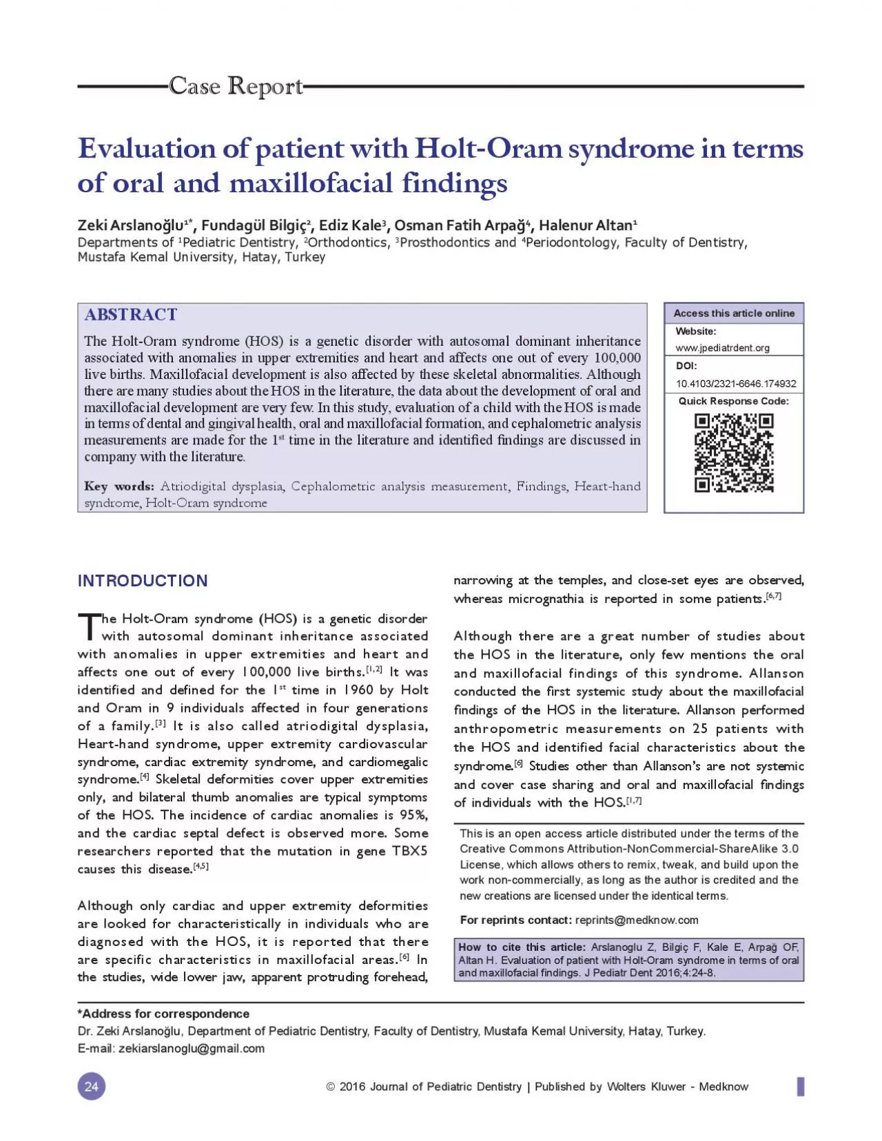
24 Evaluation of patient with HoltOram syndrome in terms Osman Fatih Arpa Halenur AltanPediatric Dentistry Periodontology Faculty of Dentistry Mustafa Kemal University Hatay Turkey INTRODUCT ID: 936880

hos patient syndrome upper patient hos upper syndrome ndings dentistry allanson heart pediatric anomalies maxillofacial dental literature analysis teeth

Share:

Link:

Embed:

Download Presentation from below link

Download Pdf The PPT/PDF document "2016 Journal of Pediatric Dentistry Pub..." is the property of its rightful owner. Permission is granted to download and print the materials on this web site for personal, non-commercial use only, and to display it on your personal computer provided you do not modify the materials and that you retain all copyright notices contained in the materials. By downloading content from our website, you accept the terms of this agreement.


Presentation Transcript

© 2016 Journal of Pediatric Dentistry | Published by Wolters Kluwer - Medknow 24 Evaluation of patient with Holt-Oram syndrome in terms , Osman Fatih Arpa, Halenur AltanPediatric Dentistry, Periodontology, Faculty of Dentistry, Mustafa Kemal University, Hatay, Turkey INTRODUCTION with autosomal dominant inheritance associated with anomalies in upper extremities and heart and It was time in 1960 by Holt and Oram in 9 individuals affected in four generations It is also called atriodigital dysplasia, Heart-hand syndrome, upper extremity cardiovascular Skeletal deformities cover upper extremities of the HOS. The incidence of cardiac anomalies is 95%, and the cardiac septal defect is observed more. Some researchers reported that the mutation in gene TBX5 Although only cardiac and upper extremity deformities are looked for characteristically in individuals who are diagnosed with the HOS, it is reported that there In the studies, wide lower jaw, apparent protruding forehead, narrowing at the temples, and close-set eyes are observed, Although there are a great number of studies about the HOS in the literature, only few mentions the oral and maxillofacial findings of this syndrome. Allanson rst systemic study about the maxillofacial ndings of the HOS in the literature. Allanson performed anthropometric measurements on 25 patients with the HOS and identified facial characteristics about the Studies other than Allanson’s are not

systemic ndings The Holt-Oram syndrome (HOS) is a genetic disorder with autosomal dominant inheritance associated with anomalies in upper extremities and heart and affects one out of every 100,000 live births. Maxillofacial development is also affected by these skeletal abnormalities. Although maxillofacial development are very few. In this study, evaluation of a child with the HOS is made time in the literature and identi ed ndings are discussed in Key words: Atriodigital dysplasia, Cephalometric analysis measurement, Findings, Heart-hand syndrome, Holt-Oram syndrome Website:www.jpediatrdent.org Dr. Zeki Arslanolu, Department of Pediatric Dentistry, Faculty of Dentistry, Mustafa Kemal University, Hatay, Turkey. This is an open access article distributed under the terms of the Creative Commons Attribution-NonCommercial-ShareAlike 3.0 License, which allows others to remix, tweak, and build upon the work non-commercially, as long as the author is credited and the reprints@medknow.com Arslanoglu Z, Bilgiç F, Kale E, Arpa OF, ndings. J Pediatr Dent 2016;4:24-8. Journal of Pediatric Dentistry / Jan-Apr 2016 / Vol 4 | Issue 1 25 in terms of dental and gingival health, dental plaque, oral and maxillofacial formation, and cephalometric analysis time in the literature ed ndings are discussed in company with the CASE REPORT the HOS applied to our clinic with complaints of ache in his decayed teeth. After the anamnesis was taken, we l

earnt that the patient has a congenital cardiac anomaly, had an associated operation 2 years ago, and had a series of operations about his forearm skeletal structure and finger structure. In the patient’s family history, no consanguineous marriage was observed, and it was ed after a genetic test performed using the amniotic uid in the 3 month of pregnancy that a sibling of the patient had the same syndrome, and the baby was aborted at the own request of the family. Forearm anomaly was found out in the extra oral examination, and the history of mild mental retardation and heart disease was learnt from the patient’s history [Figures 1 and 2]. During the ed that the patient, compared with the normal patients, has a narrower upper dental arch, more posterior cross bite, increased overjet, decreased overbite, and retrognathic and narrower mandibula [Figure 3]. Long facial structure, which the patient’s failure to make nasal breathing comfortably. In addition, an increase in the dentoalveolar height, V-shaped narrow and deep palate, small nostrils, and The cephalometric film of the patient was taken, and maxillofacial skeleton and malpositioned teeth were analyzed with the VistaDent cephalometric analysis As a result of the analysis, it was found out that vertical direction size has increased, upper and lower front facial height has increased equally, maxillary deep bite exists, mandibular hypoplasia exists, ramus length has Figure 2:

Right side view of the patient Figure 3: View of increase in the dentoalveolar height Figure 4: View of retrognathic and narrower mandibula Journal of Pediatric Dentistry / Jan-Apr 2016 / Vol 4 | Issue 1 26 shortened, mandibular plane has posterior rotation, anterior skull base has shortened, corpus length has shortened, posterior skull base has shortened, maxilla is positioned at the back compared with its skull base, upper incisors are protrusive, lower incisors are retrusive, and le.The patient was subjected to periodontal examination, and plaque index (PI) and gingival index (GI) was taken. Accordingly, an intensive plaque formation was observed 1.27 on an average. The GI value was measured 1.44. The values were observed between 2 and 7 mm in probing pocket depth. When the amount of attached gingiva was 3 and 10 mm. The highest level of attached gingiva was found 10 mm in the upper canine-premolar area. An was 6.25 mm, whereas it was found 6 mm in lower incisor area. In general, the average amount of attached ed that the teeth number 16, 36, and 46 had deep decay, and the fistula was found in soft tissue in line with the root in teeth 36 and 46. The extraction of teeth number 36 and 46 was made, after consultation with the pediatric cardiologist who followed the patient. The composite restoration was applied and curettage procedures were carried out. The patient and DISCUSSION AND CONCLUSION ned as a genetic disease with auto

somal dominant inheritance with accompanying preaxial radial anomalies in upper extremities and congenital heart Skeletal deformities cover upper extremities only, and bilateral thumb anomalies are typical symptoms of the HOS. The incidence of cardiac anomalies is 95%, Findings nition of the syndrome, and it was reported by the orthopedics and cardiology clinics that he had a series of operations and is still ndings in the myopic ed in nding of c feature of individuals with the Our case is also compatible with this data.In his article, Turleau reported mental retardation in the No data are found about mental ed that the patient has mild mental retardation and is being followed by the In his article, Lichiardopol shared the findings of micrognathia, dental malpositions, and hypoplastic mandible Arturo identiÞ ed hemifacial microsomia on the right side, depressed nasal bridge, and micrognathia in his patient In this context, Allanson conducted the most comprehensive study. In his study, Allanson made craniofacial measurements in 25 patients and presented ndings about the structure of jaw as well. Allanson and mandibula is wide in the patients with HOS. Our ndings of Allanson.Regarding the gingiva and surrounding dental tissues, there is no information in the literature about the HOS. In a lm Journal of Pediatric Dentistry / Jan-Apr 2016 / Vol 4 | Issue 1 27 Table 1: Vistadent cephalometric analysis table of patient