/
The cruciate ligaments are tough fibrous bands thatproximal tibia shin The cruciate ligaments are tough fibrous bands thatproximal tibia shin

The cruciate ligaments are tough fibrous bands thatproximal tibia shin - PDF document

audrey
audrey . @audrey
Follow
348 views
Uploaded On 2021-09-25

The cruciate ligaments are tough fibrous bands thatproximal tibia shin - PPT Presentation

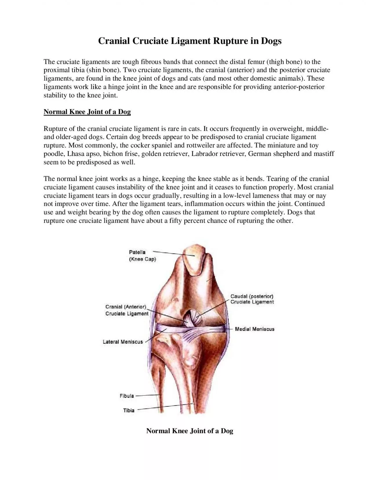
Rupture of the cranial cruciate ligament is rare in cats It occurs frequently in overweight middleeeds appear to be predisposed to cranial cruciate ligament rupture Most commonly the cocker spaniel a ID: 885592

cruciate ligament knee joint ligament cruciate joint knee cranial dogs rupture tibia surgery normal stabilization ligaments extracapsular torn sign

Share:

Link:

Embed:

Download Presentation from below link

Download Pdf The PPT/PDF document "The cruciate ligaments are tough fibrous..." is the property of its rightful owner. Permission is granted to download and print the materials on this web site for personal, non-commercial use only, and to display it on your personal computer provided you do not modify the materials and that you retain all copyright notices contained in the materials. By downloading content from our website, you accept the terms of this agreement.


Presentation Transcript

1 The cruciate ligaments are tough fibrous
The cruciate ligaments are tough fibrous bands thatproximal tibia (shin bone). Two cruciate ligaments, the cranial (anterior) and the posterior cruciate ligaments, are found in the knee joint of dogs and cats (and most other domestic animals). These ligaments work like a hinge joint in the knee and stability to the knee joint. Normal Knee Joint of a Dog Rupture of the cranial cruciate ligament is rare in cats. It occurs frequently in overweight, middle-eeds appear to be predisposed to cranial cruciate ligament rupture. Most commonly, the cocker spaniel and rottweiler are affected. The miniature and toy en retriever, Labrador retriever, German shepherd and mastiff seem to be predisposed as well. The normal knee joint works as a hinge, keeping the knee stable as it bends. Tearing of the cranial cruciate ligament causes instability of the knee joincruciate ligament tears in dogs occur gradually, resulting in a low-level lameness that may or nay not improve over time. After the ligament tears, inflammation occurs within the joint. Continued e ligament to rupture completely. Dogs that rupture one cruciate ligament ha Normal Knee Joint of a Dog ly. Similar to cranial cruciate ligament rupture in humans, resulting from athletic injuries to the knee, dogs can tear this ligament by jumping up to catch a ball or Frisbee or Symptoms usually include a h

2 istory of acute rear leg lameness. The l
istory of acute rear leg lameness. The lameness can be mild, resolve, and then appear again. Some dogs are compleKnee joint pain is usually a common symptom of cranial cruciate ligament rupture. The pain may be unapparent until someone accidentally manipulates the dog’s leg. A nip or bite from a normally in pain. The knee may appmay sound crunchy when put through a series of flexion-extension manipulations. Diagnosis of cranial cruciate ligament rupture is usually made by a positive cranial drawer sign. In posterior pressure is applied to the proximal tibia. Sliding of the distal femur over the proximal tibia (positive drawer sign) indicates cranial cruciate ligament rupture. Often, with a chronic injury, the cranial drawer sign is less effective due some joint stabilization resulting from a build-up of scar tissue in the joint capsule. A ruptured anterior cruciate ligament is not always an obvious condition. Careful manipulation and palpation of the knee is required for obtaining an accurate diagnosis. Often, heavy sedation or general anesthesia is required to conduct this examination or cruciate ligament can lead to degenerative joint disease (degenerative arthritis) of the knee. Approximately forty to fifty percent cruciate ligaments lead to injuries of the meniscus. A torn meniscus is often felt as a “click” when Surgery

3 is the treatment of choice for a rupture
is the treatment of choice for a ruptured anterior cruciate ligamenthas not significantly developed, most dogs have a good chance of recovering normal or almost normal function of the knee joint after surgery. Some small dogs; however, show signs of improvement with just rest and limited exercise. Prognosis after surgery depends on several factors. Obese dogs tend to recover more slowly than dogs in good athletic condition. The longer the interval between injury to the knee and surgery, the scar tissue formation and arthritis. Two treatments exist for CCR, surgical and non-su required. If the animal does not improve during this periodrgery. The principal of the surgery is to stabilize the femur on the tibia. There are two categories of surgery for repairing torn cranial cruciate ligaments - Intracapsular Stabilization and Extracapsular Stabilization. ization method requires replacing the torn ligament withplacing the torn ligament. Extracapsular stabilization is performed outside the joint and restores the function of the cruciate ligament rather than replacing the damaged ligament. Over the years, extracapsular stabilization has become the preferred method for repairing cranial cruciate ligament rupture in dogs. Currently there are 3 extracapsular stabilization techniques that are commonly performed - Lateral and Tibial Plateau Leveling Os

4 teotomy (TPLO). joint in a configuratio
teotomy (TPLO). joint in a configuration that restores the normal position of the femur on the tibia. The drawback implant may rupture, leading to a and development of arthritis. Fibular head transposition is an extracapsular repair technique that uses the lateral collateral ligament to stabilize the knee joint. After cranial transposition of ththe lateral collateral ligament is redirected to resemble that of the cranial cruciate ligament. The ng the attachments of the fibular head to the tibia and moving it er sign has been eliminated. Osteotomy (TPLO) Tibial Plateau Leveling Osteotomy (TPLO) is based on the premise that any kind of replacement for Instead of trying to replace the damaged ligament, this technique changes the anatomy of the knee joint by eliminating the slope in the tibial plateau. The procedure requires the surgeon to cut the tibia and rotate it so it is level to the tibia plateau. The the tibia by a custom platthe bone takes about two months alarge dogs and can also be performed on small dogs. The outcome of this surgery generally yields excellent results. Prevention ent tearing of the cranial cruciate ligament; however, there are some dogs. A good, regular exercise regime is just as important for dogs as it is for peRuptured anterior cruciate ligament is a commtment that is best for your pet. Copyright 2010 - VetNetwo