/
Neural Tissues Digital Laboratory Neural Tissues Digital Laboratory

Neural Tissues Digital Laboratory - PowerPoint Presentation

ava
ava . @ava
Follow
342 views
Uploaded On 2022-06-08

Neural Tissues Digital Laboratory - PPT Presentation

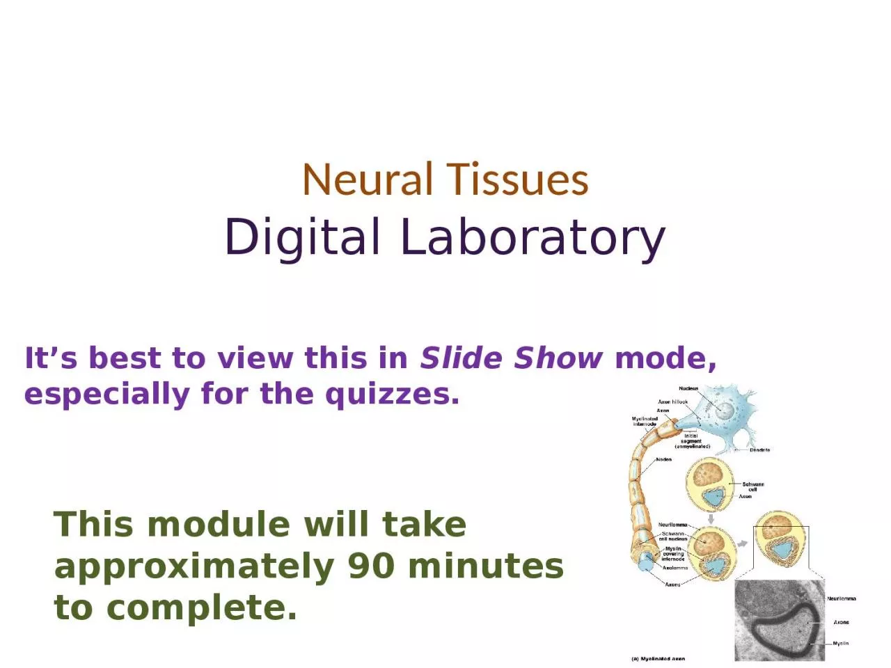
Its best to view this in Slide Show mode especially for the quizzes This module will take approximately 90 minutes to complete After completing this exercise you should be able to   Distinguish at the light microscope level each of the following components of neural tissue ID: 915079

peripheral cell axon axons cell peripheral axons axon identify nerves cord spinal cells matter slide schwann tissue arrows quiz

Share:

Link:

Embed:

Download Presentation from below link

Download Presentation The PPT/PDF document "Neural Tissues Digital Laboratory" is the property of its rightful owner. Permission is granted to download and print the materials on this web site for personal, non-commercial use only, and to display it on your personal computer provided you do not modify the materials and that you retain all copyright notices contained in the materials. By downloading content from our website, you accept the terms of this agreement.


Presentation Transcript

Slide1

Neural TissuesDigital Laboratory

It’s best to view this in Slide Show mode, especially for the quizzes.

This module will take approximately 90 minutes to complete.

Slide2

After completing this exercise, you should be able to:

 Distinguish, at the light microscope level, each of the following components of neural tissue:In spinal cord gray matter (note several stains were used)

Nerve cell bodyNucleolus

Nissl

substance (RER)

Axon

Axon hillock

Dendrites

In spinal cord white matter (note several stains were used)

Axons and dendrites (indistinguishable)

In prepared nerve specimen

Myelinated

axon

Node of Ranvier

Peripheral nerve (in any orientation)

Connective tissue of peripheral nerve

Epineurium

Perineurium

Endoneurium

Fascicle

Axon

Myelin

Schwann cell nuclei

Ganglia

Neuronal cell bodies

Satellite cells

Bundles of axons and dendrites

Distinguish, at the electron microscope level, each of the following components of neural tissue:

Peripheral nerve

Schwann cell

Myelin

Axon

Myelinated

Ensheathed

(

unmyelinated

)

Slide3

In this lab, we will examine cells, ganglia, nerves, and sensory endings of the peripheral nervous system

0This diagram shows a cross section through the spinal cord, the dorsal root and ventral root nerves, spinal ganglia (red arrow) and sensory nerve endings (green arrow).

In this lab, the major foci will be:

--the spinal cord

--peripheral nerves

--ganglia

Organization of the nervous system

Slide4

Organization of the nervous system

Illustrations of some neurons. Note the cell body, dendrites, axon. Don’t worry about the details, but appreciate the wide variety of shapes of neurons. Also, recall that glial cells (e.g. oligodendrocyte)

are support cells that are found throughout the nervous system.

Slide5

0

Back to this drawing, note that, for the most part, neuronal cell bodies gather together in specific locations (orange arrows), while axons (and long dendrites) gather together in others (pink arrows).

This slide only shows a few cells – obviously in real tissue, there are many cells or cell processes in each region.

Also note that in regions where cell bodies are gathered, there are also axons and dendrites that are “coming from and going to” the cell bodies.

Organization of the nervous system

Slide6

0

The first set of slides is from the spinal cord. The spinal cord has two major regions:--gray matter, which has neuronal cell bodies--white matter, which has axons and long dendrites

SPINAL CORD

Gray matter and white matter are shaded appropriately on this image.

Slide7

Spinal cord – gray matter

Video of spinal cord showing gray and white matter – SL181

Link to SL 181Be able to identify:

Gray matter

White matter

Note these are not in the objectives, but you need to do this to proceed

Slide8

We’re going to look first at large, multipolar neurons, which are neurons with an axon and several dendrites. An excellent example is the motor neurons of the peripheral nervous system, which have cell bodies located in the spinal cord.

The scanning view below shows a cross section through the spinal cord, with the grey matter (marked by dotted line), which contains the cell bodies, and the white matter, which contains bundles of myelinated axons.

The boxed area is enlarged at right. Now the large neuronal cell bodies are evident, as purple blotches (arrows indicate some examples)

SPINAL CORD – Gray matter

Slide9

This medium power view shows several multipolar motorneuron cell bodies (green arrows).

(The smaller, round nuclei (red arrows) are associated with oligodendrocytes, non-neuronal cells. Advance for a higher power view….

SPINAL CORD – Gray matter

Slide10

The nucleus of a neuron is large and round, with a prominent nucleolus (yellow arrow), and abundant euchromatin (pale area surrounding nucleolus). The black arrows delimit the nucleus.

The cytoplasm in the cell body, or perikaryon, is rich in clusters of rough endoplasmic reticulum, which in neurons are termed Nissl substance or Nissl bodies (the blue/purple flaky material).Dendrites (green arrows) contain Nissl substance, while axons (next slide) can be recognized because they lack Nissl substance.

SPINAL CORD – high power view of gray matter

The small, condensed nuclei (red arrows) belong to glial cell. Their cytoplasm is either not visible or barely visible.

Slide11

0

SPINAL CORD – high power view of gray matter

Although dendrites are easy to identify definitively because of their number and the presence of

Nissl

substance, axons are much more difficult to find. In this fortuitous image, an axon is indicated by the arrow. The “tapered” beginning of the axon is the axon hillock (outlined). Note the lack of Nissl substance in the axon.

Slide12

Spinal cord – gray matter

Video of spinal cord showing neuronal cell bodies and glial cells – SL181

Link to SL 181Be able to identify:

Neuronal cell bodies

Cytoplasm with

Nissl

substance

Nucleus

Nucleolus

Axon

Dendrite

Glial cells

Slide13

Spinal cord – gray matter

Video of spinal cord showing neuronal cell bodies and glial cells – SL045B

Link to SL 045BBe able to identify:

Nothing specific, appreciate density of axons and dendrites in gray matter

Slide14

0

Nissl substance

capillary

Euchromatic nucleus

Nucleolus

Spinal cord – gray matter

This transmission EM shows the cell body of a

motorneuron

.

The small, dark, ring shapes (near the capillary, for example) are myelin sheaths surrounding neuronal processes.

Slide15

0

Nissl substance

(rER)

mitochondria

Spinal cord – gray matter

This high magnification transmission EM shows the

Nissl

substance (

rER

).

Slide16

0

The white matter of the spinal cord (e.g. blue box) contains myelinated axons (and some dendrites) that are sending information up and down the central nervous system.Because the fibers are oriented cranial-caudal, they are cut in cross-section in our sections of the spinal cord.

SPINAL CORD – white matter

Slide17

Spinal cord –

WHite matter

Video of spinal cord showing white matter detail – SL181

Link to

SL 181

Be able to identify:

Nothing, since there is not much to see in the white matter on this slide

Slide18

0

Special stains can be used to visualize the components of the white matter. Here, the section was labeled with a silver stain, which stains proteins. In this section, the stain nicely highlights axons (yellow arrows), which are surrounded by pale regions representing myelin that was dissolved during preparation.

SPINAL CORD – white matter

Slide19

Spinal cord –

WHite matter

Video of spinal cord showing white matter silver stain – SL045A

Link to

SL 045A

and

SL 045B

Be able to identify:

Axons

Myelin

Slide20

0

Special stains can be used to visualize the components of the white matter. This slide has been carefully prepared to preserve and stain myelin (between arrows).

SPINAL CORD – white matter

Slide21

Spinal cord –

WHite matter

Video of spinal cord showing white matter myelin stained – SL168

Link to

SL 168

Be able to identify:

Axons

Myelin

Slide22

0

From a histological standpoint, all peripheral nerves are essentially the same, so don’t worry about whether a peripheral nerve is a dorsal root, spinal nerve, etc.

Nerves contain axons (and sometimes long dendrites) together with supporting cells, embedded within connective tissue and surrounded by a highly cellular connective tissue layer. Neuronal cell bodies are not

found in nerves.

The diagram shows several locations where nerves can be found.

Peripheral nerves

Slide23

ensheathment

myelination

Peripheral nerves – individual axons

In the peripheral nervous system, individual axons (and large dendrites) are surrounded by

Schwann cells

. During embryogenesis, the Schwann cell initially surrounds several axons with portions of its cytoplasm and plasma membrane. This arrangement is termed

ensheathment

(left image, the red arrows will be explained on the next slide).

Smaller caliber axons remain

ensheathed

. When large axons are present, the Schwann cell selects a single axon to support, and wraps its cytoplasm and plasma membrane around the axon numerous times. After the cytoplasm is squeezed out, the remaining layers of Schwann cell membrane are called

myelin

. A single axon is

myelinated

by many Schwann cells; regions of bare axon not surrounded by myelin are called

nodes of

Ranvier

(right image).

Slide24

0

Peripheral nerves – individual axons

Close observation indicates “gaps” in the Schwann cell (red arrows), indicating that the Schwann cell surrounds the axons, which remain outside the Schwann cell (see previous slide). Looking at a single axon, you see “dark-light-dark” (between green arrows) – this is actually two plasma membranes, one for the axon, one belonging to the Schwann cell. The dark-light-dark indicated by the purple arrow is actually the Schwann cell plasma membrane and its external lamina (similar to basal lamina). It might be a good idea to trace a Schwann cell plasma membrane around a few axons to help you differentiate it from the axonal membranes and from the external lamina of the Schwann cell.

The stippled structures (e.g. outlined in blue) between the Schwann cells are collagen (reticular) fibers.

Scc

Scc

Scc

TEM of ~40 axons ensheathed by several Schwann cells. Each Schwann cell may ensheath several axons.

The cytoplasm of some Schwann cells is indicated (Scc).

Schwann cell nuclei may be visible, but often are out of the plane of the section.

Slide25

0

Peripheral nerves – individual axons

The numerous membranes between the purple arrows all belong to a single Schwann cell, with the exception of the innermost one, which is the axonal membrane. The main structure of the Schwann cell, including its nucleus, is out of the plane of section; its external lamina is visible (blue arrows)

The cytoplasm of the axon contains organelles, and cross-sections of microtubules (red arrow) and neurofilaments (intermediate filaments).

Electron micrograph of a

myelinated

axon (Ax) in cross-section.

Schwann cell

myelinating

an axon

Slide26

This light micrograph shows a longitudinal view of a nerve bundle that has been teased apart to separate individual axons, and then stained with osmium tetroxide, which labels the myelin membrane. The bare portion of one axon, a node of Ranvier, is indicated.

Peripheral nerves – individual axons

Slide27

Video of teased nerve stained for myelin – SL044

Link to SL 044Be able to identify:Axons

MyelinNode of Ranvier

Peripheral nerves – individual axons

Slide28

Peripheral nerves – H&E

This light micrograph shows a large nerve in H&E. The top portion of the image shows the individual nerves in cross-section, while the bottom shows them in longitudinal section. In H&E, the axons stain eosinophilic (green arrows), though they usually look a little purple. Much of the myelin washes out in tissue preparation, so the area around the axon is pale-pink (edge of the myelin indicated by the blue arrows).

Slide29

Peripheral nerves – H&E

A light micrograph similar to the previous slide, showing nodes of Ranvier (blue circles). Note the myelin tapers near the node, and that the axon is continuous through the node. So cool!!!

Slide30

Video of axilla showing peripheral nerves – SL12

Link to SL 012Be able to identify:Axons

MyelinNode of Ranvier

Peripheral nerves – H&E

Slide31

Peripheral nerves – H&E

Smaller

peripheral nerves

(outlined) can be found throughout the body. The details of the axon and myelin sheath are lost in nerves this size. However, several distinguishing features allow us to distinguish the nerve from the surrounding connective tissue:

pale

eosinophilia

- again, due to the washed out myelin

wavy appearance – nerves are elastic, and recoil on tissue preparation

a distinct

perineurium

(arrows) – we’ll discuss this layer later in the module

numerous nuclei – most of these are Schwann cells

Slide32

Video of esophagus showing peripheral nerves – SL16

Link to SL 016 & SL 023 &

SL 024 & SL 047Be able to identify:

Peripheral nerve

Peripheral nerves – H&E

Video of esophagus showing peripheral nerves – SL23

Video of lung

hilus

showing peripheral nerves – SL24

Video of spermatic cord showing peripheral nerves – SL47

Slide33

Peripheral nerves – Connective tissue organization

This is a light micrograph taken at low power from the same slide of the axilla / brachial plexus you looked at previously. For orientation, the small image to the right is the same high magnification image you saw before; the blue box indicates the position of a similar region in the larger image.

Slide34

Peripheral nerves – Connective tissue organization

Peripheral nerves are axons bundled together by connective tissues: endoneurium

– a loose connective tissue between axons perineurium (arrows) – a cellular layer that bundles many axons into structures called

fascicles

(two fascicles, one small, one large, are on this image)

epineurium

(brackets) – a dense connective tissue, often containing adipose, that bundles fascicles together

Endoneurium is throughout this region

(see next slide)

Slide35

Back to the high power image we looked at before, of a portion of a

fascicle - you can see:--the endoneurium (green arrows) is between the myelinated

axons and is loose connective tissue--the perineurium (between black arrows) is a highly cellular layer with scant ECM, which has some characteristic features of epithelium, including

occludens

junctions

--small amounts of

epineurium

in the upper left and right corners

Peripheral nerves – Connective tissue organization

Slide36

Video of axilla showing connective tissues of peripheral nerves – SL12

Link to SL 012Be able to identify:Endoneurium

Perineurium

Fascicle

Epineurium

Peripheral nerves – Connective tissue organization

Slide37

As larger nerves, i.e. bundles of fascicles, branch, they become smaller

peripheral nerves

(outlined), which are essentially a single fascicle. Therefore, these contain only

endoneurium

and

perineurium

(black arrow); there is no epineurium. These fascicles are surrounded by connective tissues of the organ or structure in which they are located. The

perineurium

clearly delineates the peripheral nerve from the surrounding connective tissue, and is useful in distinguishing peripheral nerves from connective and other tissues you will encounter later (e.g. smooth muscle).

Peripheral nerves – Connective tissue organization

Endoneurium

is throughout here

Slide38

Link to SL 016 &

SL 023 & SL 024 & SL 047Be able to identify:

perineurium

Video of spermatic cord showing connective tissue of peripheral nerves – SL47

Peripheral nerves – Connective tissue organization

Slide39

0

Ganglia consist of clusters of neuronal cell bodies, together with supporting cells, contained within a connective tissue capsule.

Ganglia

We will be looking at several different ganglia in this module. For completeness, we will indicate which ganglion is shown. Note that we do not expect you to name specific ganglia for a histology exam - just recognizing a structure as a ganglion will suffice.

Slide40

Dorsal Root Ganglion

Ganglia

This dorsal root ganglion is adjacent to the spinal cord, and contains sensory neuronal cell bodies (black arrows). In this low-power image, you can barely see the nuclei of supporting cells. Between the cell bodies are axons and dendrites. The ganglion is contained within a connective tissue capsule (blue arrows).

Slide41

Ganglia

A high magnification image shows large neuronal cell bodies with euchromatic nuclei (black arrows mark the edge of one nucleus) and prominent nucleolus (blue arrow). The nuclei surrounding the neurons belong to supporting cells called

satellite cells

(green arrows). The tissue between the neurons is mostly axons and dendrites, with a small amount of loose connective tissue.

Slide42

Video of dorsal root ganglion – SL50

Link to SL 050Be able to identify:Ganglion

Neuronal cell bodiesSatellite cells

Axons and dendrites

Ganglia

Slide43

Ganglia

This is an image of the semilunar ganglion, a structure belonging to one of the cranial nerves, which is analogous to the dorsal root ganglion. Black arrows = nuclear membrane, blue arrow is nucleolus, green arrows indicate satellite cells.

Slide44

Video of semilunar ganglion – SL49

Link to SL 049Be able to identify:Ganglion

Neuronal cell bodiesSatellite cells

Axons and dendrites

Ganglia

Slide45

Ganglia

FYI for this module (you’ll learn more about this organization in other parts of the course)...Autonomic ganglia are located away from the central nervous system or within the wall of the organ.

Slide46

Ganglia

This is an image from a ganglion of the sympathetic chain, a structure belonging to one of the cranial nerves, which is analogous to a dorsal root ganglion. Black arrows indicate the nuclear membrane, blue arrow is nucleolus, green arrows indicate satellite cells.

Slide47

Video of sympathetic ganglion – SL51

Link to SL 051Be able to identify:Ganglion

Neuronal cell bodiesSatellite cells

Axons and dendrites

Ganglia

Slide48

Ganglia

This is an image of a parasympathetic ganglion (between green arrows). Note the perineurium (at tips of green arrows), and the neuronal cell bodies with basophilic cytoplasm.

Slide49

Video of colon showing parasympathetic ganglion – SL53

Link to SL 053 & SL 014 &

SL 016 & SL 023

Be able to identify:

Ganglion

Neuronal cell bodies

Axons and dendrites

Ganglia

Video of jejunum showing parasympathetic ganglion – SL14

Video of esophagus showing parasympathetic ganglion – SL16

Slide50

The next set of slides is a quiz for this module. You should review the structures covered in this module, and try to visualize each of these in light and electron micrographs.

 Distinguish, at the light microscope level, each of the following components of neural tissue:In spinal cord gray matter (note several stains were used)

Nerve cell bodyNucleolus

Nissle

substance (RER)

Axon

Axon hillock

Dendrites

In spinal cord white matter (note several stains were used)

Axons and dendrites (indistinguishable)

In prepared nerve specimen

Myelinated

axon

Node of Ranvier

Peripheral nerve (in any orientation)

Connective tissue of peripheral nerve

Epineurium

Perineurium

Endoneurium

Fascicle

Axon

Myelin

Schwann cell nuclei

Ganglia

Neuronal cell bodies

Satellite cells

Bundles of axons and dendrites

Distinguish, at the electron microscope level, each of the following components of neural tissue:

Peripheral nerve

Schwann cell

Myelin

Axon

Myelinated

Ensheathed

(

unmyelinated

)

Note that the term “neural tissue” is nowhere to be found on this list, except in the headings, where a term like “epithelium” would be. Just saying.

Slide51

0

Identify structures A and B

What histological characteristics of this neuron reflect its high synthetic activity?

A

B

A = dendrite

B = axon

Abundant Nissl substance

(rER) and euchromatic chromatin

Final quiz

Slide52

Final quiz

Self-check: Identify the outlined

REGION

. (advance slide for answer)

epineurium

Slide53

Final quiz

Self-check: Identify the structure on this slide. (advance slide for answers)

ganglion

Slide54

Final quiz

Self-check: Identify the tissue. (advance slide for answers)

UnilocularAdipose

tissue

Slide55

Final quiz

Self-check: Identify the structure. (advance slide for answer)

perineurium

Slide56

0

Nuclei at A might belong to what kinds of cells?What is the structure at B?

What is the dark thread-like line at C?

What occupies the space between the arrowheads?

B

A

C

Schwann cells, fibroblasts. Less likely: endothelial cells, white blood cells. NOT neurons!

A node of Ranvier

An axon

Myelin membrane

Final quiz

Slide57

Final quiz

Self-check: Identify A-D. (advance slide for answers)

A

B

C

D

Schwann cell nucleus

Schwann cell cytoplasm

Myelinated

axon

Unmyelinated

axon

Slide58

Final quiz

Self-check: Identify the tissue closest to the arrows. (advance slide for answers)

Stratified squamous (non keratinized)

Slide59

Final quiz

Self-check: Identify the structures. (advance slide for answers)

Most likely a glial cell nucleus

nucleolus

Nissl

substance (

rER

)

Slide60

Final quiz

Self-check: Identify the structure. (advance slide for answers)

Artifact – broken nerve, note the right angles

These are nodes

Slide61

Final quiz

Self-check: Identify the structure. (advance slide for answers)

Peripheral nerve

Slide62

Final quiz

Self-check: Identify the structures. (advance slide for answers)

myelin

axon

Slide63

Final quiz

Self-check: Identify the structures. (advance slide for answers)

neuron

ganglion

Slide64

Final quiz

Self-check: Identify. (advance slide for answers)

X

X

Schwann cell

axon

Slide65

Final quiz

Self-check: Identify the structures or cells. (advance slide for answers)

Nuclear membrane

Satellite cells

Axon or dendrite

Slide66

Final quiz

Self-check: Identify the structures. (advance slide for answers)

Hmmm….definitely an axon or dendrite, can’t tell which one

nucleus

Either an axon or dendrite

Slide67

Final quiz

Self-check: Identify the outlined tissue (advance slide for answers)

Multilocular

adipose

Slide68

Final quiz

Self-check: Identify the structures. (advance slide for answers)

epineurium

perineurium

Slide69

Final quiz

Self-check: Identify 1, 2, 5, 7. (advance slide for answers)

Nucleus of Schwann cellSchwann cell cytoplasm(cytoplasm of another Schwann cell)Golgi

Axons or dendrites

External lamina

Collagen

Slide70

Final quiz

Self-check: Identify the structure and cells. (advance slide for answers)

neuron

ganglion

Most likely Schwann cells

Slide71

Final quiz

Self-check: Identify the structures. (advance slide for answers)

perineurium

axon

axon

myelin

epineurium

endoneurium

Slide72

Final quiz

Self-check: Identify the structure. (advance slide for answers)

Peripheral nerve

Schwann cell nuclei (most likely)

perineurium

Slide73

Final quiz

Self-check: Identify the outlined

TISSUE

. (advance slide for answer)

Multilocular

adipose

Slide74

Final quiz

Self-check: Identify the structures. (advance slide for answers)

axon

myelin

Slide75

Final quiz

Self-check: Identify the structures or tissues. (advance slide for answers)

Dense irregular connective tissue

Peripheral nerve

Slide76

Final quiz

Self-check: Identify the structure. (advance slide for answers)

ganglion

Slide77

Final quiz

Self-check: Identify all. (advance slide for answers)

Neuron, (actually touching the nucleolus)

Loose connective tissue

Most likely Schwann cells

ganglion