/
Tibia Talus Fibula Motivation: Tibia Talus Fibula Motivation:

Tibia Talus Fibula Motivation: - PowerPoint Presentation

bery
bery . @bery
Follow
64 views
Uploaded On 2024-01-03

Tibia Talus Fibula Motivation: - PPT Presentation

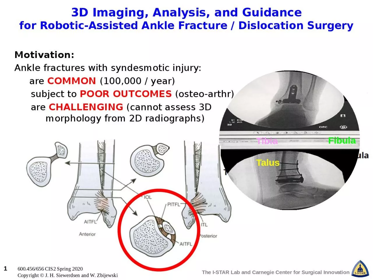
Ankle fractures with syndesmotic injury are COMMON 100000 year subject to POOR OUTCOMES osteo arthr are CHALLENGING cannot assess 3D morphology from 2D radiographs ID: 1037736

analysis ankle imaging assisted ankle analysis assisted imaging aim fracture guidancefor robotic dislocation syndesmosis minimum motorized control surgery students

Share:

Link:

Embed:

Download Presentation from below link

Download Presentation The PPT/PDF document "Tibia Talus Fibula Motivation:" is the property of its rightful owner. Permission is granted to download and print the materials on this web site for personal, non-commercial use only, and to display it on your personal computer provided you do not modify the materials and that you retain all copyright notices contained in the materials. By downloading content from our website, you accept the terms of this agreement.


Presentation Transcript

1. TibiaTalusFibulaMotivation:Ankle fractures with syndesmotic injury: are COMMON (100,000 / year)subject to POOR OUTCOMES (osteo-arthr)are CHALLENGING (cannot assess 3D morphology from 2D radiographs)3D Imaging, Analysis, and Guidancefor Robotic-Assisted Ankle Fracture / Dislocation Surgery

2. Pertinent Anatomy: Bones: Tibia, fibula, and talus Ligaments: AITFL, PITFL, IOL, and ITL A Complex Space: the syndesmosisVery challenging to assess the 3D morphology of the syndesmosis from 2D projection views  3D imaging and guidanceTibiaTalusFibula3D Imaging, Analysis, and Guidancefor Robotic-Assisted Ankle Fracture / Dislocation Surgery

3. Overall Project Goals:Develop a robot-assisted system for ankle reduction.Use intraoperative 3D imaging in concert with 2D radiographs (3D-2D registration) to guide reduction.Develop automated 3D image segmentation and analysis to precisely analyze the syndesmotic space.3D Imaging, Analysis, and Guidancefor Robotic-Assisted Ankle Fracture / Dislocation Surgery

4. Prototype JigControlled Motion(x,y,qz) of FibulaWhat Students Will Do: Aim 1Design and build a jig for controlled disruption of the syndesmosis.Minimum: Manual mechanical motionExpected: Motorized control 1 DoFMaximum: Motorized control 3 DoF3D Imaging, Analysis, and Guidancefor Robotic-Assisted Ankle Fracture / Dislocation SurgeryTurn away if squeamish.Cadaver SetupImaging + Controlled Disruptionof Syndesmosis

5. What Students Will Do: Aim 1Design and build a jig for controlled disruption of the syndesmosis.Minimum: Manual mechanical motionExpected: Motorized control 1 DoFMaximum: Motorized control 3 DoFWhat Students Will Do: Aim 2Develop automatic image segmentation and analysis of 3D ankle morphology.Minimum: Test existing (ASM) method in C-arm CBCTExpected: Develop more robust segmentation (using deep learning)Maximum: Combine with multi-body 3D-2D registration to guide reduction3D Imaging, Analysis, and Guidancefor Robotic-Assisted Ankle Fracture / Dislocation Surgery

6. What Students Will Do: Aim 1Design and build a jig for controlled disruption of the syndesmosis.Minimum: Manual mechanical motionExpected: Motorized control 1 DoFMaximum: Motorized control 3 DoFWhat Students Will Do: Aim 2Develop automatic image segmentation and analysis of 3D ankle morphology.Minimum: Test existing (ASM) method in C-arm CBCTExpected: Develop more robust segmentation (using deep learning)Maximum: Combine with multi-body 3D-2D registration to guide reductionWhat Students Will Do: Aim 3Characterize performance in laboratory experiments.Minimum: Measure accuracy of planned and executed motion (Aim 1)Expected: Measure accuracy of segmentation (Aim 2)Maximum: Integrate systems from Aims 1-2 to prescribe motion pattern for robot-assisted reduction of the syndesmosis.3D Imaging, Analysis, and Guidancefor Robotic-Assisted Ankle Fracture / Dislocation Surgery

7. Size group: 2–3Skills:Machining / instrumentation (ankle jig)Programming skills (e.g., Matlab and Python)Basic knowledge in machine learning(e.g., TensorFlow/PyTorch)Mentors:Dr. Jeffrey H. Siewerdsen(jeff.siewerdsen@jhu.edu)Dr. Wojtek Zbijewski(wzbijewski@jhu.edu)Superior ramus3D Imaging, Analysis, and Guidancefor Robotic-Assisted Ankle Fracture / Dislocation Surgery