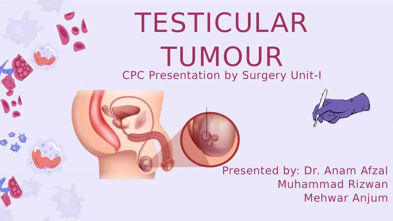
/
TESTICULAR TUMOUR CPC Presentation by Surgery Unit-I TESTICULAR TUMOUR CPC Presentation by Surgery Unit-I

TESTICULAR TUMOUR CPC Presentation by Surgery Unit-I - PowerPoint Presentation

bethany
bethany . @bethany
Follow
0 views
Uploaded On 2024-02-16

TESTICULAR TUMOUR CPC Presentation by Surgery Unit-I - PPT Presentation

Presented by Dr Anam Afzal Muhammad Rizwan Mehwar Anjum BIODATA AND PRESENTING COMPLAINT Biodata Presenting Complaint Swelling of right testicle for 2 years Muhammad Rehman so Muhammad Iqbal ID: 1046332

swelling normal lymph pain normal swelling pain lymph history testicular years complaint nodes size chest positive testicle tenderness bowel

Share:

Link:

Embed:

Download Presentation from below link

Download Presentation The PPT/PDF document "TESTICULAR TUMOUR CPC Presentation by Su..." is the property of its rightful owner. Permission is granted to download and print the materials on this web site for personal, non-commercial use only, and to display it on your personal computer provided you do not modify the materials and that you retain all copyright notices contained in the materials. By downloading content from our website, you accept the terms of this agreement.


Presentation Transcript

1. TESTICULAR TUMOURCPC Presentation by Surgery Unit-IPresented by: Dr. Anam AfzalMuhammad RizwanMehwar Anjum

2. BIODATA AND PRESENTING COMPLAINTBiodataPresenting Complaint:Swelling of right testicle --- for 2 yearsMuhammad Rehman s/o Muhammad Iqbal20 years oldUnmarried Muslim maleResident of Feroza, LiaquatpurIronsmith by professionPresented to OPD 1 month ago

3. HISTORY OF PRESENTING ILLNESSPatient was in his usual state of health, 2 years ago when he noticed swelling on right side of testicle. Patient narrates a history of trauma due to fall from an animal ride 2 years ago, with accompanying constant excruciating pain, relieved by medication from local doctors.Sudden in onset, it appeared at the size of a pea slowly progressive increased to the size of a tennis ball over 2 years.It was ovoid. Initially soft then hard in consistency. No color change in the swelling. It was associated with feeling of heaviness and no pain.

4. HISTORY OF PRESENTING ILLNESSThe swelling size doesn’t change with cough, walking or lying down. It is not associated with fever or constipation.There is no complaint of dysuria, burning micturition, and urinary incontinence or other genitourinary complaintsThere is no abdominal swelling or lower limb swellingNo swelling of the contralateral testesNo complaint of gynecomastia or undescended testes in childhoodThere is no complaint of hemoptysis, back ache or bone pain, decreased appetite, jaundice or other proofs of metastatic spread.Tunica vaginalisTestesSpermatic cordEpididymis

5. No dyspnea, chest pain, palpitations, SOB on exertion, PND, orthopnea or claudicationCARDIO-VASCULARREVIEW OF THE SYSTEMSRESPIRATORYNo complaint of hemoptysis, SOB, cough, sputum, Pleuritic chest pain, allergic rhinitis, or seasonal variation of coughNERVOUSSYSTEMNo Headache, ALOC, weakness of limbs, fits, difficulty in speech, difficulty in swallowing, vertigo, loss of hearing, numbness, paresthesiaMUSCULO-SKELETALNo bone pain, backache, joint pain/swelling, restriction of mobility, photosensitivity, oral rash or alopecia, ,TO FILLGASTRO-INTESTINALNo Nausea, vomiting, diarrhea, abdominal pain, abdominal distention, change in bowel habits, yellow discoloration of eyesGENITO-URINARYNo Oliguria, dysuria,, pyuria, , burning micturition, frothy urine, hematuria, renal colic or urinary incontinence, genital rash or dyspareunia

6. OTHER COMPONENTS OF HISTORYFAMILY HISTORYNo positive family history of similar disease or complaintPERSONAL HISTORYNaswar addict, normal sleep hygiene, normal bowel/bladder habits and diet SOCIO-ECONOMICHISTORYPatient appeared to be of average socioeconomic statusPAST MEDICALNo significant past medical history of any disease or comorbidityPAST SURGICALHistory of RTA; plaster onright forearm 5 years agoMEDICATION/ALLERGYNo history of medication except painkillers and no known allergies

7. GPE110/80 mm Hg86bpm16 breaths/min98 ℃POSITIVE FINDINGPallorNEGATIVE FINDINGSClubbingPalmer erythemaHepatic flapDupuytren's contractureKoilonychiaLeukonychiaPittingWastingOsler nodesRA deformitiesPOSITIVE FINDINGSConjunctival pallorNEGATIVE FINDINGSJaundiceExophthalmosPtosisWastingCushingoid faceOral ulcerCheilosisLeukoplakiaTongue cyanosisNEGATIVE FINDINGSNo lymph nodes palpableNo JVP distentionNo swelling in the neck

8. LOCAL EXAMINATIONOn inspection:Disparity between two testicles: right sided testicle was increased in size, left side looks normal. Skin colour normal looking, back of scrotum also normalNo other bulge examined on inspection.Penis also looks normal.01On Palpation:Normal temperatureNon-tenderWell circumscribedMobile and adherent to skinHard in consistencyTesticle could not be felt separate from swellingVas deferens and epididymis was not felt as wellSize of swelling measured to be 6x8cmGet above the swelling positiveTransillumination Positive 02Contralateral testes was normal03No inguinal lymph nodes palpable04

9. INSPECTION: No scar marks, bulging, deformity or pulsationPALPATION: Apex beat was localized at 5th IC, it was of normal character and volume, there was no tenderness, crepitus and no parasternal heaveAUSCULTATION: S1 and S2 were normal and there were no added sound or murmurSYSTEMIC EXAMINATIONHigher Mental Function: intact.Superficial reflexes: intactMotor System: IntactSensory System: intactCranial Nerves: intactSOMI: NegativeINSPECTION: No deformity, scar marks, bulging, pulsation or any apparent featurePALPATION: Chest expansion was 3 cm, trachea was central, apex beat was localized to 5th IC, No tenderness, crepitus and vocal fremitus was equivocal bilaterallyPERCUSSION: Upper border of liver was percussed at 6th ICS and anterior plus posterior percussion was normal bilaterallyAUSCULTATION:NVB with no added breath soundsINSPECTION: Poor oro-dental hygiene however the shape and symmetry of abdomen was normal, No scar marks, prominent veins or stria. Umbilicus was central and inverted and there were no hernial orificePALPATION: There was no tenderness, rebound tenderness, rigidity or guardingPALPATION OF VISCERA: No viscera was palpable. No lymph nodes palpablePERCUSSION: No shifting dullness or fluid thrill and normal span of liverAUSCULTATION: 3 bowel sounds were heard in one minute and no hepatic, renal or splenic bruit was heard

10. INVESTIGATIONS01HB: 13.0 g/dLT.L.C: 7500/nm3Platelet count: 268000/nm3PT: 14 secondsAPTT: 35 secondsAnti-HIV: NegativeAnti-HCV: NegativeHbsAg: NegativeBUN: 13mg/dLCreatinine: 1.0mg/dLBilirubin: 0.4mg/dLALT: 16U/LNa+: 140mmol/LK+: 3.7mmol/LChloride: 100mmol/LSEROLOGYTUMOR MARKERS02AFP: NormalBeta hCG: NormalLDH: 1923(high)

11. RADIOLOGICAL INVESTIGATIONSNo abnormal finding in the chestMatted enlarged lymph nodes noted adjacent to left vessels (Para-aortic) measuring approximately 4*4.3 cm.Right testicular lobulated enhancing mass with calcification measuring 6.5*5.2*8.3 cmLeft testicle is normal0304ULTRASOUNDCT-SCAN OF CHEST AND PELVISMultiple enlarged lymph nodes around Aorta and IVCLargest lymph node is measured 21mm in short axisRight testis is enlarged and replaced by neoplastic ,multiloculated mass. Mild amount of free fluid is present in epididymis Left testis and epididymis are normal

12. TESTICULAR TUMOURDIAGNOSISBased on history, examination and investigations, we reached the conclusion that the patient was a case of:

13. MANAGEMENTKeeping in view the diagnosis:Right-sided orchidectomy was planned and performedSample was sent for biopsyFurther management is awaiting the biopsy report

14. THANK YOU

15. Testicular TumorsDr. Anam Afzal1st Yr PGR Surgical Unit 1

16. Clinical FeaturesPainless Testicular SwellingTesticular HeavinessTesticular Pain Back painCough or DyspneaAnorexia , Nausea or VomitingBone PainLower Extremity Swelling

17. Risk factorsCryporchidismFamily historyEarly onset pubertyHIV/AIDSHypospadiasWhite males

18. Differential DiagnosisEpididymoorchitisHydroceleSpermatoceleHematoceleGranulomatous Orchitis

19. InvestigationsRoutine Hematology & BiochemistryTumor markersScrotal UltrasoundChest XraysAbdominal CT

20. Classification

21. Tumor MarkersHelp in Differential DiagnosisDirectly Proportional to tumor burdenIndicate histologyFollow up & RecurrenceBecome positive earlier than other investigations

22. Tumor MarkersTumor Markers Oncofetal SubstancesCellular enzymesAFPHCGPLAPLDHTrophoblastic cellsNormal below 16ng/mlHalf life 5-7 daysRaised inPure Embryonal carcinomaTeratocarcinomaYolk sac tumorCombined TumorSyncytiotrophoblastic cellsNormal <1ng/mlHalf Life 24-36 hoursRaised in100% Choriocarcinoma60% Embryonal carcinoma55% Teratocarcinoma25% Yolk sac tumor7% SeminomaLess specific than HCG or AFPSeminoma & NSGCTSpecially in seminomaReliable only in nonsmokers

23. Epidemiology1% of all malignant TumorsAffects young adults

24. Surgical anatomy

25. Staging

26. Reference: Bailey and Love

27. Treatment PolicyReference: Bailey and Love

28. Radiosensitivity and ChemosensitivityBEPBleomycin, Etoposide, CisplatinRPLND: Retroperitoneal lymph node dissection

29. SurvivalThe prognosis of testicular tumours depends on several factors, including the histological type and the stage at presentation. For seminoma, if there are no metastases, 90-95% of patients will be alive 5 years after diagnosis. If there are poor prognostic features, the survival rate drops to around 70%.For NSGCTs a 5-year survival rate of more than 90% is achievable in patients with good prognosis tumours, while for more advanced tumours, the 5-year survival rate is about 60%.

30. THANK YOUAny Questions?