/
Journal of Clinical and Diagnostic Research 2011 June Vol53 542 Journal of Clinical and Diagnostic Research 2011 June Vol53 542

Journal of Clinical and Diagnostic Research 2011 June Vol53 542 - PDF document

bety
bety . @bety
Follow
347 views
Uploaded On 2022-08-16

Journal of Clinical and Diagnostic Research 2011 June Vol53 542 - PPT Presentation

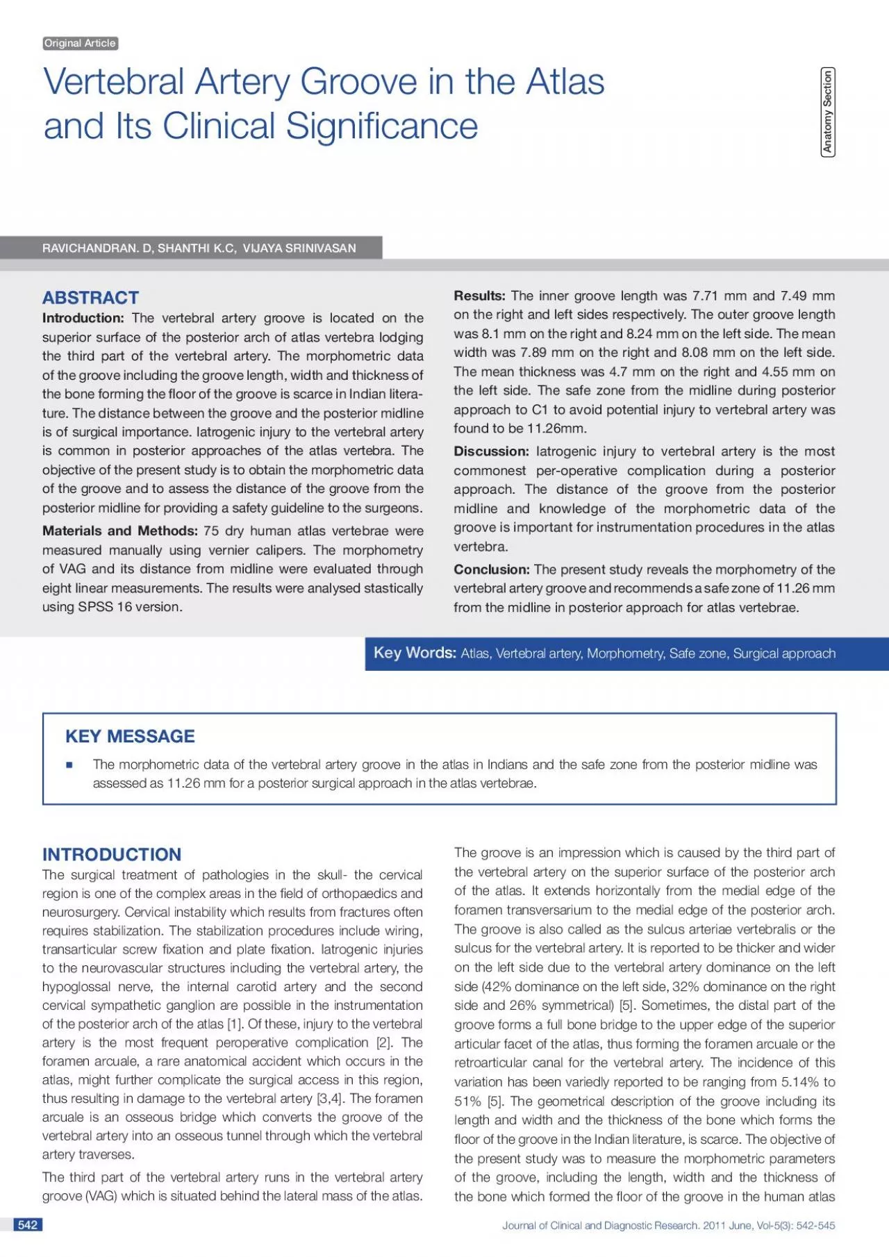
542 542 Vertebral Artery Groove in the Atlas Key WorAtlas Vertebral artery Morphometry Safe zone Surgical approachIntroduction The vertebral artery groove is located on the superior surface of t ID: 936828

artery groove projection vertebral groove artery vertebral projection posterior left side length atlas midline medial lateral width distance outer

Share:

Link:

Embed:

Download Presentation from below link

Download Pdf The PPT/PDF document "Journal of Clinical and Diagnostic Resea..." is the property of its rightful owner. Permission is granted to download and print the materials on this web site for personal, non-commercial use only, and to display it on your personal computer provided you do not modify the materials and that you retain all copyright notices contained in the materials. By downloading content from our website, you accept the terms of this agreement.


Presentation Transcript

Journal of Clinical and Diagnostic Research. 2011 June, Vol-5(3): 542-545 542 542 Vertebral Artery Groove in the Atlas Key WorAtlas, Vertebral artery, Morphometry, Safe zone, Surgical approachIntroduction: The vertebral artery groove is located on the superior surface of the posterior arch of atlas vertebra lodging RAVICHANDIJAYA SINIVA The surgical treatment of pathologies in the skull- the cervical region is one of the complex areas in the eld of orthopaedics and neurosurgery. Cervical instability which results from fractures often requires stabilization. The stabilization procedures include wiring, transarticular screw xation and plate xation. Iatrogenic injuries to the neurovascular structures including the vertebral artery, the hypoglossal nerve, the internal carotid artery and the second cervical sympathetic ganglion are possible in the instrumentation of the posterior arch of the atlas [1]. Of these, injury to the vertebral The morphometric data of the vertebral artery groove in the atlas in Indians and the safe zone from the posterior midline was assessed as 11.26 mm for a posterior surgical approach in the atlas vertebrae. Journal of Clinical and Diagnostic Research. 2011 June, Vol-5(3): 542-545 www.jcdr.netRavichandran D. et al., Vertebral artery groove in the atlas 543 vertebra in the Indian population and to provide a safety guideline for the surgeons to avoid potential injury to the vertebral artery.MATERIALS AND METHODSSeventy ve atlas (C1) vertebra without any obvious pathology and belonging to the bone banks of Vinayaka Mission’s Kirupananda Variyar Medical College, Salem, Tamil Nadu, Annapoorana Medical College, Salem, Tamil Nadu, Vinayaka Mission’s Homeopathy Medical College, Salem and the Govt. Mohan Kumaramangalam Medical College, Salem, Tamil Nadu were utilized for the study. The gender and the age of the bones were not considered in the present study. Eight linear measurements were taken bilaterally (right and left) by using a vernier caliper. The measurements were repeated by an independent second observer with the rst results being blinded to the second observer. Both the observers were qualied anatomists (rst and second authors) The anatomical parameters which were measured were groove length (inner and outer), groove width, thickness of the bone which forms the oor of the groove, proximal medial projection (D1), proximal lateral projection (D2), distal medial projection (D3) and distal lateral projection (D4).The VAG was identied. All the parameters were measured according to the instructions which were given by Max Franco et at the appropriate places [Table/Fig-1 & 2] . The length of the groove was taken as the maximum anteroposterior distance along the inner and outer edges of the groove. The width of the groove is the distance between the inner and outer edges at the middle of the groove. The thickness of the bone which formed the oor of the groove is the distance between the superior and inferior surfaces of the posterior arch at the middle of the groove. The proximal medial projection (D1) is the distance between the midline and the inner edge of the groove at the initial portion of the groove. D2 is the distance between the midline and the outer edge of the groove at the initial

portion of the groove. D3, the distal medial projection is the distance between the midline and the inner edge of the groove at the nal portion of the groove. D4 is the distance between the midline and the outer edge of the groove at the nal portion of the groove.The mean measurements of each of the parameters were compared separately between the two readers by using ANOVA paired ‘t’ tests and correlation coefcients and the ‘p’ values were calculated. For each of the measurements, the right side values and the left side values of the two observers were compared individually. The SPSS version 16 statistical package was used for RESULTSThe results of the measurements of observer 1 are tabulated in [Table/Fig-3]. The mean values ( SD) for the inner groove length Inner groove length (Rt.)Inner groove length (Lt.)Outer groove length (Rt.)Outer groove length (Lt).Width (Rt.)Width (Lt.)Proximal medial projection (D1) Rt. SideProximal medial projection (D1) Lt. SideProximal lateral projection (D2) Rt. SideProximal lateral projection (D2) Lt. SideDistal medial projection (D3) Rt. SideDistal medial projection (D3) Lt. SideDistal lateral projection (D4) Rt. SideDistal lateral projection (D4) Lt. Side[Table/Figure-3]: Vertebral artery groove measurement of right and left side average , minimum and maximum (Observer 1) [Table/Fig-1]: Shows Parameters – Groove Length (Outer and Inner) and Width of the Vertebral Artery Groove [Table/Fig-2]: Journal of Clinical and Diagnostic Research. 2011 June, Vol-5(3): 542-545 Ravichandran D. et al., Vertebral artery groove in the atlaswww.jcdr.net 544 544 parameters correlated highly with the correlation co-efcient of 0.97 to 0.99 (p10). The results of the analysis of the inter-observer variation are presented in [Table/Fig-4].The third part of the vertebral artery, after its exit from the foramen transversarium produces an impression on the superior surface of the posterior arch of the atlas, behind the lateral mass of the atlas vertebra. This groove is called as the vertebral artery groove (VAG) [5]. In the superior view, the groove appears to be curved from the lateral to the medial view [6,7].The morphology of the VAG, including the length, width and the thickness of the bone which formed its oor has scarcely been reported in the Indian literature.The length, width and the thickness of the groove did not show any statistically signicant difference between the right and left sides in the present study. The outer groove length was found to be signicantly longer on both the sides (p values, rt 0.05 and lt 0.001) ) than the inner groove length. A comparison of our morphometric results with that of another author (Max Franco de Carvalho) is presented in [Table/Fig-5].The distance between the longitudinal midline of the atlas vertebra and the VAG carries surgical importance. Surgeries involving the on the right side was 7.71 1.16 mm and on the left side, it was found to be 7.49 1.33 mm. The outer groove length on the right side was 8.1 1.58 mm and on the left side, it was 8.24 The mean width ( SD) on the right side was 7.89 1.29 mm and The mean thickness of the bone which formed the oor of the groove ( SD) on the right side was 4.7 0.98 mm and on the left The mean ( SD) D1 on the le

ft side was 11.06 1.61 mm and on the right side, it was 11.46 1.62 mm. The mean (SD) of the projection D2 on the left side was 17.821.70 mm and on the right side, it was 17.04 1.97 mm. The mean ( SD) D3 on the left and right sides were 14.3 2.03 mm and 14.26 1.69 mm respectively. The mean (SD) D4 on the left side was 23.14 1.65 The parameters D1 and D2 alone showed a statistically signicant difference between the right and left sides (p = 0.036 and 0.002 respectively). All the other parameters (length, width, thickness, D3 and D4) did not show signicant differences between the sides.The inter observer variation between observers 1 and 2 was not statistically signicant (p � 0.05). The measurements for all the Measurement of Reader 1Measurement Paired orrelation oeffecient &Inner groove length (Rt.)Inner groove length (Lt.)Outer groove length (Rt.)Outer groove length (Lt.)Groove Width (Rt.)Groove Width (Lt.)Proximal medial projection (D1)Left sideProximal lateral projection (D2) Left sideDistal medial projection (D3) Left sideDistal lateral projection (D4) Left sideProximal medial projection (D1) Right sideProximal lateral projection (D2) Right sideDistal medial projection (D3) Right sideDistal lateral projection (D4) Right side[Table/Fig-4]: Vertebral Artery Groove Measurement comparison between observers A Mean Length ( Mean Width ( (Only Dry bone results) INNER:Right – 7.58 Right – 8.49 Left – 7.96 Present study INNER:Right – 7.71 Right – 7.89 Left – 8.08 [Table/Fig-5]: A Comparison of the morphometric results of the present study with the study of Max Franco de Carvalho et al (2009) Journal of Clinical and Diagnostic Research. 2011 June, Vol-5(3): 542-545 www.jcdr.netRavichandran D. et al., Vertebral artery groove in the atlas 545 545 atlas include plate osteosynthesis, posterior cervical arthrodesis, wiring and lateral mass xation. A posterior approach through a midline longitudinal incision is the commonly advocated approach to the upper cervical region [8]. When exposing the upper cervical spine, care has to be taken to avoid injury to the vertebral arteries. Iatrogenic injury to the vertebral artery is the most commonest intra-operative complication during a posterior approach [5,9,10,11].Considering the more anterior position of the artery in the groove, the projection, D1 (proximal medial projection at the initial portion of the groove) would suggest the distance between the midline and the VAG. In our study, D1 was found to be 11.06 mm 1.61 on the left side and 11.46 1.62 on the right side. Although the numerical difference which was noted between the right and left side values was statistically signicant (p = 0.036), it does not carry much clinical signicance. However, the values which were observed on both the sides were close to 11 mm. Therefore, the mean distance between the midline and the edge of the VAG as observed in our study would be 11.26 mm, which could be taken as the safe zone from the midline. Various authors have quoted the safe zone for surgical manipulations on the posterior arch of the atlas to avoid iatrogenic injury to the vertebral artery. According to Simpson et al [12], the surgical exposure of the posterior arch should not exceed 15 mm from the midline in adults a

nd 10 mm in children. Max franco suggested that the posterior dissection of the posterior arch should be limited to a distance of 11.2 mm from the midline. Stauffer ES [13] recommends a safe zone of 10 mm from the posterior midline. Ebraheim et al [14] noted the safe zone as 10 mm for males and 9 mm for females from the posterior midline. All these data belong to the Western literature. Our results coincide with the results which were observed by Max franco [5]. However, the standard textbooks on posterior exposure suggest a safe distance of 15 mm from the the The authors recommend future studies with a larger sample size, This study which was done on 75 atlas vertebra provides morphological data about the vertebral artery groove. The knowledge of the safe zone for surgical manipulations from the posterior midline in the local population is mandatory for the surgeons who operate in this area. The present study recommends a safe zone of 11.26 mm from the midline in the posterior approach for the atlas vertebra.oach for the atlas vertebra.   Matthias Gebauer et al. Evaluation of anatomic landmarks and safe zones for screw placement in the atlas via the posterior arch. ch.    Madawi AA, Solanski G, Casey ATH, Crockard HA. Variation of the groove in the axis vertebra for the vertebral artery: implications for for    Hasan M, Shukla S, Siddiqui MS, Singh D. Posterolateral tunnels and and    Lamberty BG, Zivanovic S. The retro-articular vertebral artery ring of of    Max Franco de Carvalho, Roberta Teixeira Rocha et al. Vertebral artery groove anatomy. .    Cvadar S, Arisan E. Variations in the extracranial origin of the human vertebral artery. .    Newton TH, Mani RL. Radiol of Skull and Brain In: The vertebral artery. , New York; 1974: 1659-709.ork; 1974: 1659-709.   Maxim R. Leventhal. Campbell’s Operative Orthopaedics In: Anatomy and Surgical Approach. 9th edn; Vol. 3. Mosby, St. Louis; Louis;    Neo M, Sakamoto T, Fujibayashi S, Nakamura T. The clinical risk of vertebral artery injury from cervical pedicle screws which were inserted inserted    Madawi AA et al. Radiological and anatomical evaluation of the atlantoaxial transarticular screw xation technique. J. Neurosurg. 1997a; 1997a;    Weidner A, Wahler M, Chiu ST. Modication of C1-C2 transarticular screws xation by image guided surgery. .    Simpson JM, Ebraheim NA, Jackson WT et al. Internal xation of the thoracic and lumbar spine by using Roy-Camille plates. plates.    Stauffer ES. Posterior atlanto-axial arhtrodesis: the Gallie and Brooks Tech Orthopech Orthop   Ebraheim NA, XU R, Ahmad M. The quantitative anatomy of the vertebral artery groove of the atlas and its relationship to the posterior atlantoaxial approach. Dr. Ravichandran D. Dr. Shanthi K.C.3.Dr. Vijaya Srinivasan Corresponding Author.Assistant Professor, Department of Anatomy, Mission Kirupananda Variyar Medical College, Professor, Department of Community Medicine, Vinayaka Mission Kirupananda Variyar Medical College, Dr. D. Ravichandran MD (Anatomy)Department of Anatomy,Vinayaka Mission Kirupananda Variyar Medical College,Salem, Tamil Nadu , INDIAATINo competing Interests.Date of per revie