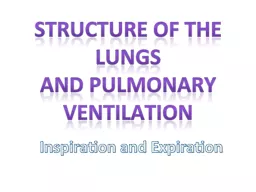
/
Structure and development of the diaphragm Structure and development of the diaphragm

Structure and development of the diaphragm - PowerPoint Presentation

bety
bety . @bety
Follow
342 views
Uploaded On 2022-06-18

Structure and development of the diaphragm - PPT Presentation

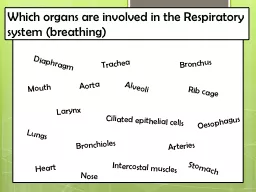
Semmelweis University Faculty of Medicine 1st year 2nd semester Department of Anatomy Histology and Embryology Katalin Kocsis 2018030910 Diaphragm thoracic ID: 920836

crus diaphragm part hiatus diaphragm crus hiatus part thoracic phrenic aorta structures abdominal passing development cava medial vena lateral

Share:

Link:

Embed:

Download Presentation from below link

Download Presentation The PPT/PDF document "Structure and development of the diaphra..." is the property of its rightful owner. Permission is granted to download and print the materials on this web site for personal, non-commercial use only, and to display it on your personal computer provided you do not modify the materials and that you retain all copyright notices contained in the materials. By downloading content from our website, you accept the terms of this agreement.


Presentation Transcript

Slide1

Structure and development of the diaphragm

Semmelweis University,

Faculty

of

Medicine

1st

year

2nd

semester

Department

of

Anatomy

,

Histology

and

Embryology

Katalin Kocsis

2018.03.09-10.

Slide2

Diaphragm (thoracic

)

diaphrassein

=

separate

(

Greek

)

Slide3

Slide4

Slide5

Diaphragm

4-5 mm

thick

striated

muscle

sheet

Separates

thoracic

cavity

from

abdominal

cavity

Most

important

respiratory

muscle

Role

in

abdominal

pressure

Important

structures

pierceing

it

:

Esophagus

Aorta

Inferior

vena

cava

Nerves

Lymph

vessel

Slide6

5th

rib

upper

edge

5th

intercostal

space

Centrum tendineum

Abdominal

cavity

(Abdomen)

Thoracic cavity (Thorax)

Muscular-tendineous

separating

sheet

between

thoracic

and

abdominal

cavities

Slide7

Parts

of

diaphragm

Tendinuous

part: centrum

tendineum

Muscular

part:

sternal

part (

xyphoid proc.)costal part (lower 6 ribs)lumbal partmedial crus(intermediate crus)laterale crus

Slide8

Lumbal

part

Medial

crus

:

Right

:

L1-L(3)4 body

Left:

L1-L(2)3 body (median arcuate lig.)Aortic hiatus (L1)Esophageal hiatus (Th11)(Intermediate crus: L2 body)Lateral crus:Medial arcuate lig. - L2 body- L2 Costal proc.

(M. psoas major)Lateral arcuate lig. - L2 Costal proc. - 12th rib (M. quadratus lumborum)

Slide9

Lumbal part

Medial crus

Lateral crus

Lateral

and

medial

arcuate

ligaments

Median

arcuate

ligament

Slide10

Diaphragm

superior

view

centrum tendineum

inf

.

vena

cava

esophagus

aorta thoracica

costal

part

pericardium

stomach

liver

abdominal

muscles

Slide11

centrum tendineum

lumbal

part

sternal

part

costal

part

inf

.

vena

cava

esophagus

aortic

hiatus

esophageal

hiatus

vertebral

column

m. psoas major

m. psoas minor

m. quadratus lumborum

Diaphragm

inferior

view

Slide12

Diaphragm

inferior

view

Slide13

Openings

Hiatus

of

inferior

vena

cava

Esophageal

hiatus

Aortic

hiatus

Sternocostal

triangle

Lumbocostal triangle

Slide14

Structures passing

through

internal

thoracic

a. (

superior

epigastric

a.

)sternocostal triangleinferior vena cava (right phrenic n., pericardiacophrenic a.)inf. v. cava hiatusesophagus

n. vagus(left gastric a. (v.), esoph. branches)esophageal hiatus aortathoracicus ductaortic hiatus v. azygos , v. hemiazygosgreater

splanchnic n.medial crus truncus sympathicuslesser splanchnic n.mediale crus - laterale crus

Slide15

internal

thoracic

a., v.

superior

epigastric

a., v.

Sternocostal

triangle

(

Larrey

,

Morgagni

foramen

)Structures passing through

Slide16

Structures passing through

Slide17

17

Sternum

manubrium

Sternum

body

xyphoid

process

heart

Thoracic

aorta

Abdominal

aorta

Aortic

arch

Structures

passing

through

Slide18

Structures passing through

Slide19

esophagus

left vagus n.

right vagus nerve

inf. vena cava

Esophageal

hiatus

Aortic

hiatus

Thoracic duct

Cysterna chyli

Descending aorta

Structures

passing

through

Slide20

Between medial and lateral crus:

sympathetic trunk and lesser splanchnic nerve

Between

the

fibers

of

medial

crus

:

greater splanchnic nerve and azygos/hemiazygos vein (ascending lumbar veins)

sympathetic

trunk

hemiazygos v.

azygos v.

greater splanchnic n.

Structures

passing

through

Slide21

Blood supply, innervation

pericardiacophrenic

a.

musculophrenic

a.

sup

.

phrenic

a.

inf

. phrenic a.

interna thoracica a. (subclavian a.)

thoracic

aorta

abdominal

aorta

Motor innervation: phrenic n. (C3-4-5)

(Cervical pl.)Sensory innervation:phrenic n.: centrum tendineum(diaphragmatica pleura, parietal peritoneum)intercostal nerves (7-12):muscular part

Blood

supply

Slide22

Slide23

http://

www.flspinalcord.us

/

innervation-of-diaphragm

/innervation-of-diaphragm-studydroid-flashcards-on-the-web-and-in-your-hand/

Slide24

Blood supply

From internal thoracic artery:

-pericardicophrenic artery

-musculophrenic artery

From descending aorta:

-superior phrenic artery

-inferior phrenic artery

Slide25

pleura

serous

pericardium

peritoneum

fibrous pericardium

diaphragm

Serous

membranes

on

the

diaphragm

Slide26

Slide27

Normal inspiration

Paralysis of right phrenic nerve

Paralysis of the phrenic nerve – paradox movement of diaphragm

Slide28

Slide29

Development

of

diaphragm

Slide30

Slide31

Development

of

diaphragm

develops

from

4

parts

:

Septum

transversum (C3-C5 cervical somites)2. Dorsalis mesentery of esophagus3. Pleuroperitoneal membrane

4. Lateral body wall

Septum

transversum

Szív

Szív

Máj

Slide32

5th

week

4th

week

Development

of

diaphragm

Slide33

Development

of

diaphragm

Slide34

Development

of

diaphragm

Slide35

Development

of

diaphragm

Slide36

D

iaphragmatic

hernia

Slide37

congenital

(

CDH

)

aquired

(

hiatushernia

)

D

iaphragmatic

hernia

Slide38

References