PPT-JEJUNUM & ILIUM
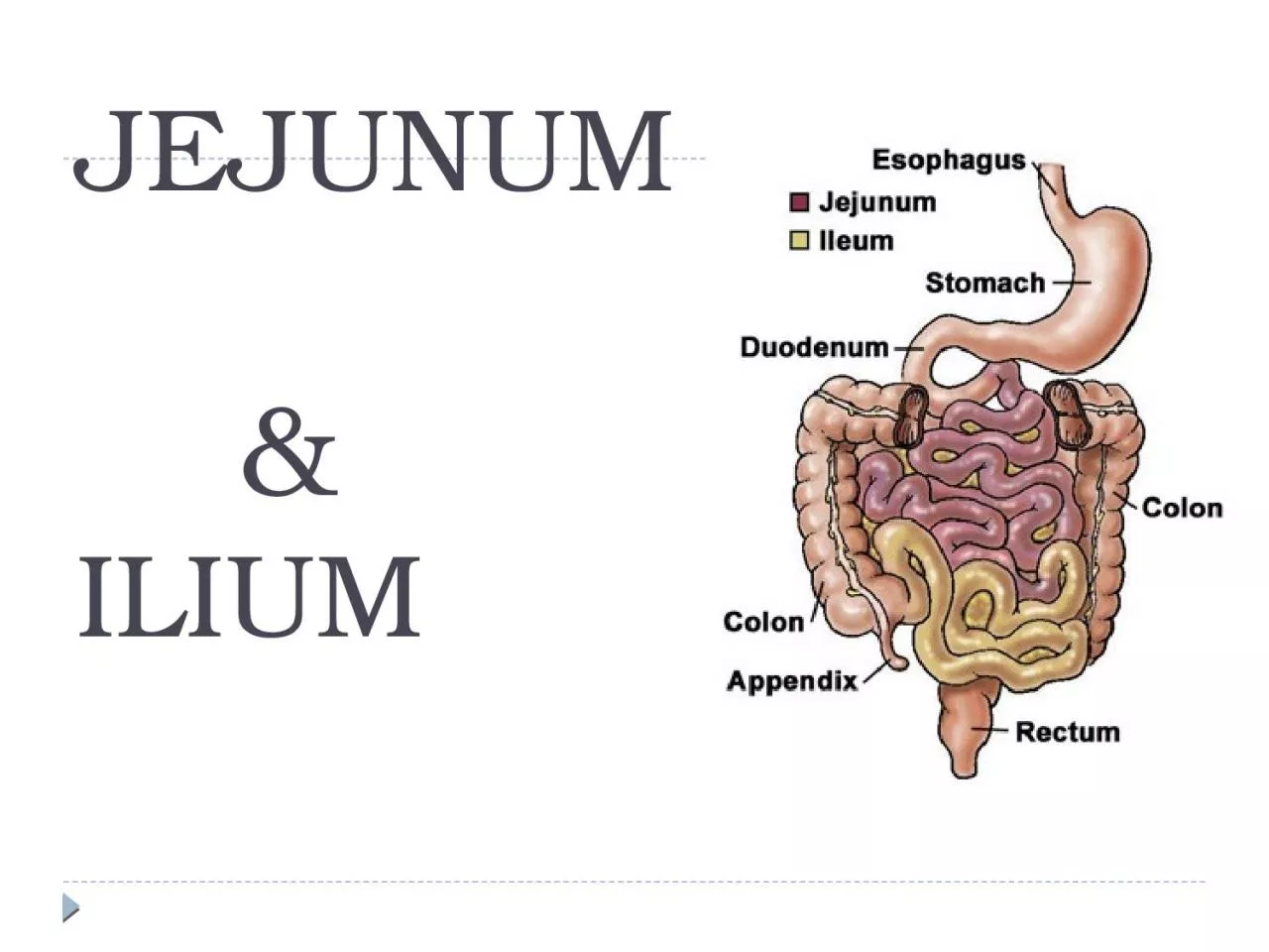
Features Jejunum Ilium Position Left infracolic compartment upto umbilical region Pelvic region ends in rtiliac fossa by opening into ilioceacal valve Median external
Download Presentation
"JEJUNUM & ILIUM" is the property of its rightful owner. Permission is granted to download and print materials on this website for personal, non-commercial use only, provided you retain all copyright notices. By downloading content from our website, you accept the terms of this agreement.
Presentation Transcript
Transcript not available.