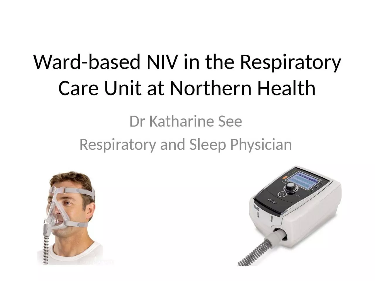
/
Ward-based NIV in the Respiratory Care Unit at Northern Health Ward-based NIV in the Respiratory Care Unit at Northern Health

Ward-based NIV in the Respiratory Care Unit at Northern Health - PowerPoint Presentation

blanko
blanko . @blanko
Follow
0 views
Uploaded On 2024-03-13

Ward-based NIV in the Respiratory Care Unit at Northern Health - PPT Presentation

Dr Katharine See Respiratory and Sleep Physician Overview RCU at Northern Health Patient selection Evaluation of response Wardbased vs critical care locations Background to development of RCU ID: 1047539

respiratory niv epap care niv respiratory care epap icu rcu ward dead case based settings space ipap oxygen paco2

Share:

Link:

Embed:

Download Presentation from below link

Download Presentation The PPT/PDF document "Ward-based NIV in the Respiratory Care U..." is the property of its rightful owner. Permission is granted to download and print the materials on this web site for personal, non-commercial use only, and to display it on your personal computer provided you do not modify the materials and that you retain all copyright notices contained in the materials. By downloading content from our website, you accept the terms of this agreement.


Presentation Transcript

1. Ward-based NIV in the Respiratory Care Unit at Northern HealthDr Katharine SeeRespiratory and Sleep Physician

2. OverviewRCU at Northern HealthPatient selectionEvaluation of responseWard-based vs critical care locations

3. Background to development of RCULimited capacity within ICU to offer NIVBed-block for patients stabilized on NIV (both ED and ICU)Likely some patients missing out on NIV – potential impact on LOS

4. RCU4-bed room on T2Able to offer NIV or HFOTNon-invasive monitoring1:2 nursing ratioNot an experienced ward (yet)ConfidenceCapability/resourcing

5.

6.

7. Patient selectionNIV may be appropriate for many indications associated with acute respiratory failure in a critical care settingWard-based acute NIV (within RCU) is more limitedHypercapnic respiratory failure due to AECOPDAndNot much else (step-down for NMD, OHS)

8. Why won’t ward-based NIV work?Not a respiratory ICU or HDULimited (non-invasive) monitoringRelatively inexperienced nursing staffNo medical staff at locationFalse reassurance of NIV (evidence demonstrates delays in escalating care)No ability to escalate care

9. RCU isn’t ICU (or ED)

10.

11. What is a respiratory care unit (RCU)?GeographyLocation of medical supportAbility to escalate careLimited monitoring capability – non-invasiveThat means that patient safety (and therefore patient selection) is criticalThose likely to benefit from RCU interventionsThose likely to require ICU care

12. Who benefits from ward-based acute NIV?COPD

13. HypercapnoeaElevation of the arterial carbon dioxide tensionDirectly proportional to CO2 productionIndirectly proportional to rate of elimination by lungsPaCO2 = K x VCO2/[Ve(1-Vd/Vt)]PaCO2 rises with:  production (fever) Minute ventilation (sedatives) Dead space ventilation (COPD)

14. Decreased minute ventilation / global hypoventilationMV = RR x TVMechanism:Central: respiratory centre in medulla responds to chemoreceptors (H+ and PaCO2 and PaO2) and mechanoreceptors (stretch in upper airway and lungs). Drive is also responsive to cognitive and behavioural inputs (anxiety/pain)Peripheral muscle: (diaphragm, intercostals etc) alter RR, depth and duration of inspiration. Shallow breathing increased VD/VT Thoracic cage dysfunction: post operatively. RR may increase to compensate but fatigue ensues. Also increases VD/VT

15. ExamplesWon’t breathe (the brain): SedativesEncephalitismajor strokecentral sleep apnoeabrain stem diseasemetabolic alkalosis hypothermiaCan’t breathe (nerves, muscles, thoracic cage)C spine injury (diaphragmatic paresis/paralysis)MNDPolioGuillian BarrePhrenic nerve injuryMyastheniaPolymyositisMyopathy (critical illness, secondary to thyroid)KyphoscoliosisAnkylosing spondylitisPectus excavatum

16. Increased dead spaceThe non-gas exchanging part of the lungAnatomical dead space: upper airways to terminal bronchiolesAnatomical dead space is a fixed volume Alveolar dead space: reduced perfusion to ventilated alveoli pulmonary capillary compression (extrinsic obstruction) from overinflated lungs (too much positive pressure) destruction of pulmonary capillaries (emphysema, interstitial fibrosis, pulmonary vasculitis)It is the proportion of the tidal volume that is dead space that we can change

17. COPDIncreased VD main mechanism of hypercapnoea in COPDSome compensate: MV or redistribute perfusion to improve V/Q matching (pink puffers)Some cannot and develop chronic hypercapnoea (blue bloaters)

18. MechanismDestruction of capillaries by emphysemaAirflow obstruction leading to hyperinflationReduced lung compliance at high lung volumesBreathing pattern in COPD: lower tidal volumes, higher RR which increases VD/VTDrive is high, inspiratory time in short ( VT) expiratory time in long ( RR)

19. Oxygen induced hypercapnoea Always aim for normoxia Don’t kill someone with untreated hypoxia for fear of hypercapnoea. Don’t believe that everyone needs oxygen and that oxygen is good – it can kill people

20.

21. Oxygen induced hypercapnoeaSome COPD pts given oxygen will develop hypercapnoeaThis is NOT due to loss of hypoxic drive (it’s only a small part)Increased dead space: worsening of V/Q mismatch due to loss of hypoxic pulmonary vasoconstriction (HPV improves matching between blood flow and ventilation which improves V/Q and decreases physiologic dead space). Worse in more hypoxic patientsHaldane effect: Right shift of CO2-Hb dissociation curve in presence of increased oxygen

22. Effects of hypercapnoeaCerebral: depressed conscious state and reduced respiratory drive, increased cerebral blood flow and intracranial pressureCardiorespiratory: dyspnoea (increased drive), reduced myocardial and diaphragmatic contractilityPhysiologic: shift in oxy-haemoglobin dissociation curve to right, acidosis and associated problemsMetabolic: buffering with bicarbonate

23. What to do with AECOPDBlood gas – if abnormal VBG MUST have ABGBronchodilator – MDI with spacer Conservative oxygen therapy – sats 88-92%Prednisolone at 0.75mg/kgSend sputumSingle agent abx – checking for previous organisms and sensitivitiesDVT prophylaxisGOPC

24. Case 1: Mrs C72F from retirement village “AECOPD”Complicated by T2RFFEV1 0.90L (38%), smokerOSA?Weight 120kg, BMI 36Examination: RR 24 SpO2 85% RABP 130/69, HR 100T 37.2GCS 14-15

25. Challenge the diagnosis:Not all ventilatory failure = AECOPDCapacityLoadChest wallLung complianceUpper airwayStrengthCNS disordersSleepDrugsNeuromuscular disordersDriveObesity Chest wall restrictionCOPDCO2

26. Case 1: Mrs CCommenced on NIV on arrivalInitial VBG: pH 7.25

27. Case 1: Mrs CCommenced on NIV on arrivalInitial VBG: pH 7.25What should happen next?Can we stop there?

28. Venous blood gasesUseful screening test for acidaemia in someone with AECOPD Valuable to confirm a clinical decision that NIV is not required Results are redundant once a clinical decision to institute NIV has been made (Opinion: Should refute this decision with the gold standard test)Example: Empirical anticoagulation for PE – wouldn’t cease therapy on the basis of a negative D-dimerPvCO2 is (often) misleadingUnable to use serial results to titrate ventilator settings

29.

30. Venous pH in AECOPDVenous pH cut-off of 7.34 correctly classified 87% of arterial pH≥7.35 (mean difference is 0.03)Arguably a higher cut-point is sensible (7.38-7.40)The aim is not perfect diagnostic accuracy – a sensitive screening test should be preferred in this situationA false negative VBG means potentially withholding an evidenced based therapy (NIV) from a patient that can benefit from it

31. Case 1: Mrs CABG on NIV (IPAP 10, EPAP 5, FiO2 21%): pH 7.25pCO2 88pO2 47HCO3 38

32. Case 1: Mrs CABG on NIV (IPAP 10, EPAP 5, FiO2 21%): pH 7.25pCO2 88pO2 47HCO3 38 What should happen next?Transfer to ward for ongoing NIVRepeat ABGRefer to ICUAdjust NIV settingsReview the maskAll of the above

33. Case 1: Mrs CABG on NIV (IPAP 10, EPAP 5, FiO2 21%): pH 7.25pCO2 88pO2 47HCO3 38 These don’t look right

34. Case 1: Mrs C72F from retirement village “AECOPD”Complicated by T2RFFEV1 0.90L (38%), smokerOSA?Weight 120kg, BMI 36Examination: RR 24 SpO2 71% RABP 130/69, HR 100T 37.2GCS 14-15

35. Case 1: Mrs CSettings adjusted:IPAP 20cmH2O, EPAP 10cmH2OBronchodilators, Prednisolone, AntibioticsRepeat ABG pH 7.32, pCO2 62, pO2 61, HCO3 31Improving -> transfer to RCU for ongoing NIV

36. Considerations with NIV settingsWhich mask?What pressures (IPAP and EPAP)?How is a breath triggered and how sensitive should the trigger be?How long should a breath be?

37. NIV masksNon-vented masks – ED/ICU/HDU Separate exhalation line or valve that avoids re-breathing of expired airVented masks – used in RCU (and all home CPAP or NIV devices)Mask has in-built vents which intentionally leak and allow expulsion of expired airUsing a non-vented mask on a ward ventilator (or CPAP device) can result in re-breathingIf in doubt – please contact on of our Respiratory CNCs or registrars for advice

38. VentedNon-ventedVentsOxygen ports – not vents!Don’t use ICU/ED/theatre masks anywhere but ICU/ED/theatre with ICU/ED/theatre machines

39. NIV pressuresEPAP – typically set to overcome upper airway resistance (and to vent CO2 from the mask)IPAP – provides the driving pressure to increase alveolar ventilationRule of thumb: EPAP should be ~1cmH2O for every 10-15kg of body weight120kg person: EPAP ~10cmH2O

40. Titrating pressuresAiming for the lowest effective pressuresIncrease IPAP-EPAP difference (=pressure support) to increase tidal volumeIncrease EPAP if O2 low or possible upper airway obstructionSerial ABG (fall in PaCO2, rise in pH) allows titrationImprovement confirms adequate settings and high probability that ward-based NIV will be successfulLack of change or deterioration implies incorrect settings, incorrect mask, incorrect patient?

41. Admission criteria for RCUED referralsAcute exacerbations of COPDMild to moderate hypoxaemic respiratory failureICU referrals (step-down care)OHS/NMD/other – on NIVStable patients requiring ongoing respiratory monitoring (single organ dysfunction)Ward referrals

42. Contraindications to ward-based NIV (RCU admission)High likelihood of failure of NIVInvasive ventilation (via tracheostomy)Acute severe asthmaAcute coronary syndromesAcute pulmonary oedemaSevere hypoxaemic respiratory failureMassive or submassive PETraumaHaemodynamic instabilityConfusion/need for iv sedationAcute surgical pathology or complex post-op care

43. Written by critical care physicians (Society of Critical Care Medicine)

44. From Ergan et al, ERS, 2018

45. Problems with this approachToo restrictive/prescriptiveDoesn’t consider response to Rx or likelihood of failureFrequent ABGs!??! (every 8hrs) Reality is that RCU sits above the ‘general ward’ but below the HDU in this paper

46. Venous blood gasesGood screening test for acidaemia in AECOPDShould confirm abnormal result with an ABG (arguably any VBG <7.40)Once a clinical decision is made to commence NIV -> ABG(should refute your clinical judgement with an accurate test)Ignore the PvCO2 – it is misleading and can’t be used to titrate settings

47. Clinical parametersComfort and toleranceRespiratory rateOxygen saturationConscious stateOnce you are considering sedation to improve NIV tolerance you have an HDU/ICU patient in front of you

48. Comfort and toleranceMask choice (size) is criticalZero leak is not the goalStarting pressures may be low (for tolerance) but should not necessarily stay lowRespiratory rate should fall with applicationCoordination with the device can be clinically assessed

49. NIV starting pressuresIPAP 10 and EPAP 5 will not work for most Reasonable to start but expect to increaseEPAP to overcome upper airway resistanceIf on CPAP use this as a guide (EPAP generally ~2-4cmH2O below CPAP)Roughly 1cmH2O for every 10-15kg body weightIPAP (or pressure support level) titrated to PaCO2 level (but can use tidal volume as interim marker)

50. Gas exchangeContinuous pulse oximetryNo data to support a particular target range but no value in SpO2 >94-95%A lower target (88-92%) has been recommended but this should be the unsupported target range in my viewOn NIV I think 90-94% is adequate

51. ABG analysisGold standard for monitoringBaseline – then 1-2hrs after commencing NIVNo data supports the use of VBG for this purposeA positive response would be >3mmHg fall in PaCO2 and increase in pH by >0.03Failure to achieve these suggests failure of NIV (on current settings)

52. SummaryRCU is not the same as HDUResponse (using PaCO2 and pH) is criticalStarting settings are rarely the final NIV settingsUse clues to guide settings (weight, CPAP, previous admission)Clinical and biochemical responseIf not responding -> review and consider if RCU appropriate