/
2001 2010 annals of neurosurgeryannals of neurosurgery 20 2001 2010 annals of neurosurgeryannals of neurosurgery 20

2001 2010 annals of neurosurgeryannals of neurosurgery 20 - PDF document

briana-ranney
briana-ranney . @briana-ranney
Follow
363 views
Uploaded On 2015-10-02

2001 2010 annals of neurosurgeryannals of neurosurgery 20 - PPT Presentation

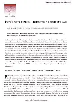
Neurosurgery unit Department of surgery Nnamdi azikiwe university teaching Hospital PmB 5025 Nnewi anambra state NIgerIa ID: 147039

Neurosurgery unit Department surgery

Share:

Link:

Embed:

Download Presentation from below link

Download Pdf The PPT/PDF document "2001 2010 annals of neurosurgeryannals o..." is the property of its rightful owner. Permission is granted to download and print the materials on this web site for personal, non-commercial use only, and to display it on your personal computer provided you do not modify the materials and that you retain all copyright notices contained in the materials. By downloading content from our website, you accept the terms of this agreement.


Presentation Transcript

© 2001-2010 annals of Neurosurgeryannals of Neurosurgery, 2010; 10(1): 1-4sir Percival Pott in the 18 century described osteomyelitis of the frontal skull bone with associated sub-periosteal abscess collection, which became known as Pott’s Puffy tumour. the incidence of the disease Neurosurgery unit, Department of surgery, Nnamdi azikiwe university teaching Hospital, PmB 5025 Nnewi, anambra state, NIgerIa. © 2001-2010 annals of NeurosurgeryPott’s Puffy tumour – a grotesque caseannals of Neurosurgery, 2010; 10(1): 1-4paranasal sinuses and the intracranial cavity, and with computed tomography, the full extent of bone infection is better de�ned. abscess drainage and craniectomy followed by 4 – 6weeks of antibiotic therapy usually cures the disease, and in most cases We report the case of a 16-year old male student who was admitted into our service with a grotesque looking Pott’s Puffy tumour, without any known predisposing factor, and was treated operatively and discharged home without neurological de�cits. the size of the lesion was as worrisome as the duration of ill health, in this era of antibiotics when the availability of medical services in Nigeria is presumed to have improved signi�cantly. is a 16-year male right handed secondary school student who was referred to our service on 5february, 2009 with complaints of recurrent fever, scalp pains and scalp swellings of 6months’ duration.there were associated headaches, neck pains, bilateral eye swellings and purulent eye discharge. the swellings had appeared serially at 2-monthly intervals in the parietal region and were treated with herbs and orthodox drugs, discharging pus but with persistence of scalp pains. the present episode started 1week earlier with a frontal scalp swelling that rapidly spread over most of the scalp. there was no history of trauma to the head, or urtI, prior to onset and no associated alteration in sensorium, sphincteric dysfunction, hemi-corporeal weakness, seizures or clinically, he was a young male, fully conscious but with a grotesque c-shaped cystic scalp collection involving the left frontal, left parietal and right parietal regions in coalescence. there was intense bi-periorbital oedema and mechanical ptosis with underlying chemosis, and moderate nuchal rigidity, but equivocal Kernig’s sign. there were no cranial nerve, gross motor, sensory or autonomic We made a clinical diagnosis of Pott’s Puffy tumour and requested for laboratory workup and skull x-rays because our centre did not have computed tomographic facilities and the closest centre with the facility was about 100km away. the skull x-rays showed osteomyelitis of the left frontal and left parietal bones but no paranasal sinus lesions. the HIV I&II screening was non-reactive, genotype was ‘as’ and total white cell count was 18.5x10/L We subsequently did a preliminary incision and drainage within six hours of admission to evacuate the abscess and relieve the pressure symptoms on the head and eyes. the procedure yielded frank pus, 1500ml, and some specimen was sent for microbiology but the culture yielded no growth. He was placed on twice-daily dressings with honey-soaked gauze packing and parenteral antibiotics. By the 12 day post-op we sent him for ct scanning with bone window and contrast which con�rmed fronto-parietal osteomyelitis but no intracranial abscess. craniectomy and delayed primary scalp closure were done and the patient was placed on post-operative antibiotics for another two weeks and subsequently, discharged home on per-oral Pott’s Puffy tumour is thought to be a disappearing disease in the antibiotic era, and most are thought to arise from contiguous paranasal sinus spread mostly from the frontal bones according to many reports [19,20,1,3,5,10,11, 12,13,15,16,17,18]. our case appears, instead, to have been a case of de novoparietal osteomyelitis that was neglected for long and extended to involve the frontal bone over a period of 6 months. there was no history suggestive of trauma prior to the onset of illness but this would not be completely ruled out in an active young male student. there was, however, a de�nite denial of any history of upper respiratory tract infection in association with the fever, scalp pains and scalp swellings which was quite unlike other reports [3,5]. But periorbital © 2001-2010 annals of Neurosurgeryannals of Neurosurgery, 2010; 10(1): 1-4oedema and orbital cellulitis had in our case have been reported by Lamoreau and fanciullo, morley, and Hayek, et al to be associated with this disease [7,8,9]. Despite the classical advocacy of urgent ct scanning and de�nitive surgical intervention, we modi�ed our protocols in the light of our technical handicaps and did a preliminary incision and drainage to relieve sepsis, reduce pains and save life, before resorting to a classic work-up and de�nitive treatment; and still got a good outcome. It would have been unwise to insist on a full work-up with ct scanning in the light of the enormous size of the abscess and painful incapacitation of this patient, thus we went ahead with a simple drainage procedure �rst, and only obtained a proper imaging in a more stable patient, following which we did craniectomy as the de�nitive procedure.In these modern times, it should be realized that sub-optimal care and mismanagement of clinical cases still pervade our health system and a case as grotesque as ours could still be found in this era of antibiotics. also, without readily available computed tomography, one could resourcefully modify the standard protocols to provide a fairly good surgical care for the patient; so long as the basic principles are strictly adhered to, thereby reducing morbidity and mortality. references1. raja V, Low c, sastry a, moriarty B. Pott’s puffy apr-Jun;53(2):114-6.2. Durur-subasi I, Kantarci m, Karakaya a, orbak Z, ogul H, alp H. J craniofac surg. 2008 Pott’s puffy tumor: multidetector computed tomogra3.Karaman e, Hacizade y, Isildak H, Kaytaz a. Pott’s puffy tumor. J craniofac surg. 2008 4. Wu ct, Huang JL, Hsia sH, Lee Hy, Lin JJ. Pott’s puffy tumor after acupuncture therapy. eur J Pediatr. 2008 Dec 5.5. Bannon PD, mccormack rf. Pott’s puffy tumor emerg med. 2008 Nov 18.6. martinez-Diaz gJ, Hsia r. Pott’s Puffy tumor after minor head trauma. am J emerg med. 2008 7. Lamoreau KP, fanciullo Lm. Pott’s puffy tumour 8. morley am. Pott’s puffy tumor. a condition still 9. Hayek Br, sitole s, andreoli m, Banich a, ahmad aZ. Bilateral eyelid edema and orbital cellulitis associated with Pott’s puffy tumor. ophthal Plast reconstr surg. 2007 mar-apr;23(2):163-5.10. shehu BB, mahmud mr. Pott’s puffy tumour: a case report. ann afr med. 2008 sep;7(3):138-40.11.Kaabia N, abdelka� m, Bellara I, Khalifa m, Bahri f, Letaief a. Pott’s puffy tumor. a case report. © 2001-2010 annals of Neurosurgeryannals of Neurosurgery, 2010; 10(1): 1-4 12. Bambakidis Nc, cohen ar. Intracranial complications of frontal sinusitis in children: Pott’s puffy tumor revisited. Pediatr Neurosurg. 2001 13. guillén a, Brell m, cardona e, claramunt e, costa Jm. Pott’s puffy tumour: still not an eradicated entity. childs Nerv syst. 2001 may;17(6):359-62.14. greenberg ms. Handbook of Neurosurgery. Infections. 5th ed, thieme New york 2001; 200-253.15. Khan ma. Pott’s puffy tumor: a rare complication of mastoiditis. Pediatr Neurosurg. 16. strony rJ, Dula D. Pott puffy tumor in a 4-year-emerg care. 2007 Nov;23(11):820-2.17. Badatolu c, güleryüz a, ersöz g, talas Du, Kandemir o, Köksel t. a rare clinical entity: Pott’s puffy tumor. a case report. Pediatr Neurosurg. 2001 18. maheshwar aa, Harris Da, al-mokhthar N, evans ra. Pott’s puffy tumour: an unusual presJun;115(6):497-9.19. mcDermott c, o’sullivan r, mcmahon g. an unusual cause of headache: Pott’s puffy tumour. eur J emerg med. 2007 Jun;14(3):170-3. 20. alsanosi a, el-seyed y. Pott’s puffy tumor. a correspondenceJude Kennedy c emeJuLu , Neurosurgery unit, Department of surgery, Nnamdi azikiwe university teaching Hospital, PmB 5025 Nnewi, anambra state, judekenny2003@yahoo.com\r\f: +234-803-the historical description of a sub-periosteal or sub-galeal purulent collection at the cephalic region, by sir Percival Pott (1714-1788), known classically as Pott’Puffy tumor, recently falls in the de�nition of a subgaleal and / or sub-periosteal abscess, with the classical characteristics of in�ammation secondary to an infection, with the presence of �ushing, heat, pain and increased volume (tumor). this purulent collection is usually due to the spread of an infection from the paranasal sinus reached by a cranial fracture, which allows the passage of germs into neighboring tissues; also is due to traumatic brain injury, with open trauma of the scalp, but contaminated, and subsequently due to an inappropriate treatment and control, infecting the skull,establishing an osteomielitis, thus allowing the collection of germs in closed areas at the sub-periosteal or sub-galeal spaces ; and �nally, that could correspond to the case reported by J.K. emejulu and I. Bc. Iloabachie , to minor trauma, accidental or provoked, some caused by sharp objects, which can cause late osteomyelitis of the calvarium. It´s necessary to comment, that noting the patient’s clinical conditions reported, the authors performed the best, that is to solve the subgaleal abscess with conventional surgical techniques, primarily to drain the pus and then studying the patient in order to search the origin of the infection, which was observed in the phase 3D computed tomography, which corresponded to a location of the osteomyelitis at the parietal bone, left posterior region. Later, it was the best performed, removing the bone with osteomyelitis and infected splinters, and starting speci�c antibiotic treatment according to the antibiogram of the evacuated pus. the craneoplasty must be made on a deferred time, once septic parameters have normalized. It is a well-described and documented case by the authors, which was resolved in an appropriate manner, despite not having some immediate accessory Prof Dr Leonidas m quintana mDValparaiso, chile © 2001-2010 annals of Neurosurgeryannals of Neurosurgery, 2010; 10(1): 1-4 12. Bambakidis Nc, cohen ar. Intracranial com-plications of frontal sinusitis in children: Pott’s puffy tumor revisited. Pediatr Neurosurg. 2001 13. guillén a, Brell m, cardona e, claramunt e, costa Jm. Pott’s puffy tumour: still not an eradicated entity. childs Nerv syst. 2001 may;17(6):359-62.14. greenberg ms. Handbook of Neurosurgery. In-fections. 5th ed, thieme New york 2001; 200-253.15. Khan ma. Pott’s puffy tumor: a rare com-plication of mastoiditis. Pediatr Neurosurg. 16. strony rJ, Dula D. Pott puffy tumor in a 4-year-emerg care. 2007 Nov;23(11):820-2.17. Badatolu c, güleryüz a, ersöz g, talas Du, Kandemir o, Köksel t. a rare clinical entity: Pott’s puffy tumor. a case report. Pediatr Neurosurg. 2001 18. maheshwar aa, Harris Da, al-mokhthar N, evans ra. Pott’s puffy tumour: an unusual pres-Jun;115(6):497-9.19. mcDermott c, o’sullivan r, mcmahon g. an unusual cause of headache: Pott’s puffy tumour. eur J emerg med. 2007 Jun;14(3):170-3. 20. alsanosi a, el-seyed y. Pott’s puffy tumor. a correspondenceJude Kennedy c emeJuLu , Neurosurgery unit, Department of surgery, Nnamdi azikiwe university teaching Hospital, PmB 5025 Nnewi, anambra state, judekenny2003@yahoo.com\r\f: +234-803-the historical description of a sub-periosteal or sub-galeal purulent collection at the cephalic region, by sir Percival Pott (1714-1788), known classically as Pott’Puffy tumor, recently falls in the de�nition of a subgaleal and / or sub-periosteal abscess, with the classical characteristics of in�ammation secondary to an infection, with the presence of �ushing, heat, pain and increased volume (tumor). purulent collection is usually due to the spread of an infection from the paranasal sinus reached by a cranial fracture, which allows the passage of germs into neighboring tissues; also is due to traumatic brain injury, with open trauma of the scalp, but contaminated, and subsequently due to an inappropriate treatment and control, infecting the skull,establishing an osteomielitis, thus allowing the collection of germs in closed areas at the sub-periosteal or sub-galeal spaces ; and �nally, that could correspond to the case reported by J.K. emejulu and I. Bc. Iloabachie , to minor trauma, accidental or provoked, some caused by sharp objects, which can cause late osteomyelitis of the calvarium. necessary to comment, that noting the patient’s clinical conditions reported, the authors performed the best, that is to solve the subgaleal abscess with conventional surgical techniques, primarily to drain the pus and then studying the patient in order to search the origin of the infection, which was observed in the phase 3D computed tomography, which corresponded to a location of the osteomyelitis at the parietal bone, left posterior region. , it was the best performed, removing the bone with osteomyelitis and infected splinters, and starting speci�c antibiotic treatment according to the antibiogram of the evacuated pus. the craneoplasty must be made on a deferred time, once septic parameters have normalized. It is a well-described and documented case by the authors, which was resolved in an appropriate manner, despite not having some immediate accessory Prof Dr Leonidas m quintana mD Valparaiso, chile © 2001-2010 annals of Neurosurgeryannals of Neurosurgery, 2010; 10(1): 1-4oedema and orbital cellulitis had in our case have been reported by Lamoreau and fanciullo, morley, and Hayek, et al to be associated with this disease [7,8,9]. Despite the classical advocacy of urgent ct scanning and de�nitive surgical intervention, we modi�ed our protocols in the light of our technical handicaps and did a preliminary incision and drainage to relieve sepsis, reduce pains and save life, before resorting to a classic work-up and de�nitive treatment; and still got a good outcome. It would have been unwise to insist on a full work-up with ct scanning in the light of the enormous size of the abscess and painful incapacitation of this patient, thus we went ahead with a simple drainage procedure �rst, and only obtained a proper imaging in a more stable patient, following which we did craniectomy as the de�nitive procedure.In these modern times, it should be realized that sub-optimal care and mismanagement of clinical cases still pervade our health system and a case as grotesque as ours could still be found in this era of antibiotics. also, without readily available computed tomography, one could resourcefully modify the standard protocols to provide a fairly good surgical care for the patient; so long as the basic principles are strictly adhered to, thereby reducing morbidity and mortality. references1. raja V, Low c, sastry a, moriarty B. Pott’s puffy apr-Jun;53(2):114-6.2. Durur-subasi I, Kantarci m, Karakaya a, orbak Z, ogul H, alp H. J craniofac surg. 2008 Pott’s puffy tumor: multidetector computed tomogra-3.Karaman e, Hacizade y, Isildak H, Kaytaz a. Pott’s puffy tumor. J craniofac surg. 2008 4. Wu ct, Huang JL, Hsia sH, Lee Hy, Lin JJ. Pott’s puffy tumor after acupuncture therapy. eur J Pediatr. 2008 Dec 5.5. Bannon PD, mccormack rf. Pott’s puffy tumor emerg med. 2008 Nov 18.6. martinez-Diaz gJ, Hsia r. Pott’s Puffy tumor after minor head trauma. am J emerg med. 2008 7. Lamoreau KP, fanciullo Lm. Pott’s puffy tumour 8. morley am. Pott’s puffy tumor. a condition still 9. Hayek Br, sitole s, andreoli m, Banich a, ah-mad aZ. Bilateral eyelid edema and orbital cellulitis associated with Pott’s puffy tumor. ophthal Plast reconstr surg. 2007 mar-apr;23(2):163-5.10. shehu BB, mahmud mr. Pott’s puffy tumour: a case report. ann afr med. 2008 sep;7(3):138-40.11.Kaabia N, abdelka� m, Bellara I, Khalifa m, Bahri f, Letaief a. Pott’s puffy tumor. a case report. © 2001-2010 annals of NeurosurgeryPott’s Puffy tumour – a grotesque caseannals of Neurosurgery, 2010; 10(1): 1-4paranasal sinuses and the intracranial cavity, and with computed tomography, the full extent of bone infection is better de�ned. abscess drainage and craniectomy followed by 4 – 6weeks of antibiotic therapy usually cures the disease, and in most cases We report the case of a 16-year old male student who was admitted into our service with a grotesque looking Pott’s Puffy tumour, without any known predisposing factor, and was treated operatively and discharged home without neurological de�cits. the size of the lesion was as worrisome as the duration of ill health, in this era of antibiotics when the availability of medical services in Nigeria is presumed to have improved signi�cantly. is a 16-year male right handed secondary school student who was referred to our service on 5february, 2009 with complaints of recurrent fever, scalp pains and scalp swellings of 6months’ duration.there were associated headaches, neck pains, bilateral eye swellings and purulent eye discharge. the swellings had appeared serially at 2-monthly intervals in the parietal region and were treated with herbs and orthodox drugs, discharging pus but with persistence of scalp pains. the present episode started 1week earlier with a frontal scalp swelling that rapidly spread over most of the scalp. there was no history of trauma to the head, or urtI, prior to onset and no associated alteration in sensorium, sphincteric dysfunction, hemi-corporeal weakness, seizures or clinically, he was a young male, fully conscious but with a grotesque c-shaped cystic scalp collection involving the left frontal, left parietal and right parietal regions in coalescence. there was intense bi-periorbital oedema and mechanical ptosis with underlying chemosis, and moderate nuchal rigidity, but equivocal Kernig’s sign. there were no cranial nerve, gross motor, sensory or autonomic We made a clinical diagnosis of Pott’s Puffy tumour and requested for laboratory workup and skull x-rays because our centre did not have computed tomographic facilities and the closest centre with the facility was about 100km away. the skull x-rays showed osteomyelitis of the left frontal and left parietal bones but no paranasal sinus lesions. the HIV I&II screening was non-reactive, genotype was ‘as’ and total white cell count was 18.5x10/L We subsequently did a preliminary incision and drainage within six hours of admission to evacuate the abscess and relieve the pressure symptoms on the head and eyes. the procedure yielded frank pus, 1500ml, and some specimen was sent for microbiology but the culture yielded no growth. He was placed on twice-daily dressings with honey-soaked gauze packing and parenteral antibiotics. By the 12 day post-op we sent him for ct scanning with bone window and contrast which con�rmed fronto-parietal osteomyelitis but no intracranial abscess. craniectomy and delayed primary scalp closure were done and the patient was placed on post-operative antibiotics for another two weeks and subsequently, discharged home on per-oral Pott’s Puffy tumour is thought to be a disappearing disease in the antibiotic era, and most are thought to arise from contiguous paranasal sinus spread mostly from the frontal bones according to many reports [19,20,1,3,5,10,11, 12,13,15,16,17,18]. our case appears, instead, to have been a case of de novoparietal osteomyelitis that was neglected for long and extended to involve the frontal bone over a period of 6 months. there was no history suggestive of trauma prior to the onset of illness but this would not be completely ruled out in an active young male student. there was, however, a de�nite denial of any history of upper respiratory tract infection in association with the fever, scalp pains and scalp swellings which was quite unlike other reports [3,5]. But periorbital © 2001-2010 annals of Neurosurgeryannals of Neurosurgery, 2010; 10(1): 1-4sir Percival Pott in the 18 century described osteomyelitis of the frontal skull bone with associated sub-periosteal abscess collection, which became known as Pott’s Puffy tumour. the incidence of the disease has been reported to have progressively dropped since the advent of antibiotics in the 20 century. most of the frontal skull infections are thought to result from contiguous spread from the paranasal sinuses, though post traumatic cases originally and staphylococc is the isolated pathogen. osteomyelitis of other skull bones ha been described with associated abscesses as well, and in some cases, there is an accompanying intracranial empyema. cranial imaging with computerized tomography is the gold standard in evaluation, and abscess drainage with craniectomy, followed by weeks of broad spectrum antibiotic therapy is the mode of treatment. We report a case of grotesque looking Pott’s Puffy tumour in a 16-year old male student who was admitted into our service and was treated operatively and discharged home without neurological de�cits. the size of the lesion was as worrisome as the duration of ill health in this era of antibiotics when the availability of medical services in Nigeria is presumed to have improved signi�cantly. re grotesqueNeurosurgery unit, Department of surgery, Nnamdi azikiwe university teaching Hospital, PmB 5025 Nnewi, anambra state, NIgerIa. ott’s puffy tumour as originally described by the British surgeon, sir Percival Pott [1714-1788], was a sub-periosteal abscess collection following an osteomyelitis of the frontal bone as a result of trauma to that bone [1,2]. It is equally known to complicate infections of the paranasal sinuses in majority of cases [3,4,5,6,7,8,9]. But osteomyelitis could also affect the other skull bones, and not the frontal bone in isolation. also, beyond the extracranial abscess collection, intracranial abscesses, orbital sepsis and periorbital oedema have been reported to complicate this disease [10,11,12,13,3]. staphylococci have been reported as the most common infective agents, but anaerobes are increasingly being cultured, especially, in the paediatric cases [14,12]. With the advent of antibiotics, Pott’s puffy tumour became increasingly rare as less number of cases became reported, but it is not yet extinct [15,16,17,18,11,12,13]. Imaging studies readily diagnose the lesion in the affected cases, and also reveal contiguous infections of the