PPT-Colocalization of synapsin I and Munc13 within presynaptic
Author : briana-ranney | Published Date : 2016-08-03
Michael P Tekin and William L Coleman Department of Biological and Allied Health Sciences Bloomsburg University 400 E 2 nd St Bloomsburg PA 17815 Abstract Synapsins
Presentation Embed Code
Download Presentation
Download Presentation The PPT/PDF document "Colocalization of synapsin I and Munc13 ..." is the property of its rightful owner. Permission is granted to download and print the materials on this website for personal, non-commercial use only, and to display it on your personal computer provided you do not modify the materials and that you retain all copyright notices contained in the materials. By downloading content from our website, you accept the terms of this agreement.
Colocalization of synapsin I and Munc13 within presynaptic: Transcript
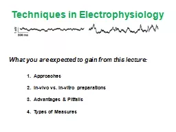
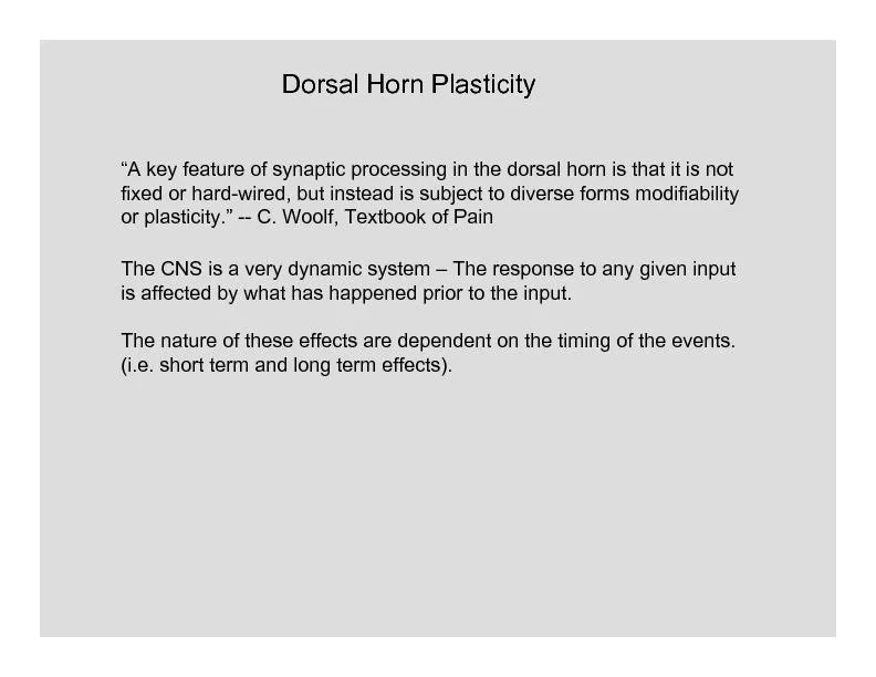
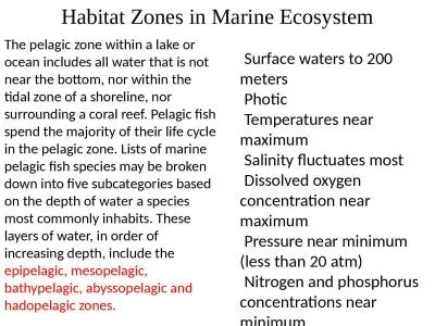
Michael P Tekin and William L Coleman Department of Biological and Allied Health Sciences Bloomsburg University 400 E 2 nd St Bloomsburg PA 17815 Abstract Synapsins are a group of proteins classically thought to regulate synaptic vesicle cycling well upstream of vesicle exocytosis Several previous studies however have suggested that synapsin I may have multiple regulatory roles including functions closer in time to the actual release event This led to the hypothesis that synapsin I may interact with the major vesicle priming protein Munc13 Synapsin I Munc13 interaction was investigated using double immunofluorescence staining at earthworm . For example if people are asked to guess the weight of a prizewinning ox Galton 1907 the error of the average response is substantially smaller than the average error of individual estimates This fact which Galton interpreted as support for democrat What was the best and smallest discount our company received and what were our reasons for taking it 2 Can you anticipate the next discount 3 What concessions would be better for our company Everybody wants a good deal but what is a deal Most unit e orals molluscs with attractive shells starfish and pufferfish are particularly popular Gastropods and bivalves have been collected for centuries both as individual or ornamental shells and for shellcraft jewellery and handicrafts that are made of sh What . you are expected to gain from this lecture:. 1. . Approaches. 2. . . In-vivo . vs. . in-vitro . preparations. 3. . Advantages & Pitfalls. 4. . Types of Measures. 5 Common . Ephys. Approaches:. presynaptic ending – . portion of the . axon . conveying information to the next neuron. . Some terms…….. presynaptic ending – . the portion of the axon that is conveying information to the next neuron. Neurons: Cellular and Network Properties. Integration: Divergence. Figure 8-25a. Integration: Convergence. Figure 8-25b. Integration: The Abundance of Synapses on a Postsynaptic Neuron. Figure 8-26. Axon terminals. NATIONAL CARGO For transportation within New Zealand domestic pets may travel on Air New Zealand �ights, provided the following container criteria are met.Kennel/Cage Container Requirements Moving sheep into and within NSW NLIS databaseThe NLIS databasemust be notified of all sheepmovements. The persons responsible for notifying the database are:The saleyard for sheepsold through a sale Presynaptic events maybe generated by certain transmitter(s).. Somorjai Dávid. N. eurotransmitters. Neurotransmitters are endogenous bioactive substances . Substances used as neurotransmitters belong to three. distinct families:. Hiking and Backpacking Hiking and backpacking Hiking is generally a day trip where departure and return occur within the same day. Backpacking involves spending one or more nights in the outdoors, covering much greater distances. Within contemporary organizations, reframing an EEO office and its functions within the Office of Human Resources and Strategic Talent Management (HRSTM). Employee & Labor Relations portfolio creates a paradigm shift. Such a shift interdependently aligns and couples the functions and offers an efficient Lecture . 5. Dr. Amar AL-. M. usawi. MD, PhD. Synaptic plasticity:. . It is the changes in the synaptic functions as a result of the history of discharging at a synapse, i.e. synaptic conduction can be strengthened or weakened on the basic of past experience. . epipelagic. , . mesopelagic. , bathypelagic, . abyssopelagic. and . hadopelagic. zones.. Habitat Zones in Marine Ecosystem. Surface waters to 200 meters. Photic. Temperatures near maximum. Salinity fluctuates most.
Download Document
Here is the link to download the presentation.
"Colocalization of synapsin I and Munc13 within presynaptic"The content belongs to its owner. You may download and print it for personal use, without modification, and keep all copyright notices. By downloading, you agree to these terms.
Related Documents