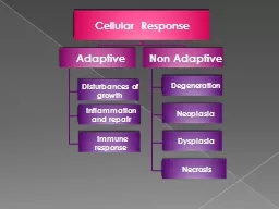
Degeneration Its a structure and function changes in the cells and intracellular substance CT due to mild and moderate injury not severe enough to cause cell death 1 Cloudy swelling ID: 259222
Download Presentation The PPT/PDF document "DEGENERATION" is the property of its rightful owner. Permission is granted to download and print the materials on this web site for personal, non-commercial use only, and to display it on your personal computer provided you do not modify the materials and that you retain all copyright notices contained in the materials. By downloading content from our website, you accept the terms of this agreement.
Slide1Slide2
DEGENERATIONSlide3
Degeneration:
It’s a structure and function changes in the cells and intracellular substance (C.T) due to mild and moderate injury, not severe enough to cause cell death.
Slide4
1- Cloudy swelling
:
Accumulation of water within the cells caused by ATP.
2-
Fatty changes
:
Pathological accumulation of excess neutral fat in the cells. Slide5
3-A
myloid
degeneration(
amyloidosis
):
Extracellular accumulation of
amyloid
(abnormal protein)
4-Hyaline degeneration:
Microscopic change in the tissues which become homogenous ,structure less & stain pink with eosin have a glassy appearance. Slide6
1- Cloudy swelling in kidney “Gross”:
The kidney are swollen due to entry of water , soft, bloodless and pale, the borders are rounded.Slide7
2- Cloudy swelling in kidney “Microscopic”:
Cells are swollen, conical in shape with fine red granular cytoplasm and normal nuclei, lumen of cell appear as star shape.Slide8
3- Fatty degeneration in liver “Gross”:
Pale Yellow color ,enlarged of size, soft and border round Slide9
4- Fatty degeneration in liver “Microscopic”:
-the cell swollen & show empty vacuoles in cytoplasm of cells .It is occupied by fat accumulation ,
-the nuclei of cell are pushed by accumulated fat against the cell membrane ,And become flattened giving the cell a signet ring appearance Slide10
5- Fatty degeneration in liver “Microscopic”:Slide11
6-
Amyloid degeneration in liver:
Homogenous pink a
myloid
deposit in:
-The branches of the hepatic artery in the portal tracts which become thickened and narrowed.
-B.M of sinusoids between lining endothelium and liver cells.
Slide12
7-
Amyloid degeneration in kidney “Gross”:
The kidney is enlarged, the cut surface is waxy with border shape &
shows light brown dots caused by
amyloid
depositSlide13
8-
Amyloid degeneration in kidney “Micro” Early stage:
Homogenous pink a myeloid deposit in wall of the arteries. The wall appear thick and narrow lumen
a myeloid deposit in the basement membrane of
glomerular
capillaries Slide14
9-
Amyloid degeneration in kidney (Advanced stage):
Homogenous pink a myeloid deposit in:
- wall of the arteries. It is appear thick and narrow lumen
the basement membrane of
glomerular
capillaries
the basement membrane of collecting tubules preventing
waterr
reabsorbing.Slide15
10- Hyaline degeneration in spleen:
Hyaline change affecting the central arterioles of the lymph follicles ,the C.T of it ,become structure less homogenous and pink with eosin and glass reflected material. The wall of arterioles become thick and narrowed lumens