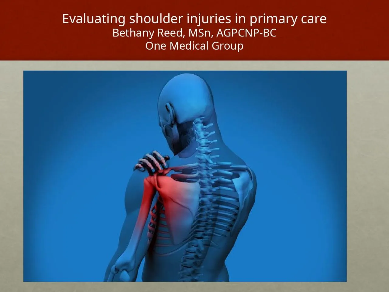
/
Evaluating shoulder injuries in primary care Evaluating shoulder injuries in primary care

Evaluating shoulder injuries in primary care - PowerPoint Presentation

byrne
byrne . @byrne
Follow
345 views
Uploaded On 2022-06-01

Evaluating shoulder injuries in primary care - PPT Presentation

Bethany Reed MSn AGPCNPBC One Medical Group Disclosures There has been no commercial support or sponsorship for this program The planners and presenters have declared that no conflicts of interest exist ID: 913239

rotator shoulder pain cuff shoulder rotator cuff pain test impingement joint rotation tears tear strength disease external physical supraspinatus

Share:

Link:

Embed:

Download Presentation from below link

Download Presentation The PPT/PDF document "Evaluating shoulder injuries in primary ..." is the property of its rightful owner. Permission is granted to download and print the materials on this web site for personal, non-commercial use only, and to display it on your personal computer provided you do not modify the materials and that you retain all copyright notices contained in the materials. By downloading content from our website, you accept the terms of this agreement.


Presentation Transcript

Slide1

Evaluating shoulder injuries in primary careBethany Reed, MSn, AGPCNP-BCOne Medical Group

Slide2

DisclosuresThere has been no commercial support or sponsorship for this program.The planners and presenters have declared that no conflicts of interest exist.

The program co-sponsors do not endorse any products in conjunction with any educational activity.

Slide3

https://www.hss.edu/pcp.asp

https://

www.hss.edu/professional-conditions_musculoskeletal-medicine-for-the-primary-care-physician-shoulder-exam.asp

Slide4

How often do you assess shoulder injuries in your practice?

Daily

Weekly

Monthly

Yearly

Never

Slide5

Session objectivesDiscuss anatomy of the shoulder joint.

Identify common injuries/conditions of shoulder injuries in the primary care setting—including typical presentation and mechanisms of injury.

Review physical examination skills and orthopedic testing.

Participation analysis case scenarios for your reference to test your skills.

Discuss operative and non-operative approaches for the treatment and rehabilitation process.

Slide6

Common shoulder injuriesAcute Onset

Fractures

Dislocations/subluxations

Sprains/strains

Contusions

Rotator cuff tears

Bicep tendon ruptures

Calcific tendonitis

Adhesive capsulitis

Chronic

Osteoarthritis

Rotator cuff impingement

Cervical disease

Slide7

Case Study38 yo female right shoulder pain worsening 3 weeks after starting CrossFit program with heavy lifting. Pain began 2/10 and has progressed to 7/10. Pain comes and goes with overhead and cross body reach.Also c/o pain w/ sleeping on right side. Denies trauma. Doesn’t recall acute injury.

Denies numbness, tingling in right extremity.

Mild improvement w/ rest, ice, Advil.

PMH: Asthma, HLD

ROS: As above, all others negative

VS: HR 78, RR 14, B/P 130/85, T 99.1

Exam: Visual inspection unremarkable for edema, erythema, deformity. Tenderness over anterior GH joint. ROM limited flexion, abduction, and external rotation. (+) painful arc. (-) empty can, neurovascular intact

(

-) imaging

Slide8

Diagnosis

Acute rotator cuff tear

Labrum tear

AC separation

Osteoarthritis

Slide9

Shoulder anatomyBonyHumerus

Scapula

Clavicle

Joints

Scapulothoracic

Glenohumeral

Acromioclavicular

Coracoclavicular

Soft tissue

Trapezius, rhomboids, serratus, pectoralis minor/major, latissimus dorsi, deltoids, SCM

Rotator cuff—supraspinatus, infraspinatus, teres minor, subscapularis

Capsule and ligaments

Labrum

Tendons

Rotator cuff tendons, long head of biceps

Subacromial and subdeltoid bursa

Slide10

http://hospitalforspecialsurgery.interactive.understand.com/view/shoulder/rotator-cuff-anatomy

Slide11

Shoulder pain: Differential diagnosesTraumatic disorders

Instability

AC joint disease

Rotator cuff disease

Tears

Bursitis/tendonitis [impingement]

Calcific tendonitis

Adhesive capsulitis (frozen shoulder)

Osteoarthritis

Cervical disease

Slide12

Rotator cuff pathologyImpingement syndromeCalcific tendonitis

Rotator cuff tears

Partial thickness tears

Full thickness tears

Massive tears

Slide13

Musculoskeletal work-upHistoryInspection

P

alpation

R

ange of Motion

Other Tests

S

trength

NeurovascularResisted wrist extension tests radial nerve, Resisted opposition of thumb test median nerve, Resisted digit abduction tests the ulnar nerve

Slide14

historyAge

Hand dominance (RHD, LHD)

Occupation

CC: pain, weakness, instability, strength

Location—where is the pain?

What is the quality of the pain?

Onset

Precipitating and alleviating factors

Associated medical conditions and social history

Most importantly smoking!

Previous treatments: surgeries, medications, PT, injections

Neurological complaints: numbness, tingling, weakness

Slide15

Shoulder disorders by ageAge 12-30

Labral tears

Instability

Traumatic disorders

Age 30-50

Rotator cuff disease

Calcific tendonitis

Adhesive capsulitis

Age 50-90

Rotator cuff

Tears

Impingement

Calcific

tendonitis

Osteoarthritis

Adhesive capsulitis

Slide16

AlgorithmHistory of severe trauma? Deformity? Severe acute pain? Fracture? Dislocation?If Yes, refer for X-ray, A&E, specialist.If No, do they have referred pain?

Cervical pathology—degenerative disc disease, costochondritis, cardiac—myocardial ischemia, pericarditis, pulmonary—pneumonia, diaphragmatic irritation—ulcer

If No, do they have systemic illness?

Polymyalgia rheumatica, malignant tumor, brachial neuritis, Herpes Zoster, Paget’s Disease,

Fibromyalgia

If Yes, refer, investigate, treat accordingly

If No. . . . .

Slide17

AlgorithmAcuteFractures of clavicle, humerus and scapula

Glenohumeral dislocations

AC joint sprain separation

Rotator cuff injury/tear

Chronic

Rotator cuff tendonitis (including biceps tendon/bursitis/tears)

Frozen Shoulder (Adhesive Capsulitis)

Arthritis of the glenohumeral joint

Slide18

Physical examinationObservation/Inspection

Erythema

Swelling

Ecchymosis

Deformity

Bony or soft tissue

Asymmetry

Atrophy

Slide19

Shoulder and physical examPalpation

C spine

Upper trapezius

AC joint

Long head of the bicep

Greater tuberosity

Slide20

Shoulder and physical examRange of Motion

Neck and shoulders

Always compare bilaterally

Active and passive

Forward elevation

External rotation

Internal rotation with adduction (to vertebral level)

Slide21

Shoulder physical exam

Slide22

The empty can test should be done with the thumbs pointing up toward the ceiling?

True

False

Slide23

Rotator cuff testing

Slide24

What is the name of this useful assessment test?

Empty Can test

Lachman test

Hawkins-Kennedy test

Neers test

Slide25

TestingInfraspinatus and teres minor muscle strength test—resisted external rotation—pain or weakness + for tendonopathy or tear

Subscapularis muscle strength test—resisted internal rotation or push off test—adduct arm and internally rotate behind back resist patient’s hand as pushes hand away from back

Hawkin’s-assesses for rotator cuff impingement—stabilize scapula, passively abduct shoulder to 90, flex shoulder to 30, flex elbow to 90 and internally rotate the shoulder--+ if painful . . . Also,

Neer’s-stabilize scapula with thumb pointing down and passively flex arm--+ if painful

C

ross arm flexion test—AC arthritis or subluxation—flex shoulder 90 and adduct across body--+ pain at AC joint.

Active ROM shoulder—flexion, extension, abduction, adduction, external rotation, internal rotation, posterior scratch test (adduction/external rotation) and (adduction/internal rotation)

Assess strength of rotator cuff muscles—drop arm test—evaluates for supraspinatus muscle tear—abduct shoulder to 90, flex to 30, and point thumbs down—test is + if patient is unable to keep arm up after examiner releases

Resistance known as Empty

C

an (Jobe’s test) test evaluates supraspinatus muscle strength--+ result indicates tendonopathy or tear

Slide26

Rotator cuff testing

Slide27

External rotationPositive findings: Decreased strength or pain on resisted testing.Significant weakness-suprascapular nerve palsy secondary to trauma, ganglion cyst or injury

Slide28

What shoulder muscle is this test assessing strength of?

Infraspinatus

and

teres

minor

Pectoralis

minor and

serratus

anterior

Supraspinatus and

teres

minor

All of the above

Slide29

Subscapularis lift-off testEvaluates the muscular strength of the subscapularisPositive findings: Inability to lift the dorsum of hand off the back

Slide30

Impingement/rotator cuff(Special Tests)Neer’s Impingement

Assesses the presence of impingement of the rotator cuff, primarily the supraspinatus, as it passes under the subacromial arch during forward flexion

Positive findings: Pain in the anterior should or reproduction of the patient’s symptoms

Slide31

Hawkins Kennedy Impingement TestEvaluates impingement of rotator cuff and subacromial bursa.Positive findings: Pain in the anterior shoulder or reproduction of the patient’s symptoms with the test.

Slide32

Rotator cuff disease impingement syndromeTendonitis/bursitis

Subacromial

Supraspinatus

History:

Pain reaching to side and back, overhead

Pain sleeping

Physical exam findings:

Little to no weakness

+ Neer’s Impingement

+ Hawkins Kennedy

+ Jobe’s/Empty can

+ Scapular retraction

Slide33

Rotator cuff diseaseimpingement syndromeTreatment

NSAID’s

Rehabilitation

Postural training, periscapular stabilization, strengthening of rotator cuff and scapular muscles

Posterior stretching

Activity modification

Injections

Lidocaine + corticosteroid

Surgical intervention

Slide34

Calcific tendonitisCalcification within rotator cuff tendon supraspinatus

ACUTE onset, very painful

Painful arc of motion

Treatment:

NSAID’s

Injection

PT

Surgery

Slide35

Slide36

Rotator cuff tearsFollow impingement

Can start small and progress

Trauma

Physical exam findings

Weakness

TREATMENT

Rehabilitation

Injections

Surgery

Arthroscopic repair

Not all tears require surgery

Slide37

Adhesive capsulitis—frozen shoulderPainful shoulder

Restricted ROM

Insidious

Active and passive

X-ray is normal

Shoulder capsule thickens r/t inflammation

Etiology

Idiopathic

Diabetes Mellitus

Post traumatic

Post

surgical

Treatment:

NSAID’s, PT, intra-articular injection, TIME.

Slide38

Ac separationVarious types

Fall on tip of shoulder

Possible bony deformity

Pain on palpation of AC joint

Pain on cross body adduction

Differential diagnoses include AC arthropathy, AC osteoarthritis

Treatment:

Sling for comfort

NSAID’s

PT

Surgery in severe cases

Slide39

Labral tear

Slide40

Speed’s test

Slide41

Suspected labral tearNo real reason for acute MRIStart PT and NSAIDIf no improvement in 6 weeks obtain MRI

MRI shows labral tear

Slide42

Glenohumeral osteoarthritisDegenerative process

Progressive pain

Limitation in ROM

Active and passive

Forward elevation

External rotation

Internal rotation

Abduction

Treatment:

NSAID’s

PT

Injections

Surgery—joint replacement

Slide43

SUMMARYWith careful history and physical examination the diagnosis can be made in most cases . . . not everyone needs an MRI.

MRI if suspected large rotator cuff tear, or in patients who fail to progress with other treatment.

Never too much of a downside to giving someone 1-2 weeks of therapy or rest and re-examining the shoulder.

At least from a surgeon standpoint . . .

Immediate MRI in everyone with work injury may lead to incidental findings

i.e. ‘What am I supposed to do with this information?’

Slide44

Slide45

Slide46

Keep in mind . . . Thoracic outlet syndromeAdditional labral tear testsSpurling’s for cervical root impingementGlenohumeral joint sulcus

Impingement signs

Slide47

Case Study38 yo female right shoulder pain worsening 3 weeks after starting CrossFit program with heavy lifting. Pain began 2/10 and has progressed to 7/10. Pain comes and goes with overhead and cross body reach.Also c/o pain w/ sleeping on right side. Denies trauma. Doesn’t recall acute injury.

Denies numbness, tingling in right extremity.

Mild improvement w/ rest, ice, Advil.

PMH: Asthma, HLD

ROS: As above, all others negative

VS: HR 78, RR 14, B/P 130/85, T 99.1

Exam: Visual inspection unremarkable for edema, erythema, deformity. Tenderness over anterior GH joint. ROM limited flexion, abduction, and external rotation. (+) painful arc. (-) empty can, neurovascular intact

(

-) imaging

Slide48