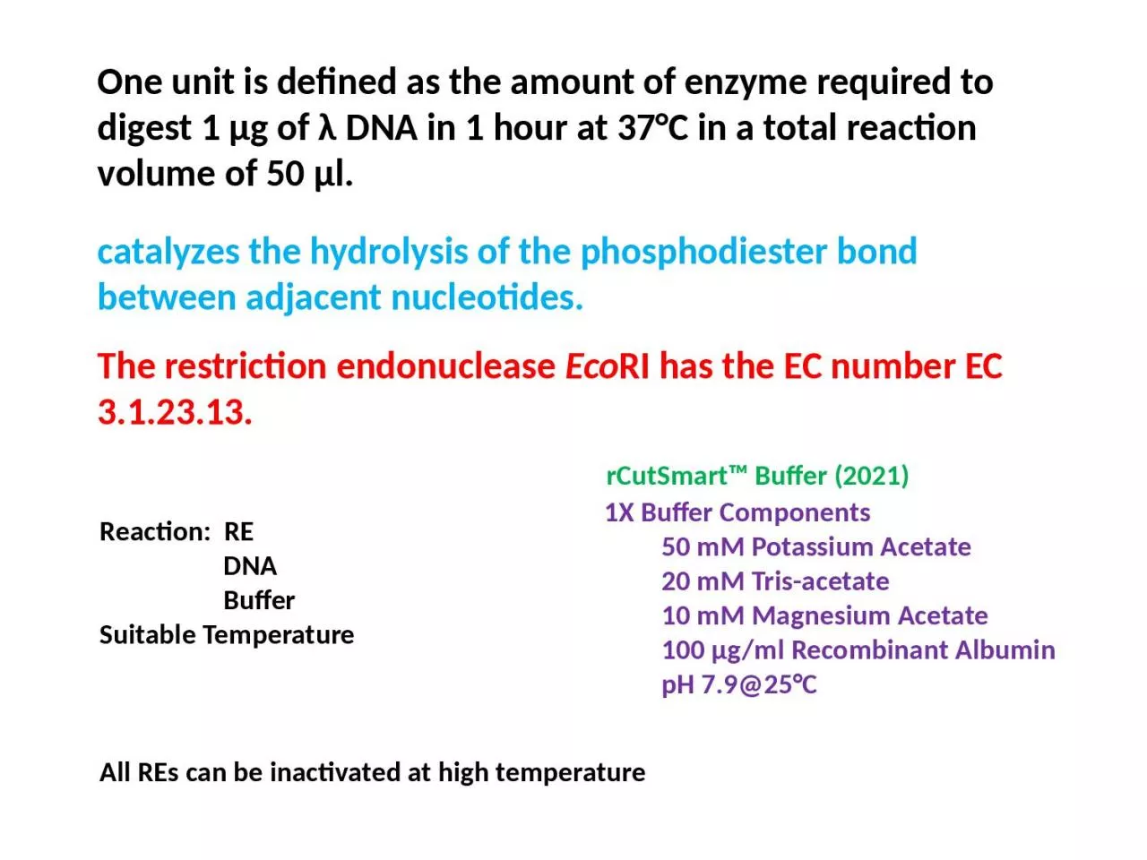
/
One unit is defined as the amount of enzyme required to digest 1 µg of λ DNA in 1 hour One unit is defined as the amount of enzyme required to digest 1 µg of λ DNA in 1 hour

One unit is defined as the amount of enzyme required to digest 1 µg of λ DNA in 1 hour - PowerPoint Presentation

byrne
byrne . @byrne
Follow
66 views
Uploaded On 2023-08-23

One unit is defined as the amount of enzyme required to digest 1 µg of λ DNA in 1 hour - PPT Presentation

Reaction RE DNA Buffer Suitable Temperature All REs can be inactivated at high temperature catalyzes the hydrolysis of the phosphodiester bond between adjacent nucleotides ID: 1014107

restriction dna fragments sites dna restriction sites fragments recognition site ecori sequence plasmid enzymes digestion lane genome human gel

Share:

Link:

Embed:

Download Presentation from below link

Download Presentation The PPT/PDF document "One unit is defined as the amount of enz..." is the property of its rightful owner. Permission is granted to download and print the materials on this web site for personal, non-commercial use only, and to display it on your personal computer provided you do not modify the materials and that you retain all copyright notices contained in the materials. By downloading content from our website, you accept the terms of this agreement.


Presentation Transcript

1. One unit is defined as the amount of enzyme required to digest 1 µg of λ DNA in 1 hour at 37°C in a total reaction volume of 50 µl.Reaction: RE DNA BufferSuitable TemperatureAll REs can be inactivated at high temperature catalyzes the hydrolysis of the phosphodiester bond between adjacent nucleotides.The restriction endonuclease EcoRI has the EC number EC 3.1.23.13.1X Buffer Components 50 mM Potassium Acetate 20 mM Tris-acetate 10 mM Magnesium Acetate 100 µg/ml Recombinant Albumin pH 7.9@25°CrCutSmart™ Buffer (2021)

2. Under extreme conditions, such as elevated pH or low ionic strength, restriction endonucleases are capable of cleaving sequences which are similar but not identical to their defined recognition sequence.This altered specificity is known as star activity.For example, EcoRI* (EcoRI star activity) cleaves the sequence N/AATTN, where N is any base, whereas EcoRI cleaves the sequence GAATTC.Star ActivityHigh-Fidelity (HF®) restriction enzymes have the same specificity as native enzymes, but have been engineered for significantly reduced star activity and performance in a single buffer (rCutSmart™ Buffer).

3. Certain restriction endonucleases show preferential cleavage of some sites in the same DNA moleculeFor example, phage λ DNA has five sites for EcoRI but the different sites are cleaved nonrandomly (Thomas & Davis 1975). The site nearest the right terminus is cleaved 10 times faster than the sites in the middle of the molecule. There are four sites for SacII in λ DNA but the three sites in the centre of the molecule are cleaved 50 times fasterthan the remaining site.There is a group of three restriction enzymes which show an even moredramatic site preference. These are NarI, NaeI and SacII and they require simultaneous interaction with two copies of their recognition sequence before they will cleave DNA. Thus NarI will rapidly cleave two of the four recognition sites on plasmid pBR322 DNA but will seldom cleave the remaining two sites.

4. A restriction map is a map of known restriction sites within a sequence of DNA. Restriction mapping requires the use of restriction enzymes. 

5. DigestionDouble DigestionPartial DigestionComplete DigestionTerminology

6. Cloning Strategy

7. M 1 2 3 4 M 6,557 23,1309,4104,3612,3222,0270.7% agarose gel showing genomic DNA preparations from Xanthomonas oryzae pv. oryzae. M denotes -Hind III molecular weight marker, Lane 1-4:genomic DNA.Credit: Alok Pandey LabCASE 1gDNA

8. M 1 2 3 1,517100600500bp 1.3% agarose gel showing PCR amplified 510bp fragment of feoB gene from Xoo genomic DNA (Lane 1-3). M denotes 100bp DNA ladder.Credit: Alok Pandey LabPreparation of Insert (Gene of Insert)

9. 10.0M 1 2 3.0Fig.5: 0.7% agarose gel showing plasmid preparations from two different recombinant clones (Lane 1-2). M represents 1 kb DNA ladder. Open circular (OC) and super coiled (SC) forms of plasmid can be seen in above plasmid preparations.Isolation of recombinant plasmidCredit: Alok Pandey Lab

10. 1.0500Ma 1 2 3 4 Mbkb10.04.03.01.50.52.0600bp1001,0001,5171.0% agarose gel showing restriction digestion analysis of recombinant plasmid pAP1. Lane 1-2: EcoR I digest of pAP1, Lane 3-4: EcoR I-Hind III digest of pAP1 .In lane 3-4, release of insert and the 2.9kb pMOSBlue band can be seen. Ma and Mb denote 1kb DNA ladder and 100bp DNA ladder respectively.ConfirmationCredit: Alok Pandey Lab

11. In above image, there are two X sites and 1 Y site;Complete digestion with X = 2 fragments of 700 and 300 bpComplete digestion with Y = 1 fragment of 1000 bp (single site cutting of a circular DNA, linearizes it).Circular DNA: no. of restriction sites n = n no. of fragmentsI IIProblem 1

12. Problem 2

13. Problem 3In Above image, there is one Sal I site and two Kpn I site.Complete digestion with Sal I = two fragments of 450 and 1200 bpComplete digestion with Kpn I = three fragments of 200, 400 and 1050 bpLinear DNA: no. of restriction sites n = (n+1) no. of fragments

14. Approximately how many fragments of human DNA will be formed if it is digested with EcoRI restriction enzyme?Human genome size = 3x10^9 bpEcoRI recognition seq = GAATTC (6bp, palindrome)Frequency of occurrence of EcoRI recognition site in given sequence considering that the all 4 bases have equal probability of occurrence:(1/4)^6 = 1/4096i.e. One EcoRI site can be found in 4096 bp.Now number of sites in human genome = 3x10^9 / 4096= 732421.875As EcoRI can not cut a sequence if it is not complete, so 732421 recognition sites in human genome can be found.As you know human genome is linear genome, and when you digest it with Restriction Endonuclease having one recognition site, it always gives two fragments.means if there are n sites, there will be n+1 fragments.So, total number of human genome fragments generated by EcoRI digestion (complete) = 732421+1 = 732422Problem 4

15. Type II Restriction Endonucleases represent the largest group of characterized enzymes owing to their usefulness as tools for recombinantDNA technology, and they have been studied extensively.Over 300 Type II REases, with >200 different sequence specificities,are commercially available.Currently, >19 000 putative REases are listed on REBASE (http://rebase.neb.com)The Restriction Enzyme Database is a collection of information about restriction enzymes, methylases, the microorganisms from which they have been isolated, recognition sequences, cleavage sites, methylation specificity, the commercial availability of the enzymes, and references - both published and unpublished observations (dating back to 1952). REBASE is updated daily and is constantly expanding.

16.

17. Q. Restriction map for the plasmid (pLSBT22) is given below. DNA length (in Kb) has given that is present between two consecutive restriction sites? Using this map, find the size (in Kb) of each restriction fragments that would result from digesting pLSBT22 with the restriction enzymes (I) EcoRI, (II) HindIII, (III) BamHI, and (IV) a combination with EcoRI + BamHI.9Kb5Kb8 Kb10 Kb6 KbEcoRIBamHIBamHIEcoRIHindIII pLSBT22

18. Digestion performed withRestriction EnzymeSize of Fragments ObtainedHindIII30 KbBamHI18 Kb, 8 Kb, 4 KbHindIII + BamHI13 Kb, 8 Kb, 5 Kb, 4 KbQ 2. An animal biotechnologist performed the following set of restriction digests on a novel plasmid, pUU214.The reaction he carried out, along with the DNA fragments obtained in single and double-digest reactions, were:Using the above information, construct a restriction map of pUU214.

19. DNA X and DNA Y were digested with restriction enzyme HindIII and analyzed by agarose gel electrophoresis. If DNA X gave three fragments and DNA Y gave four fragments, then which of the following are correct?P. DNA X has two restriction recognition sites and is circularQ. DNA Y has four restriction recognition sites and is circularR. DNA X has two restriction recognition sites and is linearS. DNA Y has two restriction recognition sites and is linear(A) Q and R (B) P and S (C) P and Q (D) R and S Q2.