/
2 Restriction Enzyme Digestion Gel Extraction and 2 Restriction Enzyme Digestion Gel Extraction and

2 Restriction Enzyme Digestion Gel Extraction and - PDF document

anya
anya . @anya
Follow
343 views
Uploaded On 2022-09-08

2 Restriction Enzyme Digestion Gel Extraction and - PPT Presentation

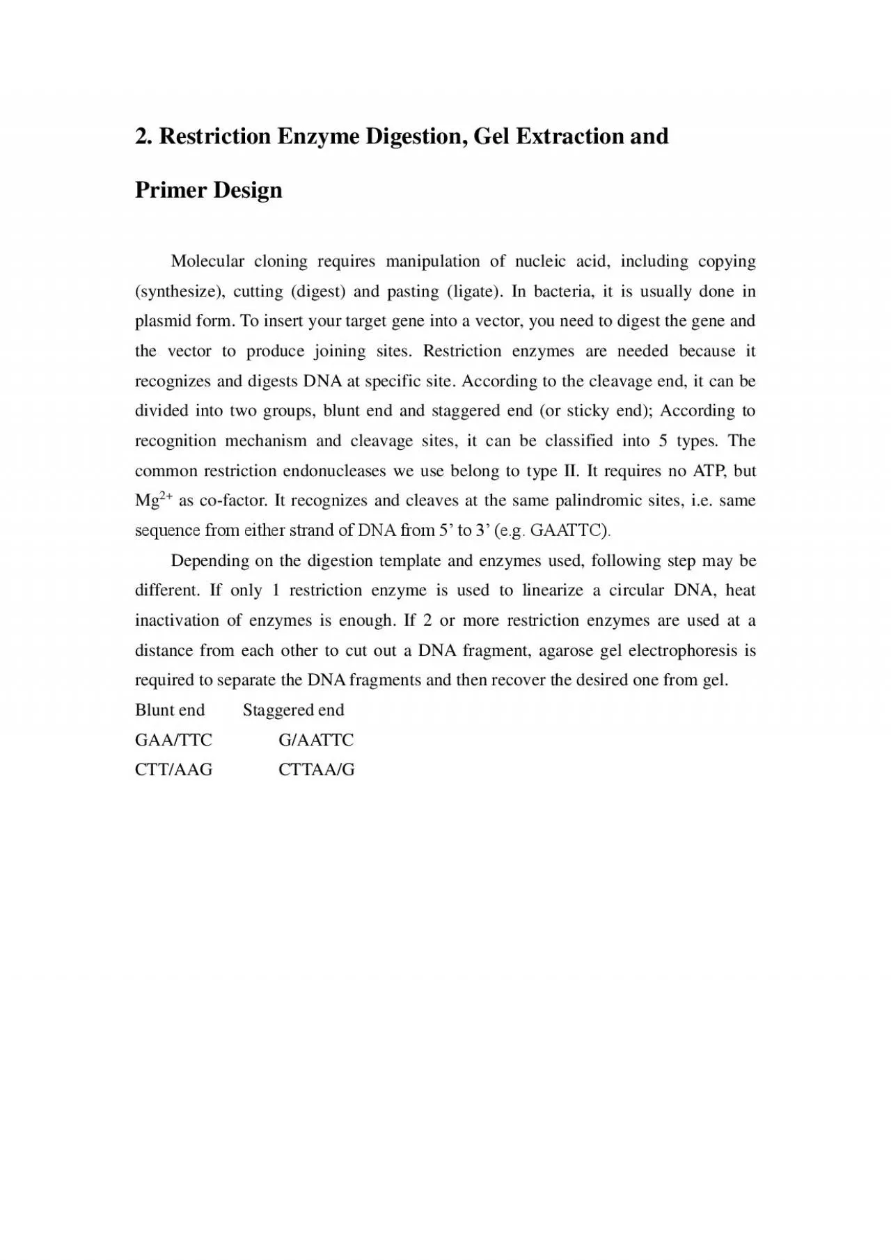
Primer Design Molecular cloning requires manipulation of nucleic acid including copying synthesize cutting digest and pasting ligate In bacteria it is usually done in plasmid form To inse ID: 953323

gel dna primer enzyme dna gel enzyme primer column quick qia buffer restriction min add volume template enzymes digestion

Share:

Link:

Embed:

Download Presentation from below link

Download Pdf The PPT/PDF document "2 Restriction Enzyme Digestion Gel Extra..." is the property of its rightful owner. Permission is granted to download and print the materials on this web site for personal, non-commercial use only, and to display it on your personal computer provided you do not modify the materials and that you retain all copyright notices contained in the materials. By downloading content from our website, you accept the terms of this agreement.


Presentation Transcript

2. Restriction Enzyme Digestion, Gel Extraction and Primer Design Molecular cloning requires manipulation of nucleic acid, including copying (synthesize), cutting (digest) and pasting (ligate). In bacteria, it is usually done in plasmid form. To insert your target gene into a vector, you need to digest the gene and the vector to produce joining sites. Restriction enzymes are needed because it recognizes and digests DNA at specific site . According to the cleavage end, it can be divided into two groups, blunt end and staggered end (or sticky end); According to recognition mechanism and cleavage sites, it can be classified into 5 types. The common restriction endonucleases we use belong to type II. It requires no ATP, but Mg 2+ as co - factor. It recognizes and cleaves at the same palindromic sites, i.e. same sequence from either strand of DNA from 5’ to 3’ (e.g. GAATTC). Depending on the digestion template and enzymes used, following step may be different. If only 1 restriction enzyme is used to linearize a circular DNA, heat inactivation of enzymes is enough. If 2 or more restriction enzymes are used at a distance from each other to cut out a DNA fragment, agarose gel electrophoresis is requi red to separate the DNA fragmen

ts and then recover the desired one from gel. Blunt end Staggered end GAA/TTC G/AATTC CTT/AAG CTTAA/G A. Restriction Enzyme Digestion This step is to generate sticky ends or blunt ends of the 5’ end/ 3’ end of the DNA, or to change a circular plasmid into a linear form. The figure below illustrates double enzyme digestion of DNA for biobrick construction. Materials Restriction enzyme(s) 100  BSA (b ovine serum albumin ) 10  NEB buffer ddH 2 O ( d eionized, sterile, DNase - free) DNA template 37 o C incubator or dry bath Procedure 1. Place the required reagents on ice. Label them. 2. Follow the table below to prepare restriction digestion reaction mixture: Reaction Volume 10 μl 25 μl 50 μl DNA template 0.1 µg 0.5 µg 1 µg 10X Buffer (1 - 4, according to enzymes) 1 2.5 5 100X BSA 0.1 0.25 0.5 Enzyme 1 U 5 U 10 U * Enzymatic units differ among enzymes and companies. Refer to the documents of the supplier to calculate the needed amount. 3. Pipette the solution up and down to ensure all reagents are mixed well. 4. Place the reaction mixture at 37 o C incubato r or dry bath fo r 2 - 4 hours. 5. Pu

rify the DNA by PCR purification kit/gel extraction kit for downstream process. Notes - Water is always added first. - Buffer is always added before that of enzyme. - Make sure that you use the relevant buffer. - Beware of the temperature for optimal enzyme digestion and DO NOT over - digest your DNA. - Enzyme dissolved in glycerol sticks to the sides of the pipette tip. Therefore, just touch the pipette tip to the surface of the enzyme solution to ensure correct volume of the enzyme being pi petted. - Total enzyme volume should be lower than 10 % of the reaction volume to avoid star activity B. DNA Purification from Gel by spin column This step is to purify the DNA (70 bp – 10 kb) by removing the nucleotides, enzymes and ions from solution, and separate different pieces of DNA for target DNA selection. The purified DNA is suitable for downstream process. Materials Gel clean kit ddH 2 O ( d eionized, sterile, DNase - free) 60 o C dry bath Procedures (Modified fr om QIAGEN Gel clean kit protocol ) All centrifugation steps are carried out at 10,000 – 13,000 rpm (17,900 x g) in a conventional tabletop micro centrifuge at room temperature. 1. Excise the DNA fragment fr

om the agarose gel with a clean, sharp scalpel. Minimize the size of the gel slice by trimming off extra agarose gel. 2. Weigh the gel slice in a colorless tube. Add 3 volumes of Buffer QG to 1 volume of gel (300 μl QG to 100 mg gel). For >2% agarose gels, add 6 volumes of Buff er QG. The maximum amount of gel slice per QIA quick column is 400 mg. (This video shows the procedures of restriction enzyme digestion) 3. Incubate the tube at 60°C for 10 min (or until the gel slice has completely dissolved). To help dissolve gel, mix by inverting the tube every 2 min during the incubation. Solubilize aga rose completely. Fo�r 2% gels, increase incubation time. 4. If the color of the mixture is orange or violet, add 10 μl of 3 M sodium acetate, pH 5.0, and mix the reagents. The color of the mixture will turn to yellow. The adsorption of DNA to the QIA quick m embrane is efficient only at pH ≤7.5. Buffer QG contains a pH indicator, which is yellow at pH ≤7.5 and orange or violet at higher pH. 5. Add 1 gel volume of isopropanol to the sample and mix the reagents well. For example, if the agarose gel slice is 100 mg, add 100 μl isopropanol. This step increases the yield of DNA fragments 500 bp and 䀀4 kb. For DNA fra

gments between 500 bp and 4 kb, addition of isopropanol has no effect on yield. Do not centrifuge the sample at this stage. 6. Place a QIA quick spin column in a provided 2 ml collection tube. 7. To bind DNA, apply the sample to the QIA quick column, and centrifuge for 1 min. The maximum volume of the column reservoir is 800 μl. For sample volumes of more than 800 μl, simply load and spin again. 8. Discard flow - thr ough and place QIA quick column back in the same collection tube. It can be reused to reduce plastic waste. 9. Add 0.75 ml of Buffer PE to wash the QIA quick column, and centrifuge for 1 min. If the DNA will be used for salt - sensitive applications, such as bl unt - end ligation and direct sequencing, let the column stand 2 – 5 min after addition of Buffer PE, before centrifugation. 10. Discard the flow - through and centrifuge the QIA quick column for one more min. Residual ethanol can inhibit downstream process. Ethanol from Buffer PE will not be completely removed unless the flow - through is discarded before this additional centrifugation. 11. Place QIA quick column into a clean 1.5 ml micro centrifuge tube. 12. To elute DNA, add 30 - 60 μl of DNA extraction buffer (10 mM Tris·

Cl, pH 8.5) or water (pH 7.0 – 8.5) (pre - warmed at 60 o C) to the center of the QIA quick membrane, let the column stand for 1 min, and then centrifuge the column for 1 min. Notes One advantage using Gel purification kit over PCR purification kit is that it can purify single type of DNA when there is any other suspected DNA contamination (such as primer - dimer, non - specific amplification, etc). You don’t have to use QIA quick from Qi agen, other brand names will do just fine but you need to follow their protocols carefully. C. Primer design To initiate nucleotide synthesis, primers are needed to specify the location. Especially in PCR, there is no topoisomerase to stabilize unwinding double helix, the specificity is defined by the complementarity of primer sequence and the annealing temperatu re of the primer:template duplex. Following requirements are suggested for good primer design:  GC content : 40 – 60%. As GC pair has 3 hydrogen bonds, while AT pair has only 2, higher GC content means higher binding strength of primer to the template. Howev er, the primer may lose its specificity if its GC content is too high.  Length : A primer usually has around 15 bp, but it can be as long as 30 or e

ven more in some cloning method, e.g. overlapping PCR. The longer the length, the higher the binding strength and melting temperature.  Melting temperature (Tm) : The temperature at which 50% of the oligonucleotide and its perfect complement are in duplex. Wallace rule gives (This video shows the procedures of gel extraction) the most basic calculation for short oligonucleotides at 0.9 M NaCl, Td = 2°C(A+T) + 4°C(G+C ). More accurate thermodynamic calculation gives the nearest neighbour melting temperature. The Tm difference between primer pair should be within 5˚C.  Secondary structure : Primers are single - stranded. It tends to for secondary structure, e.g. hairpin, to stabilize itself. PCR will fail if formation of primer hairpin is more preferable than primer:template duplex. Some online tool can do prediction, e.g. sequence manipulation suite.  Self - annealing : Primers have chance to form dimer, or even multi - mer in som e cases. It may not be “lethal”, but better avoid it.  Runs of A/T : It may cause frameshift.  GC clamp : 2 G/C at the 3’ - end is essential to ensure the primer the tightly bound to the template for elongation. More than 3 G/C at the 3’ - end may cause non - specif ic bi