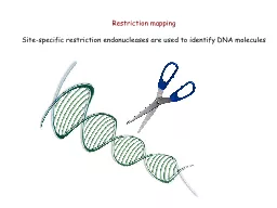
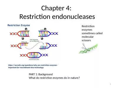
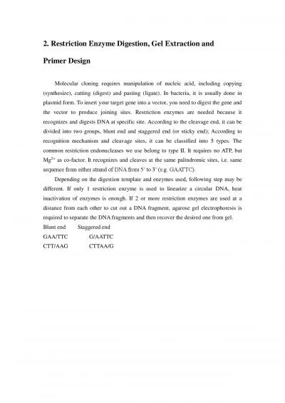
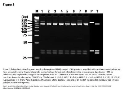
Objectives Understand how Restriction Enzymes digest DNA Know how to construct a pAMP plasmid or gel Given the size of fragments gel know how to construct a restriction map Given a restriction map know how to construct a gel ID: 911424
Download Presentation The PPT/PDF document "Restriction Enzyme digestion of DNA - Ex..." is the property of its rightful owner. Permission is granted to download and print the materials on this web site for personal, non-commercial use only, and to display it on your personal computer provided you do not modify the materials and that you retain all copyright notices contained in the materials. By downloading content from our website, you accept the terms of this agreement.
Slide1
Restriction Enzyme digestion of DNA - Exercise 8
Objectives
-Understand how Restriction Enzymes digest DNA.
-Know how to construct a
pAMP
(plasmid) or gel.
-Given the size of fragments, gel, know how to construct a restriction map.
-Given a restriction map know how to construct a gel.
Slide2NOTE: DNA IS Negatively charge
because of the phosphate groups.
Slide3DNA molecules are macromolecules that hold the genetic information of living organisms. They are extremely long, double-stranded polymers of nucleotides.
The covalent bond joining adjacent nucleotides in DNA is called a
phoshodiester
bond.
The
phoshodiester
bonds between nucleotides in DNA molecules are very stable unless they are physically stretched or exposed to enzymes name nucleases.
Slide4Enzymes are capable of breaking (hydrolyzing)
phoshodiester
bonds in DNA molecules. Nucleases can be classified into two major groups:
exonucleases
and
endonuclases
.
Exonucleases
:
If the enzyme digest nucleotides from the ends of the DNA molecules
.
Endonuclases
:
If the enzyme digest nucleotides in the interior of a DNA molecule
.
Restriction
endonuclease
: An
enzythem
that digest DNA by recognizing specific short sequences of bases that are called palindromes.
Slide5A special class of
endonucleases
from a bacteria has been isolated for this experiment. These special enzymes, termed restriction
endonucleases
(RE)
,
digest DNA by breaking bonds only within a specific short sequence of bases. These base sequences usually ran in size from 4-8 base pairs but can be as long as 23 base pairs.
Restriction
endonucleases
confer an adaptive advantage on bacteria by digesting foreign DNA usually from an invading
bacteriphage
(bacterial virus). The resulting DNA fragments can then be further degraded and destroyed by
exonucleases
. These enzymes are used to cut DNA in a precise and predictable manner. They are extensively useful in gene cloning, DNA amplification, and many recombinant DNA technologies.
Restriction
endonuclease
(RE). This RE are also attained from bacteria. In a bacteria where we get these enzymes form there protected because if a virus invades a bacteria cell these
endonuclease
will chop up the virus DNA, its like a defense system, so we can isolate these
endonuclease
for experiments, but bacteria produce these
endonuclease
to protect themselves from foreign DNA entering their cells.
Slide62 Restriction
Endonucleases
(RE)
EcoR1 &
HindIII
. Both of these recognize different nucleotide sequences.
Each strand of DNA is cut at the
phoshodiester
bond between the G and A bases
(indicated by the arrow signs). Notice that the sequence GAATTC is the same on both strands when each strand is read 5’ -> 3’. Such symmetrical sequences are called
palindromes (In a English language a palindrome reads the same thing in both directions). This enzyme cuts the double strands asymmetrically, leaving protruding ends. These protruding bases are referred to as sticky ends aka compatible cohesive ends.
Slide7EcoR1: EcoR1 recognizes palindrome on DNA, and cuts the bond between G & A, and G & A. When you do that it opens your DNA. For example, if you have plasmid and that palindrome is present once on the plasmid, you’ll get one cut.
If somewhere else that palindrome is present and you incubate it with EcoR1, you’ll get another cut. So every time EcoR1 recognizes this palindrome on your plasmid, it will cut through the DNA. So when it opens up the DNA, may get a couple of unpaired bases, and those unpaired bases are called sticky ends, and if you throw some nucleotides from different species, you can make recombinant DNA.
Slide8Like EcoRI
,
HindIII
also recognizes a
palindromic
sequence, AAGCTT, and produces sticky ends. Sticky ends can hydrogen bond together other because of complementary base pairing.
Slide9Recombinant DNA molecules are compose of DNA fragments from two or more sources. Not all RE’s produce sticky ends. Some enzymes cut DNA to produce blunt ends, as shown here.
Slide10Once the DNA has been digested, the fragments must be separated and identified.
Fragments are separated by
agarose
gel electrophoresis. Agar is a large polysaccharide.
Gel electrophoresis: You put an
agorose
gel (
agrose
is a polysaccharide) and it has spaces, your DNA can move through these spaces, you put a current against this, the negative end is up, the positive is at the bottom, and because your DNA has a negative charge, the
DNA moves down towards the positive end.
Gel is immersed in an ionic buffer. The buffer has a pH above 8.0 DNA at this pH is negatively charge because the phosphates in the DNA backbone have lost hydrogen ions.
The dye molecules serve as the indicator of the
movements of invisible DNA molecule through
the gel as an electric current is run through the gel.
The negatively charged DNA will migrate from the anode to the cathode (negative to positive) alongwith the current.
Slide11Separation of the DNA fragments occurs as they migrate through the network of
agarose
molecules.
Smaller fragments slip through the network fast than large molecules. The rate of migration is a function of fragment size, as well as the density of
agarose
.
The tightness (concentration of
agarose
). High concentration favor smaller fragments.
Low concentration favor large fragments.
Each of migration function of fragment size and density of
agarose
. Depending on what conformation a circular DNA gets, it will run differently in the gel.
So not only does the size of the DNA molecule affect migration rate, but the configuration of the DNA also affects the migration rate
. The DNA that you will electrophoresing can exist in three different conformations.
Slide121. Supercoil
circular
: 1
st
fastest. When its circular it becomes twisted and turn and be comes a little bit shorter in size. Migrates fastest down the gel. Contains small volume, more compacted.
2.
Linear
: Migrates next fastest down the gel.
3. Nicked (relaxed) circular
: One strand is intact, the other is broken and when it is nicked, it becomes extended. This one is very relaxed and faces the most difficulty making its way through the
agarose
.
Supercoil
< Linear < Nicked (relaxed) circular
Slide13In addition to conformation affecting migration rate, laboratory production of plasmid DNA can be produce very large molecules that migrate very slowly.
Two possible molecules that can be produced are
dimers
and
concatemers
. A
dimer
consists of two plasmids covalently linked in a series end to end. A
Concatemer, for example, might consist of two plasmids with one hooked through the other but not covalently linked to each other. If a purified uncut plasmid is applied to a gel, bands of super coiled plasmid, nicked circular plasmid,
dimers
, and
concatemer
can be observed.
Dimer
: Means that its link together by 2 linksConcatemer: Mean a whole bunch of plasmids linked together but not covalently linked to each other.
Slide14pAMP
- the plasmid DNA. What we did in the experiment on DNA restriction analysis is we took
pAMP
(circle) and incubated the
pAMP
this plasmid with different restriction
endonucleases
.
From your electrophoresis gel, you can estimate the size of
pAMP
.
You can also determine if
pAMP
is circular or linear. Finally, you can use the gel to draw a restriction map. A restriction map is a physical map of a piece of DNA showing recognition sites of specific restriction enzymes separated by lengths marked in numbers of bases. Separated DNA base on size
The pattern of DNA bands is characteristic for a specific DNA sample and the restriction enzymes used to cleave it. A banding pattern can be referred to as a DNA fingerprint. because it is unique to that particular DNA (and the combination of restriction fragments).
We ran a gel to see if we could determine how many DNA fragments you got
. By
electrophoresing
a series of fragments of known size (DNA ladder) along with the DNA samples of interest, the sizes of unknown fragments can be estimated.
Slide15A restriction site is a place where an enzymes cuts DNA, so there are restriction sites for EcoR1, and for
HindIII
.
When constructing the
pAMP
no restriction site where you start and where you finish.
Lane 4: A control to see what uncut plasmid looks like. How uncut DNA traveled whether they made 1 or 2 pieces. It’s your plasmid DNA
DNA
was on tube 4 which acts like a measurements and acts like a ladder. No enzyme (Lane 4).
Lane 5: DNA ladder: DNA digest, containing known base pair lengths compare with fragments in lanes 1-3. You will run DNAs of known size (DNA ladder) to help you estimate the size of your DNA fragments. Lane 5 contains DNAs of known sizes (DNA ladder).
Slide16Prokaryotic (Circular) DNA
DNA from bacteria (both chromosomal DNA and extra chromosomal plasmid DNA) and viruses is often a closed circle. If you have a circular DNA, we know that’s Prokaryotic DNA.
In Prokaryotic DNA, the number of fragments will equal the number of restriction sites.
Eukaryotic (linear) DNA
If you have one restriction site for an enzyme, you would have 2 fragments, and if you have 2 restriction sites for an enzyme, you would have 3 fragments.
In Eukaryotic DNA, the number of fragments is always going to have one more or one less than restriction sites.
In Eukaryotic DNA, there’s no reason to see multiply bands in control lane because Eukaryotic DNA is linear, it doesn’t exist as
supercoil
, relax, or
multimere
so this is a hint in lane 4. So when you have Eukaryotic DNA, you will not see multiply bands in the control lane.
Also, just because they show you multiply bands, not every time your going to have prokaryotic (circular) DNA you get multiply lanes, its only if the DNA has been damaged into a
supercoil
.
Slide17Slide18What’s going to effect the movement of the DNA
(Factors)?
Slide19Size: small pieces migrate faster, farther than bigger pieces.
Conformation (shape): Comparing 3 pieces of DNA that are the same size.
Supercoil
< Linear < Nicked (relaxed) circular
Charge: Charge (+,-)DNA is negative because of Phosphate groups (anode) to positive (cathode).
Slide20Digestion of pAMP
with
EcoRI
&
HindIII
We incubated our plasmid under several conditions. Those conditions were that we incubate
pAMP
.
Lane 1: EcoR1 - one bandLane 2: HindIII - one band Lane 3: EcoR1 &
HindIII
- two bands
Lane 4: Water - Our control. We got one main band.
Lane 5: DNA ladder, a tool to measure the size of DNA fragments.
Slide21Slide22When constructing the
pAMP
.
There’s no restriction site
where you start and where you
finish the map. You could call
this point the reference point.
Also, all your base pairs
(fragments) have to equal the
total number base pairs
of your
plasmid. For example, 6,000
Bp’s in this example.
Slide23Starting & Ending Point
Slide24Key for pAMP
KEY
Enzyme A: Light green
Enzyme
B: Pink
Enzyme
C: Orange
Slide25Enzyme A
Slide26Enzyme A
Slide27Enzyme A
Slide28Enzyme A
Slide29Enzyme B
Slide30Enzyme B
Slide31Enzyme B
Slide32Enzyme C
Slide33Enzyme C
Slide34Enzyme A + B
Slide35Enzyme A + B
Slide36Enzyme A + B
Slide37Enzyme A + B
Slide38Enzyme A + B
Slide39Enzyme A + B
Slide40Enzyme A + B
Slide41Enzyme A + C
Slide42Enzyme A + C
Slide43Enzyme A + C
Slide44Enzyme A + C
Slide45Enzyme A + C
Slide46Enzyme A + C
Slide47Enzyme B + C
Slide48Enzyme B + C
Slide49Enzyme B + C
Slide50Enzyme B + C
Slide51Enzyme B + C
Slide52Enzyme A + B + C
Slide53Enzyme A + B + C
Slide54Enzyme A + B + C
Slide55Enzyme A + B + C
Slide56Enzyme A + B + C
Slide57Enzyme A + B + C
Slide58Enzyme A + B + C
Slide59Enzyme A + B + C
Slide60Are the number of fragments correct?
Slide61Slide62Slide63Slide64Slide65How do you cut and paste DNA?
Enzymes that cut DNA at specific short sequence sites
Restriction enzymes digest DNA
Blunt end cut
Asymmetric end cut
Enzymes that paste complementary DNA fragments together
DNA ligase
Slide66Using a restriction enzyme and DNA
ligase
to make recombinant DNA
Slide67Restriction fragment analysis by Southern blotting
Characteristic pattern of bands for each sample
DNA is transferred to paper and denature to single strands
Probe complementary to the DNA sequence of interest
DNA bound to radioactive probe exposes film
Entire genome
Slide68DNA CLONING AND ITS APPLICATIONS
Most methods for cloning pieces of DNA in the laboratory share general features, such as the use of bacteria and their
plasmids
.
Cloned genes are useful for making copies of a particular gene and producing a gene product.
Slide69RESTRICTION ENZYMES
Restriction enzymes
are essentially molecular scissors that cut DNA at specific nucleotide sequences.
They originate from bacteria and function as a defense system against viral invasion. They “restrict” viral DNA.
Slide70Restriction Enzymes
Cut DNA at highly specific points
Recognize specific sequences
Four to seven bases
Each is unique
Consistent results
Slide71STICKY ENDS
Most restriction enzymes cut double stranded DNA in an asymmetrical fashion.
These cuts leave single stranded nucleotide overhangs that are competent to hydrogen bond.
These overhangs are called “sticky ends”.
Slide72Slide73Slide74AGAROSE GEL ELECTROPHORESIS
One indirect method of rapidly analyzing and comparing genomes is
gel electrophoresis
.
This technique uses a gel as a molecular sieve to separate nucleic acids or proteins by size.
Slide75Gel Electrophoresis
Separation of DNA fragments
Based on size
Slide76Cathode
Power
source
Anode
Mixture
of DNA
molecules
of differ-
ent sizes
Gel
Glass
plates
Longer
molecules
Shorter
molecules
Slide77Different
Endonucleases
Yield Different Patterns
Taq1 + Pst1
Taq1 + AvaII
E coli
clinical isolates
Slide78Questions
1. What is a nuclease?
2. How does an
endonuclease
differ from an
exonuclease
?
3. What is a restriction
endonucleases
? Write names of some restriction
endouclease
.
Slide79Questions
1. What is a nuclease?
DNA held by covalent bond joining adjacent nucleotides in DNA is called a
phosphodiester
bond. The
phosphodiester
bond between nucleotide in DNA molecules are very stable unless they are physically stretched or exposed to enzymes name nucleases.
Enzyme capable of breaking (hydrolyzing)
phosphodiester
bonds in DNA molecules and classified into
exonuclease
and
endonuclease
.
2. How does an
endonuclease
differ from an
exonuclease
?
Endonuclease
digest DNA by breaking
phosphodiester
bonds in the interior of DNA molecule.
Exonuclease
enzyme digest nucleotides from the ends of the DNA molecule.
3. What is a restriction
endonucleases
? Write names of some restriction
endouclease
.
Restriction
endonucleases
are a special class of
Endonuclease
from bacteria to cut DNA.
EcoRI
& Hind III. These are enzymes digest DNA by recognizing specific short sequences of bases called
palindromic
.
Slide80Questions
4. What are 2 restriction
endonuclease
(RE) that we used in our lab? Write DNA sequences these RE recognize. Do they produce sticky ends or blunt ends when they cut the DNA molecules?
5. How does the number of restriction sites relate to the number of fragments produced for linear DNA or circular DNA?
6. What is
palindromic
DNA sequence?
Slide81Questions
4. What are 2 restriction
endonuclease
(RE) that we used in our lab? Write DNA sequences these RE recognize. Do they produce sticky ends or blunt ends when they cut the DNA molecules?
EcoRI
& Hind III. Both produce sticky ends when cut.
5. How does the number of restriction sites relate to the number of fragments produced for linear DNA or circular DNA?
Eukaryotic DNA, always going to have one more or one less fragment than you have restriction sites.
Prokaryotic DNA, the number of fragments will equal the number of restriction sites.
6. What is
palindromic
DNA sequence?
Reading from the same thing in both direction to read the sequences bases that restriction
endouclease
recognizes. For example,
M’adam
I’m
adam
.
Slide82Questions
7. What is electrophoresis? What does
agarose
gel electrophoresis allow us to do?
8. What is the chemical nature of
agarose
?
9. What factors effect the migration rate of DNA through an
agarose
gel?
10. For DNA molecules of equal sizes, how do the different shapes (conformation) of DNA differ in terms of distance traveled through an
agarose
gel?
Slide83Questions
7. What is electrophoresis? What does
agarose
gel electrophoresis allow us to do?
It’s a gel that allows move fragment of DNA across by attracting DNA, which is negative (anode) to opposite side (cathode) positive side base on size, and conformation of DNA. It will migrate with current.
8. What is the chemical nature of
agarose
?
Polysacchirde
& sea weed.
9. What factors effect the migration rate of DNA through an
agarose
gel?
Size, shape (conformation), and charge.
10. For DNA molecules of equal sizes, how do the different shapes (conformation) of DNA differ in terms of distance traveled through an agarose gel? Supercoil
travels the fastest, follow by linear, & nicked
Slide84Questions
11. In your
pAMP
electrophoresis experiment, why did you run a DNA ladder (lane 5) and undigested
pAMP
DNA (lane 4)?
12. Write some practical applications for use of restriction end nuclease?
Slide85Questions
11. In your
pAMP
electrophoresis experiment, why did you run a DNA ladder (lane 5) and undigested
pAMP
DNA (lane 4)?
Lane 4 is control of DNA to see what uncut plasmid looks like.
Lane 5 is DNA ladder: Containing known base pair lengths and use to compare with fragments in lanes 1-3.
12. Write some practical applications for use of restriction end nuclease?
LOOK AT SLIDES 85-107 on this presentation.
Slide86Applications of DNA Technology
Diagnosis of disease
Viral genome detection (HIV)
Genetic disorders (screen for defective genes – hemophilia, CF, breast cancer)
Production of pharmaceutical products
Insulin for diabetes
Gene Therapy
Replace or supplement of a defective gene
DNA technology has revolutionized biotechnology, the manipulation of organisms or their genetic components to make useful products.
An example of DNA technology is the microarray, a measurement of gene expression of thousands of different genes.
Slide87Manipulation of DNA
Selective breeding
Domesticated animals
Dogs
Corn
Molecular Approaches
Power, precision and speed
Transfer of one gene
Transfer between species
Cloning of DNA
-Restriction
endonucleases
-Vector
-Gel electrophoresis
-PCR
Uses of DNA technology
-GMO
-Human Disease
-DNA Fingerprinting
-Bioremediation
Bioremediation
-Biological methods dealing with pollution, oil spills, pesticide residues.
-Gene responsible for breakup of harmful products (enzyme) cloned into bacteria.
-Bacteria are seeded into a contaminated area.
Slide88Other applications…
Environmental Uses
Mining minerals
Detoxifying wastes (oil, sewage, pollution)
Agricultural Uses
Transgenic organisms
Sheep with better wool
Pig with leaner meat
Genetic engineering in plants
Resistant to disease and spoilage
Delayed ripening
Forensic Investigation
Identifying criminal by DNA fingerprinting
Paternity tests
Slide89Therapeutic Cloning
Therapeutic Cloning
Creates embryonic stem cells
Produces material for organ transplants
Has been challenged on ethical grounds
Slide90Reproductive Cloning
Reproductive Cloning
Creates living child
Produces offspring identical to parents
Has been done in animals, not people
Slide91Gene Therapy
Slide92DNA Fingerprinting
Identifies individuals
Disease prevalence
Forensics
Paternity
RFLP analysis
PCR amplification
Slide93Sickle Cell RFLP
Slide9494
Detection of Sickle-Cell
Applications: Detecting mutations
Slide95RFLP – Restriction Fragment Length Polymorphism
DNA cut with Restriction Enzyme
Gel electrophoresis
DNA hybridization
Compare bands
Applications: Catching the bad guys
DNA fingerprinting
-Cut DNA with Restriction Enzymes
-Gel electrophoresis
-Compare bands
Slide96Figure 20.17 DNA fingerprints from a murder case
PCR amplify small amounts of DNA from crime scene
Digest DNA and compare pattern of bands – DNA fingerprint
Slide97MEDICAL APPLICATIONS
One benefit of DNA technology is identification of human genes in which mutation plays a role in genetic diseases.
We don’t really understand a genetic disease until we know the mutation, how the gene works, and how the protein product functions both normally and in the disease state.
Slide98HUMAN GENE THERAPY
Gene therapy
is the alteration of an afflicted individual’s genes.
Gene therapy
holds great potential for treating disorders traceable to a single defective gene.
Vectors
are used for delivery of genes into cells.
Gene therapy
raises ethical questions, such as whether human germ-line cells should be treated to correct the defect in future generations.
Slide99PHARMACEUTICAL PRODUCTS
Some pharmaceutical applications of DNA biotechnology:
Large-scale production of human hormones and other proteins with therapeutic uses
Production of safer vaccines
Slide100SOME EXAMPLES OF BIOTECHNOLOGY PRODUCTS
Tissue Plasminogen Activator- dissolves bloodclots.
Human growth hormone.
Insulin
Blood clotting factor VIII.
Recombinant vaccines such as for Hepatitis B.
Bovine Growth Hormone.
Tissue Growth Factor beta.
Platelet Derived Growth Factor.
Slide101FORENSIC EVIDENCE
DNA “fingerprints
” obtained by analysis of tissue or body fluids can provide evidence in criminal and paternity cases.
A
DNA fingerprint
is a specific pattern of bands of RFLP markers on a gel.
The probability that two people who are not identical twins have the same DNA fingerprint is very small.
Exact probability depends on the number of markers and their frequency in the population.
Slide102SOME UNUSUAL PLACES FORENSIC SCIENTISTS LOOK FOR DNA EVIDENCE.
Slide103DNA FINGERPRINTS CAN BE USED TO DETERMINE PATERNITY
Slide104ENVIRONMENTAL CLEANUP
Genetic engineering
can be used to modify the metabolism of microorganisms.
Some modified microorganisms can be used to extract minerals from the environment or degrade potentially toxic waste materials.
Slide105AGRICULTURAL APPLICATIONS
DNA technology is being used to improve agricultural productivity and food quality.
Slide106ANIMAL HUSBANDRY AND “PHARM” ANIMALS
Transgenic organisms
are made by introducing genes from one species into the genome of another organism.
Transgenic animals
may be created to exploit the attributes of new genes (such as genes for faster growth or larger muscles).
Other
transgenic organisms
are pharmaceutical “factories,” producers of large amounts of otherwise rare substances for medical use.
Slide107GENETIC ENGINEERING
IN PLANTS
Agricultural scientists have endowed a number of crop plants with genes for desirable traits.
Herbicide resistance.
Resistance to pests and disease.
Improved nutrition.
Slide108GOLDEN RICE
Genetically modified to accumulate beta carotene (vitamin A).
Over a million children a year go blind from vitamin A deficiency.
Slide109Slide110Slide111Slide112Slide113Slide114Slide115Slide116Slide117Slide118