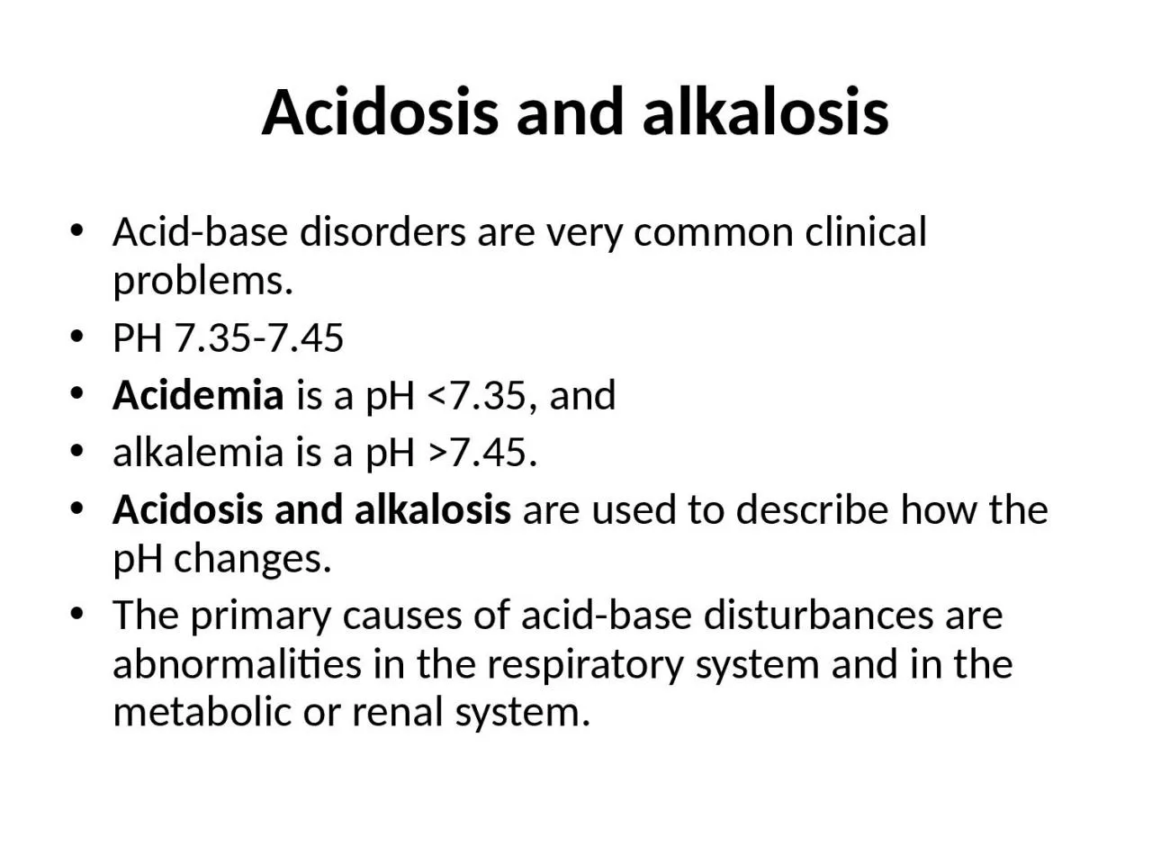
/
Acidosis and alkalosis Acid-base disorders are very common clinical problems. Acidosis and alkalosis Acid-base disorders are very common clinical problems.

Acidosis and alkalosis Acid-base disorders are very common clinical problems. - PowerPoint Presentation

cady
cady . @cady
Follow
67 views
Uploaded On 2023-05-26

Acidosis and alkalosis Acid-base disorders are very common clinical problems. - PPT Presentation

PH 735745 Acidemia is a pH lt735 and alkalemia is a pH gt745 Acidosis and alkalosis are used to describe how the pH changes The primary causes of acidbase disturbances are abnormalities in the respiratory system and in the metabolic or renal system ID: 999473

bicarbonate acidosis acid alkalosis acidosis bicarbonate alkalosis acid mmol metabolic co2 respiratory hco3 increase plasma renal fall treatment rise

Share:

Link:

Embed:

Download Presentation from below link

Download Presentation The PPT/PDF document "Acidosis and alkalosis Acid-base disorde..." is the property of its rightful owner. Permission is granted to download and print the materials on this web site for personal, non-commercial use only, and to display it on your personal computer provided you do not modify the materials and that you retain all copyright notices contained in the materials. By downloading content from our website, you accept the terms of this agreement.


Presentation Transcript

1. Acidosis and alkalosisAcid-base disorders are very common clinical problems. PH 7.35-7.45Acidemia is a pH <7.35, andalkalemia is a pH >7.45. Acidosis and alkalosis are used to describe how the pH changes. The primary causes of acid-base disturbances are abnormalities in the respiratory system and in the metabolic or renal system.

2. DISORDERS OF ACID-BASE BALANCE The pH of the arterial plasma is normally 7.40, (pH 7.36-7.44) corresponding to a H+ concentration of 40 nmol/l. An increase in H+ concentration corresponds to a decrease in pH. this parameter is under tight homeostatic regulation, such that the H+ concentration does not vary outside the range 36-44 nmol/l (pH 7.44-7.36) under normal circumstances. IN acidosis there is anaccumulation of acid or loss of abase causing afall in Ph . The converse accure in alkalosis .Abnormal acid-base balance occurs in a wide range of diseases variety of physiological mechanisms act to prevent wide swings in the pH of the ECF. The first is the action of blood and tissue buffers, of which the most important is the carbonic acid/bicarbonate buffer system. This involves the reaction of H+ ions with bicarbonate to form carbonic acid, which, under the influence of the enzyme carbonic anhydrase (c.a.), dissociates to form CO2 and water, as follows:CO2+H2O---H2CO3----H2+HCO3This buffer system is important because bicarbonate is present in relatively high concentration in the ECF (21-28 mmol/l), and two of its key components are under physiological control: the CO2 by the lungs, and the bicarbonate, by the kidneys.

3.

4.

5. Metabolic acidosis :decrease HCO3<24mmol ,decrease CO2<5.33kpa ,PH>40 Metabolic alkalosis increase HCO3>24mmol ,increase CO2>5.33kpa ,PH<40Respiratory acidosis: increase HCO3>24mmol ,increase CO2>5.33kpa ,PH>40Respiratory alkalosis: increase HCO3<24mmol ,decrease CO2<5.33kpa ,PH<40

6.

7. ACIDOSIS 1-METABOLIC ACIDOSISMetabolic acidosis occurs when an acid other than carbonic acid (due to CO2 retention) accumulates in the body, resulting in a fall in the plasma bicarbonate. The pH fall which would otherwise occur is blunted by hyperventilation, resulting in a reduced PCO2. If the kidneys are intact (i.e. not the cause of the initial disturbance), renal excretion of acid can be gradually increased over days to weeks, raising the plasma bicarbonate and hence the pH towards normal in the new steady state

8. CausesBase loss(Bicarbonate losses ) Extra renal = (   Small bowel drainage    Diarrhea )Renal = renal tubular acidosis  (  Urinary loss of HCO3 in proximal RTA  Carbonic anhydrase inhibitors     Primary hyperparathyroidism Failure of bicarbonate regeneration Distal renal tubular acidosis (impaired tubular acid secretion )Aldosterone deficiency (Addison's diseaseAldosterone insensitivity  ( Interstitial renal disease,   Aldosterone antagonists)Ureteroileostomy (ileal bladder) Acidifying salts  ( Ammonium chloride  , Lysine or arginine hydrochloride )Diabetes mellitus Reduced excretion of acids  ( Renal failure: Accumulation of organic acids Overproduction of acids Ketoacidosis1- Diabetic(Accumulation of ketones1 with hyperglycaemia) 2-Alcoholic     3-Starvation (Accumulation of ketones without hyperglycaemia) Lactic acidosis( Tissue hypoxia (e.g. shock) or liver disease) Toxin ingestion    ( Methanol     Ethylene glycol     Salicylates)

9. Clinical featuresRapid deep breathing ?stimulation of respiratory centre by reduction in PH of blood &to eleminat acid substanceThe urin strongly acidic

10. Management The first step in management of metabolic acidosis is identifying and correcting the cause when possible . This may involve control of diarrhoea, treatment of diabetes, correction of shock, cessation of drug administration, or dialysis to remove toxins. Since metabolic acidosis is frequently associated with sodium and water depletion, resuscitation with appropriate intravenous fluids will often be needed. Use of intravenous bicarbonate in this setting is controversial. Because rapid correction of acidosis has some inherent risks (e.g. induction of hypokalaemia or reduced plasma ionised calcium), use of bicarbonate infusions is best reserved for situations where the underlying disorder cannot be readily corrected and where the acidosis is severe (H+ > 70 nmol/l, pH < 7.15) and associated with evidence of tissue dysfunction. The acidosis associated with RTA can sometimes be controlled by treating the underlying cause . Usually, however, supplements of sodium and potassium bicarbonate are necessary to achieve the target of a plasma bicarbonate level above 18 mmol/l with normokalaemia in types 1 and 2 RTA, while diuretics or corticosteroids (as appropriate) may be effective in reversing the underlying disturbance in type 4 RTA.

11. RESPIRATORY ACIDOSIS acondition where the PCO2 is above the normal range is caused by:1-impairment in the rate of alveolar ventilation. 2-a sudden depression of the medullary respiratory center (narcotic overdose),3-paralysis of the respiratory muscles(M.G,G.B ,toxines ,anasthesia 4-airway obstruction.(infection ,allergy ,tumoures ,F.B…. 5-Chronic respiratory acidosis in chronic airway disease (emphysema), with extreme kyphoscoliosis, and with extreme obesity (pickwickian syndrome). The serum bicarbonate concentration is increased, the magnitude of which depends on the acuity and the severity of the respiratory disorder. Acute increases in the Pco2 result in somnolence, confusion, and, ultimately, carbon dioxide narcosis. Asterixis may be present. Because carbon dioxide is a cerebral vasodilator, the blood vessels in the optic fundi are often dilated, engorged, and tortuous. Frank papilledema may be present in patients with severe hypercapnic states. treatment 1-The only practical therapy of acute respiratory acidosis involves treatment of the underlying disorder2-O2 and3- ventilatory support. In patients with chronic hypercapnia who develop an acute increase in the Pco2, attention should be directed toward identifying factors that may have aggravated the chronic disorder. Alkalinizing salts should be avoided in patients with chronic

12. METABOLIC ALKALOSIS Aetiology and clinical assessment Metabolic alkalosis is characterised by an increase in the plasma bicarbonate concentration and the plasma pH . There is a compensatory rise in PCO2 due to hypoventilation. CAUSES 1-Hypovolaemic metabolic alkalosis is the most common pattern, such as sustained vomiting where acid-rich fluid is lost directly from the body. during treatment with most diuretic drugs (other than carbonic anhydrase inhibitors and potassium-sparing drugs), since the diuretic action involves increased acid loss into the urine.2- Normovolaemic (or hypervolaemic) metabolic alkalosis occurs in settings where both bicarbonate retention and volume expansion occur simultaneously. Classical causes include corticosteroid excess states such as primary hyperaldosteronism (Conn's syndrome, Cushing's syndrome and corticosteroid therapy. Occasionally, overuse of antacid salts for treatment of dyspepsia can produce a similar pattern.

13.

14. . Clinically, apart from manifestations of the underlying cause, there may be few symptoms or signs related to alkalosis itself. When the rise in systemic pH is abrupt, plasma ionised calcium falls and signs of increased neuromuscular irritability such as tetany may develop

15. Management In metabolic alkalosis associated with hypovolaemia, treatment involves provision of adequate intravenous fluid, specifically isotonic sodium chloride, which interrupts the volume-conserving mechanisms and allows the kidney to excrete the excess alkali in the urine. Replacement of potassium helps correct the hypokalaemia and its consequences in the kidney. In metabolic alkalosis associated with normal or increased volume, treatment should focus on correcting the underlying cause, specifically the removal or blockade of excess mineralocorticoid activity. The approach depends on the underlying endocrinological diagnosis .

16. RESPIRATORY ALKALOSISoccurs when hyperventilation reduces the arterial Pco2(31-42) and consequently increases the arterial pH. Acute respiratory alkalosis is most commonly a result of the hyperventilation syndrome. It may also occur in damage to the respiratory centers,hystericalacute salicylism, in fever and septic statesassociation with various pulmonary processes (pneumonia, pulmonary emboli, or congestive heart failure). iatrogenically by injudicious mechanical ventilatory support. Chronic hyperventilation occurs in the acclimatization response to high altitudes (a low ambient oxygen tension), in advanced liver disease, and in pregnancy. Acute hyperventilation is characterized by lightheadedness, paresthesias, circumoral numbness, and tingling of the extremities. Tetany occurs in severe cases. When anxiety provokes hyperventilation, air rebreathing with a paper bag generally terminates the acute attack

17. DisturbanceBlood H+Primary changeCompensatory responsePredicted compensationMetabolic acidosis> 401HCO3 < 24 mmol/lPCO2< 5.33 kPa2P CO2 fall in kPa = 0.16 ×HCO3 fall in mmol/lMetabolic alkalosis< 401HCO3> 24 mmol/lPCO2> 5.33 kPa2,3PCO2 rise in kPa = 0.08 ×HCO3 rise in mmol/lRespiratory acidosis> 401PCO2> 5.33 kPa2HCO3> 24 mmol/lAcute:HCO3 rise in mmol/l = 0.75 ×PCO2rise in kPaChronic: HCO3 rise in mmol/l = 2.62×P CO2 rise in kPaRespiratory alkalosis< 401PCO2< 5.33 kPa2HCO3< 24 mmol/lAcute:HCO3 fall in mmol/l = 1.50 ×CO2fall in kPaChronic: HCO3 fall in mmol/l = 3.75×P CO2 fall in kPa