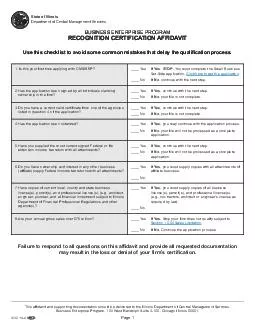
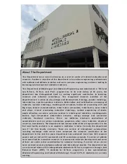
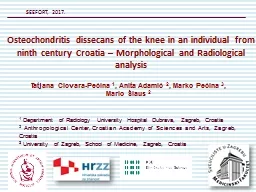
Radiology University Hospital Dubrava Zagreb Croatia 2 Anthropological Center Croatian Academy of Sciences and Arts Zagreb Croatia 3 University of Zagreb ID: 604547
Download Presentation The PPT/PDF document "1 Department of" is the property of its rightful owner. Permission is granted to download and print the materials on this web site for personal, non-commercial use only, and to display it on your personal computer provided you do not modify the materials and that you retain all copyright notices contained in the materials. By downloading content from our website, you accept the terms of this agreement.
Slide1
1 Department of Radiology University Hospital Dubrava, Zagreb, Croatia2 Anthropological Center,Croatian Academy of Sciences and Arts, Zagreb, Croatia3 University of Zagreb, School of Medicine, Zagreb, Croatia
Tatjana Cicvara-Pećina 1, Anita Adamić 2, Marko Pećina 3, Mario Šlaus 2
SEEFORT, 2017.
Osteochondritis
dissecans
of the knee in an individual from
ninth
century Croatia – Morphological and Radiological analysisSlide2
IntroductionWe present and describe an example of osteochondritis dissecans identified in the Osteological Collection of the Croatian Academy of Sciences and Arts. The case of osteochondritis dissecans described in this report was recovered from a cemetery in the Dalmatian hinterland, north-east of Split. Osteochondritis dissecans was noted in the distal femur of a subadult individual. ‘‘Although osteochondritis dissecans of the knee has been known for a long time, we still do not fully understand why it develops.’’ Ejnar ErikssonSlide3
Case ReportThe individual exhibiting osteochondritis dissecans was recovered from grave number 16 that belongs to the younger phase of the cemetery which lasted during the second half of the 9th century A.D. Osteochondritis dissecans was noted in the subadult individual. Preservation of the osseous material was generally good. The bones are of a homogeneous light brown-yellow color, and are reasonably well preserved.Slide4
The pathological changes consistent with osteochondritis dissecans are present on both medial femoral condyles. The lesion on the right femoral condyle is an oval crater-like defect with well defined margins and a porous floor of rough trabecular bone. The lesion on the left femoral condyle is basically, with two small provisions, identical to the one on the right side. The first is that it is slightly smaller, while the second is that unlike its antimere, it has a well preserved bone fragment that perfectly fits into the ostechondritic pit. A radiographic analysis of the femoral condyles supports a diagnosis of osteochondritis dissecans and shows a well-demarcated radiolucent defect in the articular surfaces of both joints surrounded by a thin sclerotic repair zone. According to classification systems this degree of change corresponds to stage 3 or grade 3 osteochondritis dissecans – a detached but non-displaced fragment. As is evident from this example it is clear that during the last 1100 years there have been no significant morphological or radiological changes in the characteristics of osteochondritis dissecans.Slide5
Figure 1. The lesions characteristic of osteochondritis dissecans are present on the medial femoral condyles of both knees.ResultsSlide6
Figure 2. The lesion on the left femoral medial condyle has a well-preserved bone fragment that fits perfectly into the osteochondritic pit.ResultsSlide7
Figure 3. The radiological appearance of the lesions made with computed tomography in horizontal direction on both medial femoral condyles is typical for osteochondritis dissecans. The osteochondral lesions are marked with arrows.ResultsSlide8
Figure 4. Radiological presentations made with computed tomography in sagittal direction of the medial femoral condyles of both knees. The osteochondral lesions are marked with arrows.ResultsSlide9
ConclusionsERIKSSON E. Long term prognosis of osteochondritis dissecans of the knee. Knee Surg Sports Traumatol Arthrosc 2008;16:435.PAGET J. Of the production of some of the loose bodies in joints. St.
Bartholomew’s Hospital Reports 1870;6:1-4.KÖNIG ?. Über freie Körper in den Gelenken. Dtsch Z Chir 1888;27:90-109.ORTNER DJ. Identifi cation of pathological conditions in human
skeletal remains. New York:
Academic Press
, 2003.PEĆINA M, HAŠPL M. Koljeno i potkoljenica. In: Pećina M, et al., editors. Ortopedija, 3rd
ed. Zagreb: Naklada Ljevak, 2004.ŠLAUS M. Bioarheologija. Demografi ja, zdravlje, traume i prehrana starohrvatskih
populacija. Zagreb: Školska knjiga, 2006.ŠLAUS M. Osteological and dental markers of
health in
the transition from the Late Antique to the Early
Medieval
period in
Croatia. Am J
Phys
Anthropol
2008;136:455-69.
References
On the
basis
of the
presented
case
we
can
conclude
that
during
the last 1100 years there have been no
significant
morphological or radiological changes in
the
characteristics
of osteochondritis
dissecans
. A
future
goal
is to
analyze
the
frequency and distribution
of
this
disease in Croatian archeological
populations
from
an epidemiological point of view.