/
Enterohaemorrhagic  E. coli Enterohaemorrhagic  E. coli

Enterohaemorrhagic E. coli - PowerPoint Presentation

callie
callie . @callie
Follow
342 views
Uploaded On 2022-06-20

Enterohaemorrhagic E. coli - PPT Presentation

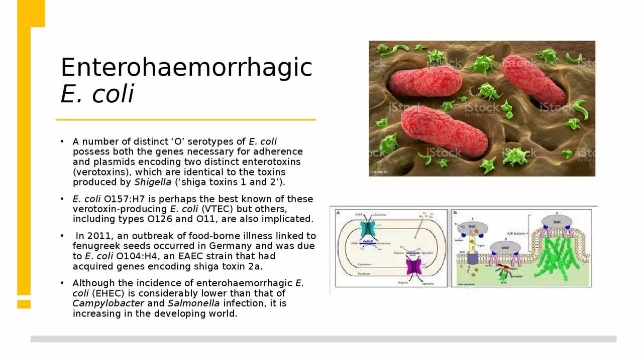
A number of distinct O serotypes of E coli possess both the genes necessary for adherence and plasmids encoding two distinct enterotoxins verotoxins which are identical to the toxins produced by ID: 920993

difficile diarrhoea severe water diarrhoea difficile water severe coli infection fluid days cholera occur toxin cases therapy illness antibiotic

Share:

Link:

Embed:

Download Presentation from below link

Download Presentation The PPT/PDF document "Enterohaemorrhagic E. coli" is the property of its rightful owner. Permission is granted to download and print the materials on this web site for personal, non-commercial use only, and to display it on your personal computer provided you do not modify the materials and that you retain all copyright notices contained in the materials. By downloading content from our website, you accept the terms of this agreement.


Presentation Transcript

Slide1

Enterohaemorrhagic

E. coli

A number of distinct ‘O’ serotypes of

E. coli

possess both the genes necessary for adherence and plasmids encoding two distinct enterotoxins (

verotoxins

), which are identical to the toxins produced by

Shigella (‘shiga toxins 1 and 2’). E. coli O157:H7 is perhaps the best known of these verotoxin-producing E. coli (VTEC) but others, including types O126 and O11, are also implicated. In 2011, an outbreak of food-borne illness linked to fenugreek seeds occurred in Germany and was due to E. coli O104:H4, an EAEC strain that had acquired genes encoding shiga toxin 2a. Although the incidence of enterohaemorrhagic E. coli (EHEC) is considerably lower than that of Campylobacter and Salmonella infection, it is increasing in the developing world.

Slide2

The reservoir of infection is in the gut of herbivores.

The organism has an extremely low infecting dose (10–100 organisms).

Runoff water from pasture lands where cattle have grazed, which is used to irrigate vegetable crops, as well as contaminated milk, meat products (especially hamburgers that have been incompletely cooked), lettuce, radish shoots and apple juice have all been implicated as sources

Slide3

Slide4

The incubation period is between 1 and 7 days.

Initial watery

diarrhoea

becomes uniformly blood-stained in 70% of cases and is associated with severe abdominal pain.

There is little systemic upset, vomiting or fever.

Slide5

Enterotoxins have both a local effect on the bowel and a distant effect on particular body tissues, such as glomerular apparatus, heart and brain.

The potentially life-threatening

haemolytic

uraemic

syndrome occurs in 10–15% of sufferers from this infection, arising 5–7 days after the onset of symptoms.

It is most likely at the extremes of age, is heralded by a high peripheral leucocyte count, and may be induced, particularly in children, by antibiotic therapy.HUS is treated by dialysis if necessary and may be averted by plasma exchange. Antibiotics should be avoided since they can stimulate toxin release.

Slide6

Clostridium difficile

infection

C. difficile

is the most commonly diagnosed cause of antibiotic associated

diarrhoea

& is an occasional constituent of the gut microbiome. C. difficile can produce two toxins (A and B).

C. difficile infection (CDI) usually follows antimicrobial therapy, which alters the composition of the gastrointestinal flora and may result in colonisation with toxigenic C. difficile, if the patient is exposed to C. difficile spores. The combination of toxin production and the ability to produce environmentally stable spores accounts for the clinical features and transmissibility of CDI.

A hypervirulent strain of C. difficile, ribotype 027, has emerged, which produces more toxin and more severe disease than other C. difficile strains.

Slide7

Clinical features

Disease manifestations range from

diarrhoea

to life-threatening pseudomembranous colitis.

Around 80% of cases occur in people over 65 years of age, many of whom are frail with comorbid diseases.

Symptoms usually begin in the first week of antibiotic therapy but can occur at any time up to 6 weeks after treatment has finished.

The onset is often insidious, with lower abdominal pain and diarrhoea that may become profuse and watery. The presentation may resemble acute ulcerative colitis with bloody diarrhoea, fever and even toxic dilatation and perforation. Ileus is also seen in pseudomembranous colitis.

Slide8

Investigations

C. difficile

can be isolated from stool culture in 30% of patients with antibiotic-associated diarrhoea and over 90% of those with pseudomembranous colitis, but also from 5% of healthy adults and up to 20% of elderly patients in residential care. The diagnosis of CDI therefore rests on detection of toxins A or B in the stool. Current practice in the UK is to screen stool from patients with a compatible clinical syndrome by detection either of glutamate dehydrogenase (GDH), an enzyme produced by

C. difficile, or of C. difficile nucleic acid (e.g. by PCR); if screening is positive, a C. difficile toxin ELISA or a tissue culture cytotoxicity assay is performed.

Slide9

The rectal appearances at sigmoidoscopy may be characteristic, with erythema, white plaques or an adherent

pseudomembrane

, or may resemble ulcerative colitis.

In some cases, the rectum is spared and abnormalities are observed in the proximal colon.

Patients who are ill require abdominal and erect chest X-rays to exclude perforation or toxic dilatation. CT may be useful when the diagnosis is in doubt.

Slide10

Slide11

Management

The precipitating antibiotic should be stopped and the patient should be isolated.

Supportive therapy includes intravenous fluids and bowel rest.

First-line antimicrobial therapy involves metronidazole (500 mg orally 3 times daily for 10 days) or vancomycin (125 mg orally 4 times daily for 7–10 days).

Although vancomycin is more effective than metronidazole against hypervirulent

C. difficile

strains (e.g.

ribotype

027), it is more expensive and may drive the emergence of vancomycin resistance in other organisms (e.g. enterococci,

Staph. aureus

).

Slide12

Slide13

Yersinia enterocolitica

infection

Yersinia enterocolitica

, commonly found in pork, causes mild to moderate gastroenteritis and can produce significant mesenteric adenitis after an incubation period of 3–7 days.

It predominantly causes disease in children but adults may also be affected.

The illness resolves slowly.

Complications include reactive arthritis (10–13% of cases), which may be persistent, and anterior uveitis

.

Slide14

Cholera

Cholera, caused by

Vibrio cholerae

serotype O1, is the archetypal toxin-mediated bacterial cause of acute watery

diarrhoea

.

The enterotoxin activates adenylate cyclase in the intestinal epithelium, inducing net secretion of chloride and water. V. cholerae O1 has two biotypes, classical and El Tor, and each of these has two distinct serotypes, Inaba and Ogawa.

Slide15

El Tor is more resistant to commonly used antimicrobials than classical

Vibrio

, and causes prolonged carriage in 5% of infections.

An atypical serotype, O139, has been responsible for localised outbreaks in Bangladesh.

Infection spreads via the stools or vomit of symptomatic patients or of the much larger number of subclinical cases.

Organisms survive for up to 2 weeks in fresh water and 8 weeks in salt water.

Transmission is normally through infected drinking water, shellfish and food contaminated by flies, or on the hands of carriers.

Slide16

Clinical features

Severe

diarrhoea

without pain or colic begins suddenly and is followed by vomiting.

Following the evacuation of normal gut

faecal

contents, typical ‘rice water’ material is passed, consisting of clear fluid with flecks of mucus. Classical cholera produces enormous loss of fluid and electrolytes, leading to intense dehydration with muscular cramps.

Shock and oliguria develop but mental clarity remains. Death from acute circulatory failure may occur rapidly unless fluid and electrolytes are replaced. Improvement is rapid with proper treatment.

Slide17

The majority of infections, however, cause mild illness with slight

diarrhoea

.

Occasionally, a very intense illness,

‘cholera sicca’,

occurs, with loss of fluid into dilated bowel, killing the patient before typical gastrointestinal symptoms appear.

The disease is more dangerous in children.

Slide18

Diagnosis and management

Clinical diagnosis is easy during an epidemic.

Otherwise, the diagnosis should be confirmed bacteriologically.

Stool darkfield microscopy

shows the typical

‘shooting star’ motility of

V. cholerae. Rectal swab or stool cultures allow identification.

Cholera is notifiable under international health regulations.

Slide19

Maintenance of circulation by replacement of water and electrolytes is paramount.

Ringer-Lactate is the best fluid for intravenous replacement.

Vomiting usually stops once the patient is rehydrated, and fluid should then be given orally up to 500 mL hourly.

Early intervention with oral rehydration solutions that include resistant starch, based on either rice or cereal, shortens the duration of

diarrhoea and improves prognosis.

Severe dehydration, as indicated by altered consciousness, skin tenting, very dry tongue, decreased pulses, low blood pressure or minimal urine output, mandates intravenous replacement.

Slide20

Total fluid requirements may exceed 50 L over a period of 2–5 days.

Accurate records are greatly facilitated by the use of a ‘cholera cot’, which has a reinforced hole under the patient’s buttocks, beneath which a graded bucket is placed.

Three days’ treatment with tetracycline 250 mg 4 times daily, a single dose of doxycycline 300 mg or ciprofloxacin 1 g in adults reduces the duration of excretion of

V. cholerae

and the total volume of fluid needed for replacement.

Slide21

Prevention

Strict personal hygiene is vital and drinking water should come from a clean piped supply or be boiled.

Flies must be denied access to food. Oral vaccines containing killed

V. cholerae

with or without the B subunit of cholera toxin are used in specific settings.

In epidemics, improvements in sanitation and access to clean water, public education and control of population movement are vital. Mass single-dose vaccination and treatment with tetracycline are valuable. Disinfection of discharges and soiled clothing, and scrupulous hand-washing by medical attendants reduce spread.

Slide22

Vibrio parahaemolyticus

infection

This marine organism produces a disease similar to enterotoxigenic

E. coli

. It is very common where ingestion of raw seafood is widespread (e.g. Japan). After an incubation period of approximately 20 hours, explosive diarrhoea

, abdominal cramps and vomiting occur. Systemic symptoms of headache and fever are frequent but the illness is self-limiting after 4–7 days. Rarely, a severe septic illness arises; in this case, V. parahaemolyticus

can be isolated using specific halophilic culture.

Slide23

Bacillary dysentery (shigellosis)

Shigellae

are Gram-negative rods, closely related to

E. coli

, that invade the colonic mucosa.

There are four main groups:

Sh. dysenteriae,

flexneri, boydii and sonnei. In the tropics, bacillary dysentery is usually caused by Sh. flexneri, while in the UK most cases are caused by Sh. sonnei. Shigellae are often resistant to multiple antibiotics, especially in tropical countries. The organism only infects humans and its spread is facilitated by its low infecting dose of around 10 organisms.

Slide24

Spread may occur via contaminated food or flies, but person to person transmission by unwashed hands after

defaecation

is the most important factor.

Outbreaks occur in psychiatric hospitals, residential schools and other closed institutions, and dysentery is a constant accompaniment of wars and natural catastrophes, which bring crowding and poor sanitation in their wake.

Slide25

Clinical features

Disease severity varies from mild

Sh.

sonnei

infections that may escape detection to more severe

Sh. flexneri infections, while those due to

Sh. dysenteriae may be fulminating and cause death within 48 hours.In a moderately severe illness, the patient complains of diarrhoea, colicky abdominal pain and tenesmus. Stools are small, and after a few evacuations contain blood and purulent exudate with little faecal material. Fever, dehydration and weakness occur, with tenderness over the colon. Reactive arthritis or iritis may occasionally complicate bacillary dysentery

Slide26

Management and prevention

Oral rehydration therapy or, if

diarrhoea

is severe, intravenous replacement of water and electrolyte loss is necessary.

Antibiotic therapy is with ciprofloxacin (500 mg twice daily for 3 days)

Azithromycin and ceftriaxone are alternatives but resistance occurs to all agents, especially in Asia.

The use of

antidiarrhoeal medication should be avoided.The prevention of faecal contamination of food and milk and the isolation of cases may be difficult, except in limited outbreaks.Hand-washing is very important.