/
Neuronal   architecture  of Neuronal   architecture  of

Neuronal architecture of - PowerPoint Presentation

callie
callie . @callie
Follow
66 views
Uploaded On 2023-07-28

Neuronal architecture of - PPT Presentation

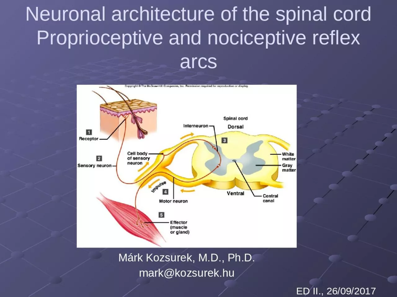
the spinal cord Proprioceptive and nociceptive reflex arcs Márk Kozsurek MD PhD mark kozsurekhu ED II 26092017 Terms white and gray matter ID: 1012677

reflex spinal fibres muscle spinal reflex muscle fibres nucleus horn dorsal muscles motoneurons nervous alpha proprioceptive ventral spindle gamma

Share:

Link:

Embed:

Download Presentation from below link

Download Presentation The PPT/PDF document "Neuronal architecture of" is the property of its rightful owner. Permission is granted to download and print the materials on this web site for personal, non-commercial use only, and to display it on your personal computer provided you do not modify the materials and that you retain all copyright notices contained in the materials. By downloading content from our website, you accept the terms of this agreement.


Presentation Transcript

1. Neuronal architecture of the spinal cordProprioceptive and nociceptive reflex arcsMárk Kozsurek, M.D., Ph.D.mark@kozsurek.huED II., 26/09/2017

2. Terms: white and gray matter, spinal nerve, dorsal and ventral roots, spinal ganglionINPUT: sensory fibresOUTPUT: motor fibres

3. I. Fine neuronal structureof the spinal cord

4. Laminar organization of the spinal cord: Rexed, 1952

5. Dorsal hornlamina II: substantia gelatinosa Rolandi

6.

7. High-threshold, slow-conducting, thin-myelinated Aδ and unmyelinated C fibres convey predominantly nociceptive information (pain, heat) to the lamina I-III. Low-threshold, high-conducting, thick-myelinated Aβ fibres carry non-painfull stimuli and terminate in deeper laminae of the spinal dorsal horn. Dorsal spinal horn plays a crutial role in sensory information processing. Local circuit neurons are more numerous, projection neurons forwarding stimuli to higher levels of the CNS constitute only 5-10%.Lamina II is densly packed with mu-opioid receptors binding morphine and other opioid analgesics.

8. Intermediate substanceIMLIMMCLARK-STILLINGNote, that at the lower thoracic and upper lumbar levels the IML nucleus is situated more laterally and constitutes the lateral horn!IMM: intermediomedial nucleusIML: intermediolateral nucleus

9. Lateral horn exists only in the thoracic and upper lumbar segments. At these levels appearance of the lateral horn can be explained by the lateral transposition of the IML nucleus.IML nucleus contains preganglionic neurons of the sympathetic (thoraco-lumbar levels) and parasympathetic (sacrral segments) nervous system, with other words visceromotor fibres arise from this nucleus.IMM nucleus is considered as an autonomic spinal reflex centre.Dorsal or thoracic nucleus (of Clarke-Stilling) found in the Th9-L3 segments is the source of the dorsal spinocerebellar tract (of Flechsig). Downward continuation of this nucleus in L4-S3 segments is the approximate site of origin of the ventral spinocerebellar tract (of Gowers). Both of these tracts convay proprioceptiv information to the cerebellum.

10. Ventral hornIn the ventral horn instead of laminae rather groups of neurons are observable. Together with many local interneurons, alpha- and gamma-motoneurons directly innervating skeletal muscles are found here in somatotopic arrangement.

11. A typical neuron in the microscope: prominent nucleolus in the large, oval or rounded, poorly stained nucleus, surrounded by dark, basofilic, granulated cytoplasm, which is always darker then the nucleus.

12. II. Spinal reflex arcs

13. Reflex: involuntary and automatic response to a stimulusA (1) receptor is responsible for detecting a stimulus and converting it into a neuronal signal. Evoked action potentials reach the central nervous system through the (2) afferent limb. Decission is made and response is generated by the (3) central processing neuronal networks. (4) Efferent limb exits the central nervous system and terminates on the (5) effector, which might be a muscle or a gland. (1) to (5) components altogether constitute the reflex arc!Spinal reflex: In the case of spinal reflex arcs the stimulus is received and processed by the spinal cord and the response is organized and performed by the spinal neuronal circuits.Under normal circumstances stimuli evoking spinal reflexes also reach the brain and descending fibres from higher centres may modulate spinal reflexes but experimentally spinal reflexes can be evoked even after the removal of the brain!

14. Fundamentals of the nervous systemcentral nervous systemskin, muscles, joints, tendonssomatosensoryvisceromotorsmooth muscles , glandsinternal organsviscerosensoryskeletal musclessomatomotorreceptors effectors

15. Fundamentals of the nervous systemcentral nervous systemskin, muscles, joints, tendonssomatosensoryvisceromotorsmooth muscles , glandsinternal organsviscerosensoryskeletal musclessomatomotorSENSORY GGL.AUTONOMIC GGL.vreceptors effectors

16. 1. Nociceptive reflex arcSynonyms: flexor reflex, withdrawal reflex, polysynaptic spinal reflexAncient reflex that protects the body from potentially damaging, noxious stimuli.

17. Nociceptors of the skin and mucous membranes → Adelta and/or C fibres → spinal ganglion → spinal dorsal horn → numerous collaterals: excitacion of flexors and inhibition of extensor on the side of stimulus and stimulation of extensors together with inhibition of flexors on the contralateral side (flexor reflex with crossed extensor response).

18. 2. Proprioceptive reflex arcSynonyms: stretch reflex, monosynaptic spinal reflex, myotatic reflexStretching a muscle results in its contraction providing automatic regulation of skeletal muscle length and the posture of the body. The „stretch receptors” are the muscle spindles.

19. alpha-motoneurongamma-motoneuronflower-sprayanulospiral terminalsMUSCLE SPINDLE

20. Muscle spindles are special stretch receptors enclosed by a connective tissue capsule. Within the capsule the intrafusal fibres are present, while outside the extrinsic muscle fibres of the host muscle can be found. Intrafusal fibres are innervated by gamma-motoneurons and extrafusal fibres receive stimuli from alpha-motoneurons.Within the muscle spindle two types of sensory terminals, the anulospiral endings and the flower-spray terminals can be distinguished. These inform the CNS about the actual length and movement of the muscles.

21.

22. Muscle spindle → spinal ganglion → dorsal root → alpha-motoneuron in the ventral horn → peripheral nerve → skeletal muscle fibres surrounding the muscle spindle. Collaterals through inhibitory interneurons will avoid simultaneous contraction of the antagonist muscle groups.

23. Object held in hands stretches the flexors of the arm and due to the proprioceptive reflex arc the flexors will contract and hold the weight.Knee joints tend to flex under the weight of the body, but stretching of the quadriceps femoris muscle activates the proprioceptive reflex which increases the tone of the extensors avoiding the collapse of the human body.Proprioceptive reflex is characteristic for antigravity muscles (flexors of the upper and extensors of the lower limb)!

24. Further spinal mechanisms contributing to the control of fine movements Renshaw cells of the ventral horn Gamma-loop Golgi tendon organ

25. VVV-+motoneuronsRenshaw-cellPyramidal tractRenshaw-cell: recurrent inhibition (with red on the image).Discharging motoneurons inhibit adjacent motoneurons via their recurrent axon collaterals and the Renshaw-neurons.

26. gammamotoneuronGamma-loopdescending pathwaysalpha motoneuron

27. A muscle contracton can beevoked in two different ways:Directly, through alpha-motoneuronsIndirectly, through the activation of the gamma-loop and the proprioceptive reflex arc.Descending pathways can stimulate gamma-motoneurons which innervate intrafusal muscle fibres. Contracting intrafusal fibres shorten the muscle spindle itself which has a similar effect to that when the extrafusal fibres around the spindle are passively stretched. This stretch(-like) signal evokes the spinal proprioceptive reflex activating alpha-motoneurons, and finally contracting extrafusal fibres.

28. Golgi tendon organ

29. In GTOs axons among collagen bundles can be activated by transformations of the musculotendinous junction.GTO is connected in series to the muscle fibres.Inhibits overstimulation of muscles and prevevents bone, tendon or joint injuries.

30. Thank you for your attention!