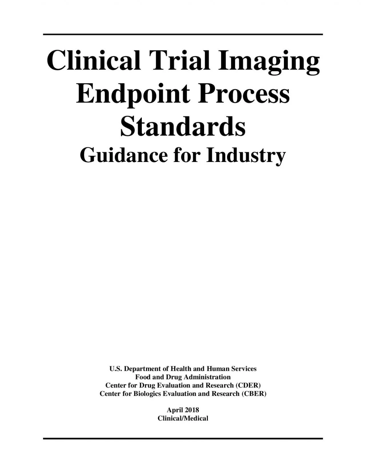
/
Clinical Trial Imaging Endpoint Process StandardsGuidance for Industry Clinical Trial Imaging Endpoint Process StandardsGuidance for Industry

Clinical Trial Imaging Endpoint Process StandardsGuidance for Industry - PDF document

carla
carla . @carla
Follow
345 views
Uploaded On 2021-09-25

Clinical Trial Imaging Endpoint Process StandardsGuidance for Industry - PPT Presentation

x0000x0000 xAttxachexd xBottxom xBBoxx 7x2 21x036x 75 x368x04 xSubxtypex Foxoterx Tyxpe xPagixnatixon 0xAttxachexd xBottxom xBBoxx 7x2 21x036x 75 x368x04 xSubxtypex Foxoterx Tyxpe xPagix ID: 885705

image imaging trial clinical imaging image clinical trial process charter images interpretation x0000 data endpoint 146 standards reader important

Share:

Link:

Embed:

Download Presentation from below link

Download Pdf The PPT/PDF document "Clinical Trial Imaging Endpoint Process ..." is the property of its rightful owner. Permission is granted to download and print the materials on this web site for personal, non-commercial use only, and to display it on your personal computer provided you do not modify the materials and that you retain all copyright notices contained in the materials. By downloading content from our website, you accept the terms of this agreement.


Presentation Transcript

1 Clinical Trial Imaging Endpoint Process
Clinical Trial Imaging Endpoint Process StandardsGuidance for IndustryU.S. Department of Health and Human ServicesFood and Drug AdministrationCenter for Drug Evaluation and Research (CDER)Center for Biologics Evaluation and Research (CBER)April Clinical/Medical �� &#x/Att;¬he; [/; ott;&#xom ];&#x/BBo;&#xx [7; 21;&#x.036;&#x 75 ;6.8; ];&#x/Sub;&#xtype;&#x /Fo;&#xoter;&#x /Ty;&#xpe /;&#xPagi;&#xnati;&#xon 0;&#x/Att;¬he; [/; ott;&#xom ];&#x/BBo;&#xx [7; 21;&#x.036;&#x 75 ;6.8; ];&#x/Sub;&#xtype;&#x /Fo;&#xoter;&#x /Ty;&#xpe /;&#xPagi;&#xnati;&#xon 0; &#x/MCI; 0 ;&#x/MCI; 0 ;Clinical Trial Imaging Endpoint Process StandardsGuidance for IndustryAdditional copies are available from:Office of Communications, Division of Drug InformationCenter for Drug Evaluation and ResearchFood and Drug Administration10001 New Hampshire Ave., Hillandale Bldg., 4th Floor Silver Spring, MD 209930002Phone: 5433784 or 3013400Fax: 3014316353; Email: druginfo@fda.hhs.govhttp://www.fda.gov/Drugs/GuidanceComplianceRegulatoryInformation/Guidances/default.htmand/orOffice of Communication, Outreach, and Development Center for Biologics Evaluation and ResearchFood and Drug Administration10903 New Hampshire Ave., Bldg. 71, rm.3128Silver Spring, MD209930002Phone: 8354709 or 2408010; Email: ocod@fda.hhs.govhttp://www.fda.gov/BiologicsBloodVaccines/GuidanceComplianceRegulatoryInformation/Guidances/default.htmU.S. Department of Health and Human ServicesFood and Drug AdministrationCenter for Drug Evaluation and Research (CDER)Center for Biologics Evaluation and Research (CBER)April Clinical/Medical �� &#x/Att;¬he; [/; ott;&#xom ];&#x/BBo;&#xx [7; 35;&#x.316;&#x 75 ;Q.0;„ ];&#x/Sub;&#xtype;&#x /Fo;&#xoter;&#x /Ty;&#xpe /;&#xPagi;&#xnati;&#xon 0;&#x/Att;¬he; [/; ott;&#xom ];&#x/BBo;&#xx [7; 35;&#x.316;&#x 75 ;Q.0;„ ];&#x/Sub;&#xtype;&#x /Fo;&#xoter;&#x /Ty;&#xpe /;&#xPagi;&#xnati;&#xon 0; &#x/MCI; 0 ;&#x/MCI; 0 ;TABLE OF CONTENTSINTRODUCTIONBACKGROUNDLOGISTICAL AND TECHNICAL CONSIDERATIONSChoice of Imaging ModalityIs Centralized Image Interpretation Important for an ImagingBased Primary Endpoint?Should Image Interpretation Be Blinded to Clinical Data?How Often Should Imaging Evaluations Be Performed?How Soon After Acquisition Should Images Be Interpreted?What Procedures Should Be Standardized for an ImagingBased Clinical Trial Primary Endpoint?IV.THE EXTENT OF IMAGING PROCESS STANDARDSAre Existing Medical Practice Imaging Process StandardsSufficient for the Trial&

2 #146;s Primary Endpoint?What Should Be C
#146;s Primary Endpoint?What Should Be Considered When Augmenting Existing Medical Practice Imaging Process Standards to Create TrialSpecific Imaging Process Standards?REFERENCESAPPENDIX A: BEFORE IMAGING: CHARTER CONSIDERATIONSAPPENDIX B: DURING IMAGING: MONITORINGPLANS AND CHARTER MODIFICATIONSAPPENDIX C: AFTER IMAGING: DATA TRANSFER, ARCHIVING,AND ANALYSIS OF IMAGING INFORMATION ��Contains Nonbinding Recommendations 1 Clinical Trial Imaging Endpoint Process StandardsGuidance for Industry This guidance represents the current thinking of the Food and DrugAdministration (FDA or Agency) on this topic. It does not establish any rights for any person and is not binding on FDA or the public. You can use an alternative approach if it satisfies the requirements of the applicable statutes and regulations. To discuss an alternative approach, contact the FDA office responsible for this guidance as listed on the title page. INTRODUCTIONThe purpose of this guidance is to assist sponsors in optimizing the quality of imaging data obtained in clinical trials intended to support approval of drugs and biological products.This guidance focuses on imaging acquisition, display, archiving, and interpretation process standards that we regard as important when imaging is used to assess a trial’s primary endpoint or a component of that endpoint. Considerable standardization already exists in clinical imagingThere are a variety of sources, including icture rchiving and ommunication ystemand the Digital Imaging and Communications in Medicine (DICOM) formafor the handling and transmissionof clinical imaging informationthat describe the standards generally employed by clinical practitionersThis guidance recommends additional imaging endpoint processstandards thatare more specific to clinical trials. Imaging process standards help sponsors ensure that imaging data are obtained in a manner that complies with a trial’s protocol, that the quality of imaging data is maintained within and among clinical sites, and that a verifiable record of the imaging processis createdMinimization of imaging process variability may importantly enhance a clinical trial’s ability to detect drug treatment effects. Standardization, whileimportant for all clinically used measures, becomes essential for an imaging endpoint used in a clinical trial to reduce variability and to ensure interpretability of the resultsThe extent of trialspecific standardizationmay vary depending upon how standardized the local imaging procedures are (e.g., routine bone Xrays re

3 latively standardizedversusbone mineral
latively standardizedversusbone mineral density more variability across siteshis guidance does not address approachfor This guidance has been prepared by the Division of Medical Imaging Products in the Center for Drug Evaluation and Research in cooperation with the Center for Biologics Evaluation and Research at the Food andDrug Administration. For the purposes of this guidance, all references to drugsinclude both human drugs and biological products unless otherwise specified. ��Contains Nonbinding Recommendations 2 selecting an imaging endpoint for a clinical trialmaging endpoint, even if standardized and considered to be precise and accurate, might not be interpretable if it does not have clinical meaningfulness (e.g., hip fracture versus changes in local blood flow in the central nervous system detected by positron emission tomography (PETimaging)This guidance does not address whether specific imaging measureswould be acceptable in submissions used to supportapprovalof a drugor biologic. These considerations should be discussed with the FDA review division responsible for drug development. Many of the imaging process standard considerations for clinical trials of therapeutic drugs can also be applied to clinical trials that evaluate the performance of diagnostic drugs. For considerations involving the development of diagnostic imaging drugs, see the guidance for industry veloping Medical Imaging Drug and Biological Products (Parts 1, 2, and 3)In general, FDA’s guidance documents do not establish legally enforceable responsibilities. Instead, guidances describe the Agency’s current thinking on a topic and should be viewed only as recommendations, unless specific regulatory or statutory requirements are cited. The use of the word shouldin Agency guidances means that something is suggested or recommended, but not required. BACKGROUNDAlmost all medical imaging processes involve some aspects of standardizationestablished by those within the practice of medicine. In medical practice, images of human anatomy and/or physiology typically are acquired and interpreted, often with limited or no formal quantification, y a single facility’s imaging professional staff. The images typically achieve the medical practicediagnostic purposes even though the acquisition, display, and interpretation methods may vary somewhat among imaging facilities and imaging professionals. This variability may have little or no impact on the ability to provide a diagnosis in medical practice, yet in a clinica

4 l trial, imaging process variability may
l trial, imaging process variability may result in increased variability in endpoint measurements and may compromisethe ability of the trial to achieve its objectives. Although the medical practice of diagnostic imaging already follows many standardized procedures, we recommendthat some trials augment these existing standards to create trialspecificimaging process standards. r purposes of this guidance, we define these trialspecific imaging process standards as standards that extend beyond those typically performed in the medical care of a patient (i.e., the process standards are implemented solely for the purposes of the clinical trial). The extent of trialspecific imaging process standards can range from minimal processes that are described solely in the clinical protocol, such as obtaining noncontrasted and contrasted images in all subjects, to more detailed imaging process standards for image acquisition, display, interpretation, and archiving that are detailed in an imaging charter (see Appendixes A throughC). We update guidances periodically. To make sure you have the most recent version of a guidance, check the FDA guidance web page at https://www.fda.gov/RegulatoryInformation/Guidances/default.htm ��Contains Nonbinding Recommendations 3 A clinical trial’s design and clinical context are critical determinants of the extent that imaging process standards could be important tothe trial. In a clinical trial where the imaging process and imaging primary endpoint measures are not well standardized in medical practice, trialspecific standards become important. The following sections outline the imaging process topics that sponsors should address when an imaging primary endpoint is used within a clinical trial intended to support the approval of a proposed drug.LOGISTICAL AND TECHNICAL CONSIDERATIONSThe following subsections describeme of the factors a sponsor should assess when considering the use of an imagingbased primary endpoint in a clinical trial intended to support approval of a drug. he sponsor should contact the assigned FDA review division to discuss the clinical meaningfulness of the primary endpoint imaging information to be obtained in the trial.Choice of Imaging ModalityIf an imagingbased primary endpoint is chosen for a phase 3 trial, the choice of the imaging modality (such as echocardiography versus single photon emission computerized tomography) may prove to be an especially important consideration. Imaging modality upgrades and malfunctionare sometimes unpredictableli

5 nical sites mayalsoexperience unforeseen
nical sites mayalsoexperience unforeseen limitations on the use of the modality or modalityspecific imaging drugs and processes, such as the interchange of certain contrast agents that may not affect typical diagnostic imaging but may alter trialspecific quantitative imaging measures. Is Centralized Image Interpretation Important for an ImagingBased Primary Endpoint?In clinical trials, images areinterpreted either at the clinical site or at a centralized facility that receives images from the clinical sites. Sometimes, both site and centralized imaging interpretation may be performed. Image interpreters are sometimes referred to as readers, and we use that term in this guidanceThe usefulness ofa centralized image interpretation process is determined by the role, variability, and susceptibility to bias of imaging within the trial as well as modalityspecific image quality considerations and overall trial design features. Centralized image interpretation is not always critical, even for a phase 3 trial primary endpoint that uses some aspects of quantitative imaging, if the quantitative measures are widely performed and reported in clinical medicine, little imaging acquisition or interpretation variability is anticipated, and potential Evidentiary standards for diagnostic imaging products are addressed in parts 2 and 3 of the guidance for industry Developing Medical Imaging Drug and Biological Products (Parts 1, 2, and 3)Terms such as image interpretationimage review, or image readare used interchangeably in this guidance, and in literature and protocols, image readers are sometimes referred to as image reviewers ��Contains Nonbinding Recommendations 4 biases in image interpretationare controlled by the trial design features. However, these characteristics may not apply to some clinical trials, such as those that might be subject to bias and cannot be blinded, those that use imaging modalities vulnerable to image quality problems, and those that use specialized imaging measures. In openlabelclinical trials, availability of clinical information might influencea sitebased image interpretation because the expected relation of clinical features to outcome is knownandtherefore,a sitebased image interpretation couldraise concern about potentialbias. A centralized image interpretation process, fully blinded, may greatly enhance the credibility of image assessments and better ensure consistency of image assessments. Some imaging modalities also may prove vulnerable to sitespecific image quality problems

6 , and a centralized image assessmentproc
, and a centralized image assessmentprocess may help minimize these problems. For example, the National Lung Screening Trial’s experience with computed tomography of the chest suggested that centralized image quality monitoring was important to the reduction of imaging defects (Gierada, Garg, et al. 2009). Hence, a centralized image interpretation process may be used to help control image quality as well as to provide the actual imagingbased endpoint measurements.Some image interpretation methods, such as trialspecific measures of bone or joint disease in arthritic diseases, rely uponcentralized imaging because of the extent of reader training recommendedfor the specialized image interpretation. As compared to sitebased image interpretations in multicenter clinical trials, a centralized image interpretation process mayprovide more verifiable and uniform reader training as well as ongoing management of reader performance, helping to ensure quality control of the images and e interpretationand to decrease variability in image interpretationsleading to a more precise estimate of treatment effect. Nevertheless, the overarching trial design features and the other previously described features may justify the use of sitebased imaging interpretations even in large phase 3 multicenter clinical trials, so long as blinding of image interpretation to treatment can be assured or bias is otherwise controlled.Should Image Interpretation e Blinded to Clinical Data?The extent of blinding image readers to clinical data depends upon the role of imaging in the clinical trial, the specific disease and clinical setting, and the potential for unblinding effects (e.g., toxicity) of the investigational drug. In a randomized controlledtrial, we expectthat clinical trial primary endpoint image readers will be blinded to a subject’s treatment assignment because knowing the assignment would be presumed to create bias. Further, we anticipate that many, if not most, clinical trials using imagingbased primary endpoints will be conducted with no reader knowledge of individuallevel clinical data because this knowledge also may bias the reader. In unique situations, a primary endpoint mightrely upon integration of clinical data into an image interpretation (Sargent, Rubinstein, et al. 2009). To determine whether image readers should be blinded to clinical information, sponsors should have knowledge of the underlying clinical condition, an understanding of the precedent for the use of imaging as a trial’s primary endpoint, and detailed insight into the trial’s uni

7 que image interpretation procedures (suc
que image interpretation procedures (such as a plan for sequential lockedreadimage interpretation where ��Contains Nonbinding Recommendations 5 an assessment cannot be altered versus an option for modification of prior image interpretations). In certain disease conditions, readers also shouldbe blinded to the image acquisition date and/or knowledge of prior imaging observations. Again, we note that even if the image reader is aware of individuallevel clinical information, blinding to treatment assignment is almost always critical.How Often Should Imaging Evaluations e Performed?When a medical image serves as a trial’s primary endpoint, its timing and frequency of ascertainment depends upon the underlying condition being studied, the feasibility of the imaging scheduleand the overarching trial design features. For a trial using time pointbased imaging measures as a primary endpoint, the frequency of imaging evaluations should be the same in all trial arms.Asymmetric imaging evaluation time points can introduce bias in the treatment effect assessment. For a primary endpoint that uses a timeevent analytical approach, imaging evaluations should be performed at baseline and at sufficient frequency to provide a reasonably precise measure of the time to the expected clinical event. How Soon After Acquisition Should Images e Interpreted?In diagnostic medical imaging practice, images typically are interpreted onsite within several hours following acquisition. In contrast, in clinical trials using centralized imaging interpretation, the interpretationmay require alonger timeframe.Therefore, image interpretation timing typically is more of a consideration when clinical trials use centralized imaging interpretation. When planning a clinical trial that uses an imaging primary endpoint, the turnaround timeby a central image interpretation facility shouldbe appropriate for the anticipated trial design. For example, prompt image interpretation may be an important consideration for trials that use centralized image interpretations as componentof interim analyses, which may occur when imagingbased analyses are important to accommodate prespecified sample size adjustment plans. Similarly, image interpretation expediency may prove critical when centralized imaging interpretation is used to help control imaging quality; in this situation, the centralized imaging readers or an appropriately prespecified centralized imaging quality control process shouldpromptly identify technical flaws that necessitate repeat imaging of a subject. In other circum

8 stances, interpretation of batches of ra
stances, interpretation of batches of randomized images at specified intervals during a trial may be appropriate. Sponsors should consider the timeliness of entralized image interpretation when developing a clinical trial protocol that uses an imagingbased primary endpoint.What Procedures Should e Standardized for an ImagingBased Clinical Trial Primary Endpoint?No single set of detailed imaging process standards is readily applicable to every clinical trial because thetrialsdiffer in design and objectives. When usual medical practice imaging process standards are acceptable in a trial, thplans for the use of such standards should be stated in theclinical protocol. Determinations what to standardize beyond theseexpectationshould be driven by consideration of the imaging processes that might introduce variability and inaccuracy to the endpoint andby consideration ofthe other items outlinedbelow. When determining the ��Contains Nonbinding Recommendations 6 extent of imaging process standardization critical for a phase 3 clinical trial that uses an imagingbased primary endpoint, sponsors should consider the following factors:Imaging modality availability and the modality’s technical performance variation across trial sitesPerformance features of the imaging modality at the trial sites or any other locations where subjects may undergo imagingQualifications of the imaging technologists and any special technological needs for the trialroposed imaging measures’ reliancephantoms and/or calibration standards to ensure consistency and imaging quality control among clinical sitesAny unique image acquisition features of the trialdesignincluding subject positioning, anatomical coverage of imaging, use of contrast, timing of imaging, importance ofsubject sedation, and scanner settings for image acquisitionImage quality control standards, includingthose specifyingthe need forrepeimaging to obtain interpretable imagProcedures for imaging display and interpretation, including technical variations in reader display stationsature of the primary endpoint image measurement, including the importance of training image readers in trialspecific quantification methodsxtent that image archivingcould beimportant to the trial’s conduct, monitoring, and data auditingotential for imaging modality upgrades or modality failures, as well as the potential variation in imaging drugs (such as contrast agents) across trial sitesrecedent for use of the imagingbased primary endpoint measure in investigational drug development, especially previously observed im

9 aging methodological problems If the ex
aging methodological problems If the existing medical imaging practice standards should be augmentto create trialspecific imaging process standards and those trialspecific standards are too lengthy to bedescribed in the clinical protocol, then we encourage sponsors to develop an imaging charter that details the trialspecific imaging process standards (see Appendix A). An imaging charter can consist of a single document or aseriesof technical documents. Overall, the charter should describe how potential sources of imaging bias and variability are controlled and how imaging process standards are implemented to a level appropriate to the trial design. ��Contains Nonbinding Recommendations 7 IV.THE EXTENT OF IMAGING PROCESS STANDAThe extent of imaging process standards in a phase 3 clinical trial runs on a continuum fromthe standards already in place in imaging medical practice to trialecific considerations of image acquisition, display,transmission, interpretation, database development, and archiving. Similarly, the effort associated with clinical trial imaging standardization may range from relatively minor to challenging. At a minimum, the sponsor should consider the following questions when developing primary endpoint imaging process standards for a clinical trial.Are Existing Medical Practice Imaging Process Standards Sufficient for the Trial’s Primary Endpoint?To rely solely on existing medical practice imaging process standards to support an imagingbased clinical trial primary endpoint, the sponsor should anticipate the variability inherent in clinical sitebased image acquisition, display, interpretation, and archiving. Consequently, for sponsors to rely solely on existing imaging process standards, the trial’s imaging endpoint measure or outcome should be readily apparent on an image that is acquired, displayed, and interpreted using imaging processes that do not importantly vary among clinical sites. For example, existing medical practice imaging process standards may be reasonable for an endpoint focused upon the detection of a long bone fracture or intracranial hemorrhage an indisputable outcome that is readily determined with wellaccepted and widely implemented imaging methods that do not importantly vary among clinical sites. With medical practice imaging process standards, the images might be interpreted and archived at the clinical site such that the imagingoutcome may be documented in the clinical trial records solely by an investigator’s response on a case report form and/or appended clinical imaging re

10 port. This type ofimage documentation t
port. This type ofimage documentation typically is insufficient for a clinical trial endpoint that relies upon quantitative imaging assessmentunless the quantitative measure is widely accepted as reliable and consistently reported to clinicians as a component of medical practice. The more complex the image endpoint quantification process, the more likely the existing medical practice imaging process standards will not be sufficient and will necessitatetrialspecific standards thatminimize variability and document image endpoint measurements. What Should e Considered When Augmenting Existing MedicalPractice Imaging Process Standards to Create TrialSpecific Imaging Process Standards?We anticipate that most phase 3 clinical trials using an imagingbased primary endpoint will benefit from some aspect of trialspecific imaging process standardization. In some situations, this standardization may be confined to a clinical protocol’s brief statement about the nature of the imaging to be performed in the trial and the frequency of the imaging evaluations. For example, the protocol may include a statement that all subjects will be imaged both with and without contrast enhancement. In other situations, an imaging charter shouldbe developed to standardize an array of imaging procedures among the clinical sites, such as the timing of imaging during the trial, details of contrast injection, subject sedation and positioning, image display and interpretationandimage archiving. Appendix A describes the various components ��Contains Nonbinding Recommendations 8 of imaging process standards a sponsor should consider when augmenting medical practicimaging processes. In comparison to medical practice imaging process standards alone, trialspecific standards may provide better assurancethat the imaging methods for the assessment of a trial imaging endpoint are well defined and reliable. See 21 CFR 314.126(b)(6). ��Contains Nonbinding Recommendations 9 REFERENCESDogan, S, Y Plantinga, J Crouseet al., 2011, Algorithms to Measure Carotid IntimaMedia Thickness in Trials: A Comparison of Reproducibility, Rate of Progression and Treatment Effect, J Hypertens, 29(11):2181Eisenhauer, E, P Therasse, J Bogaertset al., 2009, New Response Evaluation Criteria in Solid Tumors: Revised RECIST Guideline (version 1.1), Eur J Cancer, 45(2):228Gierada, D, K Garg, H Nath, D Strollo, R Fagerstrom, and M Ford, 2009, CT Quality Assurance in the Lung Screening Study Component of the National Lung Screening Trial:

11 Implications for Multicenter Imaging Tri
Implications for Multicenter Imaging Trials, Am J Roentgenol, 193(2):419Keen, H, P Mease, C Bingham, J Giles, G Kaeley, and P Conaghan, 2011, Systematic Review of MRI, Ultrasound, and Scintigraphy as Outcome Measures for Structural Pathology in Interventional Therapeutic Studies of Knee Arthritis: Focus on Responsiveness, J Rheumatol, 38(1):142Krupinski, E, K Berbaum, R Caldwell, K Schartz, M Madsen, and D Kramer, 2012, Do Long Radiology Workdays Affect Nodule Detection in Dynamic CT Interpretation?J Am Coll Radiol, 9(3):191Sargent, D, L Rubinstein, L Schwartz, J Dancey, C Gatsonis, L Dodd, and L Shankar, 2009, Validation of Novel Imaging Methodologies for Use as Cancer Clinical Trial Endpoints, Eur J Cancer, 45(2):290 ��Contains Nonbinding Recommendations 10 APPENDIX A: BEFORE IMAGING: CHARTER CONSIDERATIONSThis ppendix outlines some of the imaging methodology factors FDA recommends for sponsors who may be considering usingspecificimaging standards for a particular clinical trial. We refer to these augmented imaging process standards as trialspecific standards. Relatively uncomplicated trialspecific standards can be described in the body of a clinical protocol. If the trialspecific standards are too tensivefor a clinical protocol, a separate imaging charter may more appropriate. An imaging charter (hereafter, charter) can consist of either a single document or aseries of documentsThe chartershoulddescribe the clinical trial imaging methodology,includingmodalityspecific technical details, image interpretation, and image archiving procedures. Sponsors should develop the documentor documentswith the same rigorous standards typically applied to the clinical protocol. Indeed, sponsors can choose to develop the charter as an appended component of a clinical protocol. In this situation, the charter can be attached to a clinical protocol as an appendix or cited as a supplementary document. The charter can also consist of a freestanding, overarching summary of the imaging methodology with references to multiple other imagingspecific documents that form a component of the charter, such as imaging acquisition protocoldata transfer plan, or image submission guidelinesThese documents ould provide detailed information on the methodology for acquiring images and for transferring and archiving the images and the image interpretation data.Generally, we do not regard the charter as part of the protocol unless the sponsor specifically designates it as a component. e encourage sponsors to submit the charter for FDA reviewand to do soas soon as poss

12 ible. In the unusual situation where re
ible. In the unusual situation where review ofcharteris critical for completing either a special protocol assessment or review of a trial’s clinical protocol, the review division can specifically request submission of the charter along with the trial’s clinical protocolSubmission of a charter for FDA review helps to support the plan for verification of the trial’s data integrity because compliance with the charter may form an important aspect of the trial conduct verification process as well asthe data quality assessment procedure.When imaging forms an important part of a phase 3 clinical trial’s primary endpoint (orsomeother important part of a trial), we encourage sponsors to briefly discuss at an endphase 2 meeting whether or not imaging standardization procedures are appropriateThere is no specific format or content recommended for a charter. When developing a chartersponsors should define the requirements for standardization based on the trial’s imaging objectives and the sponsorrequired imaging methods at the participating clinical sites. Consequently, sponsors should specify key requirements for imaging equipmenandimage quality, as well asthe processforimage acquisition, display, interpretation, storage, and data transfer. Imaging technology evolves rapidlyandcan be highly technicalThe complexity of technical standardization may markedly limit theuse of imaging in a multicenter clinical trial even if the imaging methods have wellrecognized value in clinical medicine (Keen, Mease, et al. 201). ��Contains Nonbinding Recommendations 11 Listed below are the suggested headings and subheadings for the elements within a charter. Some of these elements may not apply to a particular clinical trial. We encourage sponsors to addresseach of theelements within thecharterin a manner most appropriate for the trial. Executive Summary of the Trial Design and the Role of Imaging in the TrialThe charter should summarize the role of imaging within the clinical trial and provide a description of the imaging database variables (deliverables) to be incorporated into the analysis of the primary endpoint. It should addressimportant trial design featuresdetailed in the protocol that mightaffect the proposed imaging database variables (e.g., procedures to minimize missing data, plans for the use of offprotocol images). The xecutive ummary should also provide an overview of the major aspects of the image acquisition, interpretation, and readerdefined deliverables. Presentation of a flow chart that identifies the specific st

13 eps in the process can be especially use
eps in the process can be especially useful in summarizing the flow of the imaging information.Standards for Image Acquisition Development of image acquisition standards involves a broad knowledge of imaging modalities, includingknowledge ofanticipatimaging equipment upgrades or malfunctionduring the conduct of the clinical trial. In some situations, exploratory clinical trials may be critical to identify the most important imaging technical details, including those vulnerable to technical factors and charter noncompliance. For example, an explicit description of the imaging acquisition time may be criticalwhen rapid dynamic cardiac arteriography is used to assess coronary artery disease; in this situation, the Xray energy (kVp) should be standardized and be considered appropriate for imaging iodinated contrast agent within the heart. Similarly, optimization of Xray energy is essential for breast imaging because a high kVp will obscure the signal intensity differences between adipose, glandular, or cancerous tissueand variations in kVp among clinical sites may increase variability in the imaging endpoint. In addition to equipment settings, other imaging acquisition parameters critical to the trial imaging endpoint (e.g., the number, angle, and magnification level of radiographic views in assessing arthritis) shouldbe standardized in the charter. The feasibility of maintaining technical consistency within and among clinical sites is particularly important when choosing and optimizing the imaging modality. Equipment tandardization and perationThe charter should typically identify the followingVendorspecific equipment/platforms (e.g., injectors, scanners, ultrasound probes, software). The charter should identify the use of any investigational equipment. For sites in the United States, we recommend the use ofimaging equipmentthat hareceiveFDA marketing authorizationUse of investigational devices subject to the requirements 21 CFR part 812governing investigational deviceexemptions ��Contains Nonbinding Recommendations 12 The charter should specify the important imaging equipment for the trial, including the imaging drug (contrast) injectors, scanners, ultrasound probe selection/settings, and software. This is particularly important for certain functional and quantitative imaging tasks that benefit from the use of specific scanners and models that are able to performthe imaging and allow the control that ensures the standardization. The importance of the equipment specifications varies with the role of imaging in the trial and may limi

14 t the number of qualifying clinical site
t the number of qualifying clinical sites. For example, imaging scanners may differ in technical details that can influence image quality, such as image reconstruction software programs and techniques for respiratory and cardiac gating, subject positioning, scan times, probe positioning, and techniciandependent procedures. In trials using quantitative imaging procedures, an ongoing site qualification process may be important to maintain control of changes in imaging hardware and software. In other trials (e.g., large multinational trials using standard computed tomography or magnetic resonance imaging modalities), the ongoing control of hardware and software versions across all sites may be applicable or impractical. The charter should provide a justification for the approach proposed by the sponsor. The sponsor can provide routine extensive technical details in companion manuals (e.g., imaging acquisition and processing manuals) rather than in the charter.In situations where minimizingvariability of imaging datais critical, we encourage the use of a tabular listing of the acceptable imaging equipment, including the key characteristics of the acquisition, processing, and display components of each scanner or review workstation. Another approach couldbe toidentify the physical benchmarks and testing parameters that should met by the imaging equipment in accordance with a prespecified protocol for the acquired images to be used in the trial. When developing these specifications, sponsors are encouraged to perform exploratory analyses of imaging outcomes grouped by imaging platforms.Most threedimensional imaging technology relies uponraw data processingusing proprietary software algorithms. Software upgrades within the trial period may affect how images are generated. Changes in an image may be caused by these softwarchanges and be incorrectly attributed to actual clinical changes. The charter should specify minimum requirements for important software and also identify any situations when alternatives are acceptable. Occasionally, requisite imaging equipment may become unavailable at a qualified site because of equipment malfunction or unavailability of technical support. In these situations, a clinical site might choose to substitute one imaging modality for another (such as magnetic resonance for computed tomography). The charter should identify the situations when these changes are acceptable. We anticipate that, in many situations, modalities will not prove interchangeable (such as arteriography for ultrasound) when the endpoint assessment in

15 volves a quantitative imaging measuremen
volves a quantitative imaging measurement. Ad hoc, unplanned interchange of modalities (including substitution of film for digitized imaging data) may ��Contains Nonbinding Recommendations 13 compromise trial objectives if changes occur for a given subject during the course of the clinical trial.Equipment technical settings to be used at each site. The charter should summarize the technical settings for image acquisition for each type of important imaging equipment and should identify any acceptable deviations from these settings. We encourage sponsors to identify these settings based upon the findings from exploratory clinical trials or other trials that attempted to standardize the technology among multiple clinical sites. Typically, such information is included in site imaging manuals. Details critical tquantitative imaging, such as tomographic slice thickness, pulse sequence, and contrast agent injection time (especially for dynamic imaging),shouldbe specified in the charter.Role of site imaging technicians in equipment operation, including the identification of faulty or unacceptable images and the importance ofrepeatingimaging. The charter should describe the role of the imaging technician in the image acquisition process, including the recommended qualifications and the role of the technician,if any, in the initial assessment of image quality. Situations should be identified when repeat imaging is critical (and exposure to additionalradiation doseis justified) because technical failure has made the images uninterpretable. In some situations, such as ultrasound imaging, detailed procedures should describe the technician’s role in manipulation of the imaging probe. Depending upon the imaging modality and the technical demands, the charter can describe or reference the trial documents describing a technician training process that will help ensure consistency in image acquisition. In this situation, the charter should specify that administrators of trial siteswill document modalityspecific training standards and maintain records that show the technologists participating in the trial have met all training requirementsspecified by the charterPhantoms to be used for site qualification and image quality monitoring. In many situations, the use of phantoms (i.e., prespecified objects for scanning) is a critical part of site qualification and image quality monitoring during the conduct of a clinical trial. In other situations, phantoms may not be important if the equipment and imaging acquisition parameters are well standardiz

16 ed. Phantoms cansimulate a variety of c
ed. Phantoms cansimulate a variety of conditions and have been developed for a range of imaging modalities (e.g., magnetic resonance, nuclear medicine, radiography). The choice of the specific phantom type depends upon the imaging objectives as well asuponthe specificimaging modality. Standardization of image acquisition using imaging and dosimetry phantoms often enhances the consistent performance of the imaging equipment during the course of the trial. Subject preparation, positioning, and comfort measures. Many imaging modalities require specific subject preparation (e.g., fastingspecial dietary limitations), positioning (e.g., supine, right lateral decubitus, weightbearing status for lower extremity radiographs), preparation (e.g., removal of jewelry and eyeglasses), and comfort measures (e.g., ear plugssedation). These common aspects of imaging could vary markedly among clinical sites. The charter’s specifications for these items may prove especially useful because significant sitesite variations insubject preparation can ��Contains Nonbinding Recommendations 14 result in unacceptable levels of image variability. Subject preparation also might be based on subjectspecific factors, such as age, weight, and physical condition; the importance of standardization of these aspects may widely vary. For example, a trial conducted among pediatric subjects may call for some form of sedation. A description of the acceptable sedatives (including doses, route of administration, and potential for repeat dosing) may prove essential to quality imaging as well as to the avoidance of missing images. Schedule for imaging and alternatives. Typically,a trial’s clinical protocol would outline the schedule for imaging evaluations; hence, the charter mightnot include the schedule. In other situations, theclinical trial protocol might include only a superficial description of the imaging schedule, so the charter shouldinclude more details about imaging times. For example, in certain circumstancessubjects shouldbe imaged at a specific time of day or night or following the development of certain clinical features (such as pain in a joint) that prompt imagingspecific procedures (such as regioninterest imaging). The charter should describe these expectations and also identify date and time windows that represent acceptable alternatives to the planned imaging evaluations.Offprotocol imaging. Subjects in a clinical trial lasting many months are likely to undergo imaging examinations in addition to the ones intended to assess the response t

17 o therapy or to detect disease progressi
o therapy or to detect disease progression. These offprotocol images may or may not be made available to image readers and considered as part of the trial’s imagingbased outcomes. The clinical protocol may address some of the offprotocol imaging plans, but we ecommend that the charterelaborate upon these plans to clarify whether and in what way the offprotocol image interpretation would contribute to the trial’s endpoint assessment.Imaging risks. Imaging may involve risks to subjects, such as exposure to radiation and contrast agents. These risks should be described in the trial’s clinical protocol so that they can be considered by institutional review boards and appropriately described within consent documentsee 21 CFR arts 50 and 56Therefore, we anticipate that most charters will not contain a section that describes imaging risks. Occasionally, clinical trial imaging detects incidental findings that may be important for further clinical evaluation. Incidental image findings may result in healthbenefits as well as risks for subjects. For example, some incidental findings may needlessly prompt invasive diagnostic evaluations. If the detection of incidental findings is likely with clinical trial imaging, the clinical protocol and consent documents should describe the procedures for handling the incidental findings. The charter also shouldsummarize how these incidental findings will be handled based upon the description within the clinical protocol. A description of these plans within the clinical protocol is important to ensure that the process is reviewed by institutional review boards and, as necessary, summarized within trial consent documentsee 21 CFR arts 50 and 56 ��Contains Nonbinding Recommendations 15 Site qualification process. The charter should address the process used to qualify clinical sites for trial participation, specifically describing and/or referencing the tests to be performed to verify equipment performance, technical support, and capability for compliance with charter expectations. We anticipate thatphantom imaging, onsite inspection, and/ortraining will provide sufficient site qualification for many trials. In some situations, the site qualification process shouldbuild upon these expectations by imaging subjects as part of a qualifying clinical trial. These types of site qualificationcan be particularly important for highly technical imaging modalities or international trials that include countries where the imaging technology might be uncommon in clinical practice. Acquisition quality c

18 ontrol monitoring process. The charter
ontrol monitoring process. The charter should describe the plan for periodic, quality control monitoring of imaging acquisition, storage, and transfer, including the plan for repetitive phantom imaging and the correction of deviations from the quality expectations. The importance and nature of this type of monitoring vardepending upon the nature of the imaging technology, but at a minimum, should involve some form of episodic imaging quality reporting from clinical sites. In instances where imaging is particularly complex or novel, we recommend periodic onsite inspection by the trial’s imagingspecific monitors to assess the imaging technical compliance of each clinical site or a subset of all the sites. Situations should be identified in which sites wibe requalified or terminated because of failure to comply with image quality expectations. All requalification procedures should be described. Data storage and transfer. The charter should describe the expectations for imaging data storage and transfer to any separate facility from the imaging site (e.g., centralized laboratorythe sponsor). In general, the charter shoulddo the followingSpecify the storage of imaging data at the clinical site Describe any and all plans for transfer and storage of imaging data outside the clinical siteDescribe any image alteration procedures to be performed at the site (such as removal of all subjectidentifying information (i.e., anonymizationSpecify the time period for storage of images at clinical sites and the format for data storageImaging rug tandardization Drugs are commonly used as a component of imaging and often require administration procedures related to the scanning of a subject. Most notable drugsused as components of imaging include the followingPreparative drugsContrast agents ��Contains Nonbinding Recommendations 16 Radiopharmaceutical agentsDepending upon the nature of the imaging evaluation, the charter shouldidentify the important aspects of drug selection, dosage, and administration for each of these drugs, asexemplified below. Preparative drugs. In situations where preparative (or other) drugs are important for the planned imaging evaluations, the charter should identify acceptable and/or requisite preimaging drugs, including sedatives, stimulants, betablockers, vasodilators, intravenous fluids, or contrast agents. The drugs shouldbe identified by their nameand by dosages and routes of administration. These specifications can be particularly important for trials in whichpediatric subjectsare enrolled and for the imaging of

19 subjects following administration of dr
subjects following administration of drugs that will affect images (such as drugs essential for cardiac stress testing). Contrast agents. Many modalityspecific contrast agents are not interchangeable and differ importantly in doses, techniques for administration, and risks. If critical to the imaging evaluation, the charter should identify acceptable and/or requisite contrast agents, including specific names. The charter should also identify the doses, routes of administration, rates of administration, and any special administration procedures (such as automatic injectors or administration times that may trigger scanning). Some contrast agents can be safely administered only to subjects with acceptable renal function or other characteristics. The charter should identify any laboratory tests and outcomes critical for supporting the administration of contrast agents. Risks associated with imaging, including those associated with contrast agents, are best described in the linical protocolRadiopharmaceutical agents. In addition to specification of the administered activity, mass, and route of administration, the charter shouldbriefly identify the major drug quality features for any clinical trial radiopharmaceutical manufactured at clinical sites. Unlike preparative drugs and contrast agents, some radiopharmaceuticals (e.g., PET agents) are commonly produced at clinical sitesand the quality of these drugs may vary from site to site. Standardization of these drug attributes may be important in achieving the trial’s imaging objectives. The charter should identify any sitespecific production considerations for site qualification. Standards for Image InterpretationImage interpretation generally is carried out by trained readers, such as qualified radiology, nuclear medicine, and/or clinical specialists, who review and interpret, or read, images obtained in the course of a clinical trial. The following elements pertain predominantly to the use of a centralized facility for image interpretation in a clinical trial. Whether images are interpreted solely at the clinical site or at ��Contains Nonbinding Recommendations 17 both the clinical site and a centralized facility, we regard these elements as important aspects to address within the charterwhen a centralized facility is usedImage ransfer, eceipt ocumentation, and nitial uality ssessment The charter should identify the process for transfer of imaging data from each clinical site to the centralized image interpretation facility, including theplanforthe followingVerification of

20 the image technical adequacy Transfer of
the image technical adequacy Transfer of images and supportive information to the centralized facilityThe centralized facility process for querying sites for missing images, data, or imaging technical problemObtaining repeat images of subjectsogging of images received at the centralized facility, including the subjectspecific tracking systemormattingfor image data transfer Digitization of received images or dataAny technical evaluation (or prenterpretation) or alteration of images, including deidentification of subject information, biasing marks, or other undesired image signals Monitoring compliance with the transfer, receipt, and initial image assessment process Correction of deficiencies and failures in the transfer, receipt, or initial image assessment processThe process should be highlighted for removal of all subjectidentifying information from images relayed through electronic communication (e.g., Internetlaptop computers) or other pathways that are vulnerable to a security breach (e.g., courier or postal transfer of hard copy images or digital images on disk). Image isplay and nterpretation The paradigm shift from filmbased to filmless imaging has redefined clinicians’ processes of image display, and interpretation of images within a clinical trial may critically depend on the quality of the displayed image. Image display in many digital systems is a flexible and dynamic process whereby radiologists directly interact with the softcopy image, which is displayed on a computer workstation. The hardware component of a display system usually is composed of a display device and a display driver or graphics card. The specifications given for a system are valid only for that particular combination of devices. Another important aspect of the display ��Contains Nonbinding Recommendations 18 system is the hardware and software components used for maintaining the display presentation mapping between image values and luminance levels under a desired calibration model. formation regarding the calibration hardware, software, and procedures, including frequency and nature of the performed tests, should be identified in the charter and referenced as appropriate to a standard operation manual. Selection of images for interpretation, display sequence, and randomization. The charter should identify the nature and extent of images to be interpreted (e.g., all scheduled imagesoffprotocol images) andany important sequence aspects (e.g., baseline images followed by subsequent time point images). The appropriateness of excluding images or por

21 tions of images from the interpretation
tions of images from the interpretation (read) process should be emphasized and justified. The charter should prespecify the following:Criteria for classifying an image asuninterpretable based ontechnical failureor other considerations that lead to the exclusion of an image from the interpretation process ualificationof an individualor individualswho are to make the determination of whether an image is included or excluded in the reading queue Determination ofand criteria needed to determine whether image readers can also decide ifan image is uninterpretable when an individual or individuals other than the actual image readers have the responsibility of excluding certain images from the interpretation processCriteria for how uninterpretable or missing imaging data will be accounted for (imputation scheme)in the data analysesotential for reader interpretation drift (i.e.deviation from studyspecific image interpretation criteriaon which readers have been trainedif images are assessed on an ongoing basis for a trial that includes multiple images obtained over timeThe process of randomized image presentation is often a key component of the overall image presentation plan. If images (or image sets for a subject at any specific time point) are to be randomized for display to readers, the charter should describe the randomization process. For each type of image presentation, the charter should describe the data lock(i.e.procedures that preventmodification of a reader’sfinalinterpretation) to be used at the subject image set level. These locks generally are critical to evaluatingthe contribution of each image examination to the overall assessment of a subject’s image set. The following are someexamples of a trial’s image presentation process:In a timesequential presentation, a subject’s complete image set (from baseline through the followup evaluations) is shown in the order in which the images were obtained. In this process (unless prespecified and justified in the charter), the reader does not initially know the total number of time points in each subject’s image set. ��Contains Nonbinding Recommendations 19 In the simultaneous image presentation, a subject’s complete image set is displayed(there is no blinding of date, sequence, or total number of images).In the simultaneous, randomized temporal image presentation, a subject’s complete image set is shown at the same time in a random order with respect to the date (there is no blinding tototal number of images).In the simultaneous time point presentation,

22 a subject’s single time point imag
a subject’s single time point image set is randomized among many other subjects’ image sets. In a hybrid, randomized image presentation, a subject’s complete image set (or only the postbaseline images) is shown fully randomized. After the read results have been locked for each time point, the images are shown again in known chronological order for reread. Changes in any of the randomized assessments are tracked and highlighted in the final assessment. In withinsubjectcontrol trials (e.g., comparative imaging), images obtained before and after use of the investigational drug should be presented in fully randomized unpaired fashion and in randomized paired fashionin two separate mage evaluations. The minimum number of imagesin each randomized blocknecessary to minimize recall should be considered. Readers and their background qualifications. When developing the charter, sponsors should identify the number of image readers and their requisite background qualifications. The charter developers should considerthe followingThe extent of technical knowledge essential to image interpretation.The avoidance of any other reader involvement in the clinical trial(e.g.participation as an investigator)that might bias the interpretation of the images.The importance ofconfidentiality of image reads and/or the reading process.The potential for reader fatigue and the use ofsubstitute readers. Decline in the quality of image interpretation caused by reader fatigue has been reported in clinical medicine (Krupinski, Berbaum, et al. 2012). Sponsors should also determine whether image readers should be considered to be clinical investigators. Under the applicable regulations (21 CFR parts 54, 312, 314, 320, 330, 601, 807, 812, 814, and 860), a sponsor is required to submit to the FDA a list of clinical investigators who conducted covered clinical trials and to certify and/or disclose certain financial arrangements. Additional information is available in the guidance for clinical investigators, industry, and FDA staff Financial Disclosure by Clinical InvestigatorsWe update guidances periodically. To make sure you have the most recent version of a guidance, check the FDA guidanceb page at https://www.fda.gov/RegulatoryInformation/Guidances/default.htmSimilarly, in clinical trials, reader performance may be compromised if the readers are assigned an excessivenumber of images to examine within a short period of time. The charter developers should consider whether to specify a maximum number of images to be read in a s

23 ession if reader fatigue is an important
ession if reader fatigue is an important consideration for image interpretation. ��Contains Nonbinding Recommendations 20 The time commitment of readers and reader availability.Any unique considerations for identification of areader that adjudicates discordancin image interpretation between readers and that determines the final assessmentThe importance, if aclinical readers (i.e., image interpretation by clinicians aware of nonimaging clinical trial information).Reader training and qualification. The reader training process should be described, emphasizing the training documentation process and the use of any specific training materials (e.g., a training manualtraining images), image display training sessions, and image read testing process. The origin (e.g., other clinical trials) of training images should be described. In addition, the charter should prespecify whether any performance criteria will be used to qualify readers after training and during the course of the trial. Reader training manuals are key documents that contain more details of the reader training procedures and shouldbe provided for FDA review.Sponsors should consider the importance of the following items in the development of the reader training process:An overview of the major goals of the image interpretation.In general, reader training should emphasize only the imagespecific aspects of the image interpretation process unless the process also integrates the use of clinical information. The process should also minimize the potential for introduction of bias into image interpretation through knowledge of any potential image signatures that may break the desired blindtreatment assignment (e.g., if a PET ligand uptake is more common among the elderly, the coregistration of PETcomputed tomography may bias the PET assessment because of recognition of agingrelated cerebral atrophy on the tomogram).An overview of the major expectations for image manipulation, lesion measurement, and other image evaluations.Readersmay benefit from special training in computerassisted interpretation, measurement, or other analysis tools as well as in the process for performing and recording measurements, especially if this process involves unique software data lock features and passwordprotected features. The reading process may assume knowledge of unique assessment tools, such as Response Evaluation Criteria in Solid Tumors (RECIST) outcome expectations (Eisenhauer, Therasse, et al. 2009). The charter should describe these expectations in detail and address situations when images may no

24 t be conducive to the requisite lesion m
t be conducive to the requisite lesion measurement or other tool expectations.Identification of any unique read definitions and/or criteria, including the use of image case report forms.Some clinical trials may benefit from predefined criteria for reads (e.g., identification of the specific basis for an unreadable image)and these criteria may differ from commonly used clinical criteria. Training and verification of ��Contains Nonbinding Recommendations 21 training (with mock image reads) may be important in documenting reader proficiency.Description of any reader retraining procedures.Some image interpretation processes may include the use of test images intermixed among the clinical trial images such that readers are intermittently tested as to the proficiency and/or consistency in their reads. Failure to sustain proficiency may result in replacement of a reader with another trained and qualified reader. The charter should describe the reader testing and retraining or replacement procedure.Timing of image reads and the read process. The charter should describe the timing of image reads with respect to the clinical trial conduct. In some situations, prompt interpretation of images is important (e.g., for determining trial eligibilityconfirmingdisease progression in trial subjects). In other situations, images are interpreted only following completion of all subject evaluations. Perhaps most commonly, readers can interpret images in batches periodically during the trial. If readers interpret images in batches, the size of the batches should be specifiedand the batch size should be justified to inimize recall bias. The allowable time interval between the batch sessions also should be predefined. The charter should provide a detailed description of the image review process. We recommend that the following be identified:The review settingWhether readers interpret images independent of any other individualsOtherindividuals who may be present during the read should be specified and their rolein image interpretation describedny consensus read process should be detailed.A description of any image adjudication process.etailed description of the use of any clinical information in the read. description of the read outcome informationto be included incase report formsand any special procedures in this process (e.g., an initial read followed by a redisplay of images to form a global reassessment).The assessment toolsto be usedand qualitative and/or quantitative measurements to be performed during the image read (e.g., modified RECIST criter

25 ia assessment of each image set).A descr
ia assessment of each image set).A description of any computer software or other electronic processes involved in image interpretation, such as an automatic calculation of progression. ��Contains Nonbinding Recommendations 22 Any lesion tracking system (e.g., certain requisite target lesions), particularly any nuances related to the appearance of new lesions for tracking, or inability to identify any previously tracked lesions (e.g., imaging problemslesion resolution).The options and/or requirements for image manipulation, including application of calipers, zoom, pan, adjustment of window/level, contrast inversion, and application of image enhancement features.A description of any process forreread of images. For example, a reader may experience a sudden illness that results in an incomplete image interpretation. A prespecified plan for the rereading of incompletely read images helps to verify the integrity of the read process.The reader’s role in citation of missing images or technical deficiencies within the images. A description of the plan to ensure that all original read outcome information is locked and available for subsequent verification and comparison to any reread outcomes.hen developing the image display process, sponsors should consider, as appropriate for the chosen modality, the key performance characteristics of medical displays such as luminance range; contrast ratio; reflection coefficients; grayscale; spatial, temporal (for image stacks), and color resolution; and spatial and temporal noise. The charter shouldspecify these details as well as other modalityspecific items, such as the process for displaying dynamic images in relation to static images and any softwaremanipulation of images for the minimization of degradations that may occur along the imaging process or transfer chain. Computerassisted image interpretation may form an important component of the read process. In general, the extent of computer assistance should be described explicitly within the charter, including a plan for qualitycontrol checks upon any critical software functions. For example, the image interpretation may be driven primarily by a reader who uses a computerassisted analysis tool to complement the reader’s initial assessment. Such reliance on computer assistance can be algorithmic with prespecified parameters for the use of a toolor can be elective. In either case, such use should be defined within the charter in a manner that results in a sufficient audit trail and assessment of the roles of reader and reading tool. To ev

26 aluate for systematic errors, we suggest
aluate for systematic errors, we suggest that a subset of computergenerated analyses be verified by blinded external readers.If interpretation tools areto be used, the charter should specify the use of computerassisted interpretation toolsthat FDA has authorized for marketing. Alternatively, an investigational interpretation tool justified for use with a given imaging modality can be used in some situationsif the tool is compliant with all applicable FDA regulations, including the investigational device exemption requirements under 21 CFR part harter developers should review information regarding the software development ��Contains Nonbinding Recommendations 23 process and testing forinvestigational interpretation tools.The same computerassisted interpretation tool should be available to all readers at a site or centralized facility. Imaging case report forms. We anticipate thatspecific imaging interpretation case report formscould be importantfor many clinical trials, particularly trials that involve quantitative imaging within endpoint construction. The charter should briefly describe the content of the case report form and emphasize the specific data content or notations hat will be subsequently transferred to the sponsor to form the imaging database for the trial’s endpoint analyses. We encourage the attachment of a case report form example to the charter. On this case report form, sponsors should denote the specific items to be transferred to the sponsor to form the imaging analytical database. In some situations, the case report form may consist of a tabular display of numbers (such as lesion measurements) or categories (such as predefined categories of bone erosion).An example of the tabular display within the charter may help lessen the potential for errors during the imaging flow process.Imaging data lock process. At a predetermined point during the image review process, the image interpretation data (case report form information and any other important reader notations, including notations on images) generated by the readers should be locked. Locking data means that no further modification of image assessment is allowed. Thedata locking process and timing should be closely linked with the image read process. Data can be automatically locked by the imaging display equipment or triggered in response to reader notations. In some situations, the reading process may include reread of previously interpreted images, including access to locked data. In all situations, the charter should describe the locking of data and

27 any potential rereads. We encourage the
any potential rereads. We encourage the use of an image lock approach to the read process whereby readers interpret the assigned image (or image set) and lock their read(e.g., lesion measurements, response category, lesion severity) such that the contribution of each image read to the read outcome in each image set is documented and not altered. Quality control of the image display and interpretation process. The charter should describe the process for monitoring compliance with the image display and interpretation process. This monitoring should include technical assessment of equipment, such as display systems and data locking software, as well as the reader interpretation process.Digital test patterns for quality control purposes can be used on a daily basis to ensure consistent performance and to detect changes in the hardware or software that can degrade image quality. In some instances, automatic luminance corrections might compensate for the reduction in luminance that is expected over time. Some of these quality control approaches offer the convenience of centralized reporting that facilitates the comparison of different display systems used in a given trial. In some circumstances, these automatic adjustment features may actually complicate measurements if the featuresare unaccounted for. In either case, information about such automatic compensation should be known and accountedfor. ��Contains Nonbinding Recommendations 24 In some clinical trials, evaluation of reader interpretation performance will be of valueto help assess the extent to which readers are consistent in image interpretation and comply with the trial specifications. We recommend evaluating intraandinterreader performance with defined and prespecified metrics based upon evaluations that are ongoing during the image interpretation process. In many situations, intrareader variability as a measure of reader performance should be assessed by periodic blinded testing of the reader with a preselected or predefined set of images interspersed with the clinical trial images. These reader testing imagescan come from a source external to the trial or, with proper data locking methods, from reinterpretation of selected clinical trial images. If clinical trial images will be used as reader testing images, then prespecified methodology should ensure that the original image interpretation is considered final and, for intrareader variability, thata sufficienperiod occurredbetween the reading and rereading of the images to minimize recall bias. A change in reader performa

28 nce is not infrequently observed in clin
nce is not infrequently observed in clinical trialsand depending upon the role of imaging in the trial, periodic reader retraining and requalification may be critical. All details of reader testing, retraining, requalification, and possible replacement should be prespecified within the charter and/or supporting trial documents. Image interpretation is inherently subjectiveand readersof the same image may justifiably disagree in their interpretations of the image. In these situations, a strategy for reconciliation of discordant image interpretation to provide a final image outcome should be detailed in the charterIn some situations, the frequency of threader adjudication may provide a sense of the inherent subjectivity within the image outcomenowledge of the adjudication rate may be important for interpreting the results. When developing clinical trial protocols and the charter/supporting documents, the trial developers should consider the potential effect of reader interpretation variability upon the clinical trial outcomesand if the image adjudication rate is regarded as an important consideration, then the trial documents (protocol and statistical analysis plan) should prespecify this rate determination process. Charter Modifications Before ImagingThe charter should briefly describe the process for modifying the charter in response to potential deficiencies within the imaging process or in response to need to improve the process. Sponsors should describe the plan for submitting charter modifications to the FDA and other regulatory authorities. In general, we anticipate charter revisions to be uncommon, particularly if imaging has been used in exploratory clinical trials and the imaging processes follow precedents. To assess the sufficiency of the imaging plans for a phase 3 trial, sponsors can conduct pilot reading studies to test the reading and image lock process, the report form, and other important aspects of imaging. If so, these pilot studies should not involve images or data that will be used in the phase 3 trial. ��Contains Nonbinding Recommendations 25 Imaging Data Transfer Process to the SponsorImage interpretation should result in the completion of a case report form and/or tabular display of numbers, measures, or categories of responses. The charter should describe the process for transfer of this information to the sponsor and the time pointor pointsfor transmission of this information. The charter should describe how the sponsor will use the transferred information to establish the variables used in the analysis o

29 f the primary endpoint.Archiving of Imag
f the primary endpoint.Archiving of Images and Image InterpretationsImages should be archived as a usual component of ubject care as well as for use as the source documentation in clinical trials. Electronic source data should meet the same elements of data quality that are expected of paper records and should comply with all applicable statutory and regulatory requirements. The FDA’s acceptance of data from clinical trials for decisionmaking purposes relies upon verification of the quality and integrity of data, generally based upon the findings from audits and inspections.In addition to images themselves, the imageinterpretations (case report forms or assessment tabulations) represent source data and should be retained for potential inspection and auditing. All required source records, whether electronic or paper, must be retained for a period of no less than 2 years following approval of a marketing application or discontinuation of shipment and delivery of the drug for investigational use, as described in 21 CFR 312.57(c) and 21 CFR 312.62(c). The charter should describe the process for archiving imaging information by the site investigator as well as by the sponsor. In some situations, the sponsor may choose to archive the imaging at a centralized contractual facility or institution. Regardless of the physical storage route, the archiving process should address the following items:Limiting access to ensure images and data are retained in their original formProviding bup storageArchiving in a manner conducive to a clear audit trail, including date and time stampsAdditional information regarding systems and personnel controls for computerized source data is described in the guidance for industry Computerized Systems Used in Clinical InvestigationsVerification of Fidelity of Charter Documents With the Clinical Protocol and Statistical Analysis PlanBecause the charter may consist of an ensemble of technical documents, the developers of the charter should include a final step in which all the documents are reviewed to ensure that the charter’s technical specifications do not contradict or modify the protocolspecified imaging endpoints. Many individuals and organizations may be involved in the development of a charter. These entities may offer perspectives and proposals that may be thought to enhance or functionally adapt technical specifications inresponse to clinical protocol imaging expectations. These alterations and interpretations may, in fact, redefine important trial endpoints via the See t

30 he guidance for industry Computerized Sy
he guidance for industry Computerized Systems Used in Clinical Investigations ��Contains Nonbinding Recommendations 26 proposed imaging technical details. We encourage sponsors to include in their charters a brief section that states all imaging technical documents will be reviewed to ensure that the imagingspecific details produce imaging outcomes consistent with the trial’s clinical protocol and statistical analysis plan. ��Contains Nonbinding Recommendations 27 APPENDIX B: DURING IMAGING: MONITORING PLANSCHARTER MODIFICATIOA plan for ongoing monitoring of the imaging process is critical to ensure the quality of the acquired images. Revisions to the imaging procedures might be recommended if unanticipated technical issues arise. Monitoring Planshe charter should outline the complete plan for monitoring the imaging process. The extent of monitoring is anticipated to vary widely, dependent upon the use of imaging within a trial. In some situations, monitoring will be minimalin other trials, intense monitoring (to include requalification of equipment with phantoms and periodic retesting of readers) will be critical. Sponsors should comply with the monitoring plan described within the chartererification of this compliance may prove an important component of the assessment of imaging data integrity. Charter ModificationsDuring the clinical trial, circumstances may prompt modification of the imaging procedures. For example, unanticipated technical features may obscure a portion of an imageor preclude one of the expected quantitative assessments. In these situations, we recommend that the sponsor revisethe charter to correct the problem and maintain a record of the modification. The revision should identify any potential effect of the modification upon the trial’s important endpoint analyses. In some situations, modification of the charter may affect the definition of the primary endpoint (e.g., alteration of the method for lesion measurement may call into question the clinical meaningfulness of any size changes) and result in reconsideration of the role of imaging in the trial premature termination of the trial. To avoid these difficulties, we encourage sponsors to thoroughly consider the role of imaging (including the technical aspects) in a clinical trial, especially if the imaging is highly technical and/or relies upon quantitative assessments that require vigilant subject and site cooperation with the imaging process. The use of imaging in early phases of drug development may help lessen the challe

31 nges associated with wider use of the te
nges associated with wider use of the technology in trials intended to support a drug’s approval. ��Contains Nonbinding Recommendations 28 APPENDIX C: AFTER IMAGING: DATATRANSFER, ARCHIVING, ANDANALYSIS OF IMAGING INFORMATIONA description of the procedures for the transfer of imaging data (e.g., from a trial site to a centralized facility for image interpretationfrom a centralized facility to a sponsor for analysis) is critical for verification of data quality and integrity.Data TransferIt is important for sponsors to document fidelity to the charterspecified process of imaging information transfer from a site to a centralized facility and from the centralized facility to the sponsor throughout the clinical trial. Many clinical trials are likely to involve transfer of imaging data to the sponsor only following completion of all image assessments and interpretations, and some may involve image annotations before this transfer (such as a documented caliperbased lesion measurement). In these situations, documentation of image and imaging information transfer may greatly facilitate the completion of audits of the clinical trial’s imaging procedures.ArchivingSponsors and investigators should comply with the charterspecified plan for imaging source data archiving.Deviations from this plan and/or loss of imaging information may compromise the ability of the FDA to verify data quality and/or prompt reassessment of images. We do not accept images as a component of new drug applications or biologics license applications. However, we may requestthat sponsors display images, as part of records and reports relating to a clinical investigation; upon our request, sponsors must permit the FDA to have access to these recordssee CFR 312.58(a)). Analysis of Image InformationWe anticipate that most analyses of imaging information will be performed by the sponsor (or the sponsor’s designated analysts) in accordance with the clinical protocol specifications. In some situations, clinical sites or a centralized facilitymay analyze certain aspects of imaging as a quality control measure (such as the determination of reader interpretation consistency). Sponsors should specify these site and centralized facility roles in the charter. Clinical trial imaging data should not be analyzed in an ad hoc, unplanned manner.Imaging processes that tookplace during the conduct of the trial such as image acquisition, image interpretation, data transfer, and other processes described in the guidanceshould be thoroughly presented in the final report submitte