/
The lacrimal system Eslam shamailah The lacrimal system Eslam shamailah

The lacrimal system Eslam shamailah - PowerPoint Presentation

carny
carny . @carny
Follow
64 views
Uploaded On 2024-01-03

The lacrimal system Eslam shamailah - PPT Presentation

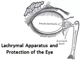
Naba jehad Dania Al Tarawneh Anatomy Introduction Disorders of the lacrimal system are common and may produce chronic symptoms with a significant morbidity The lacrimal glands normally produce about ID: 1037848

tear eye tears system eye tear system tears nasolacrimal dry obstruction drainage symptoms lacrimal sac ocular treatment deficiency conjunctiva

Share:

Link:

Embed:

Download Presentation from below link

Download Presentation The PPT/PDF document "The lacrimal system Eslam shamailah" is the property of its rightful owner. Permission is granted to download and print the materials on this web site for personal, non-commercial use only, and to display it on your personal computer provided you do not modify the materials and that you retain all copyright notices contained in the materials. By downloading content from our website, you accept the terms of this agreement.


Presentation Transcript

1. The lacrimal systemEslam shamailahNaba jehad Dania Al Tarawneh

2. Anatomy

3.

4.

5. IntroductionDisorders of the lacrimal system are common and may produce chronic symptoms with a significant morbidity. The lacrimal glands normally produce about 1.5 µl of tears per minute. Some tears are lost by evaporation, while the remainder drain via the nasolacrimal system. The tear film is re - formed with each blink. Abnormalities are found in: • tear flow and evaporation; • the drainage of tears.

6. Abnormalities in tear flow and evaporation – dry eye Dry eye is a condition of the ocular surface due to a deficiency of tear quantity or composition or excessive evaporation, characterized by hyperosmolarity leading to ocular surface damage, inflammation and symptoms of discomfort and visual loss. An alternative term is keratoconjunctivitis sicca ( KCS ).

7. 1.Aqueous - deficient dry eye A deficiency of lacrimal secretion occurs with age and may be sufficient to cause dry eyes. When dry eyes occur in combination with a dry mouth and dryness of other mucous membranes the condition is called primary Sjögren ’ s syndrome (an autoimmune exocrinopathy ). Other organ systems may be affected. When Sjögren ’ s syndrome occurs in association with a specific autoimmune connective tissue disorder the condition is called secondary Sjögren ’ s syndrome. Rheumatoid arthritis is the commonest of these associated disorders. Diagnosis of Sjögren’s syndrome is confirmed by the demonstration of reduced lacrimal and salivary function, the presence of raised titers of anti - Rho and anti - La antibodies and the demonstration of T -cell infiltration of biopsied minor salivary glands.

8. Symptoms Patients have non - specific symptoms of grittiness, burning, photophobia, heaviness of the lids and ocular fatigue. Symptoms are worse in the evening because the eyes dry progressively during the day. In more severe cases visual acuity may be reduced by corneal damage.

9. SignsIn mild cases there are few obvious signs. Staining of the eye with fluorescein will show small dots of fluorescence ( punctate staining ) over the exposed corneal and conjunctival surfaces. In severe cases tags of abnormal mucus may attach to the corneal surface ( filamentary keratitis ), causing pain due to tugging on these filaments during blinking.

10.

11. Treatment Supplementation of the tears with tear substitutes helps to reduce symptoms, and a humid environment can be created around the eyes with shielded spectacles. In severe cases it may be necessary to occlude the puncta with plugs, or more permanently with surgery, to conserve the tears. Topical anti - inflammatory agents are also in use (e.g. cyclosporin).

12. Prognosis Mild disease usually responds to artificial tears and tear conservation. Severe disease, such as that in rheumatoid Sjögren’s, can be very difficult to treat.

13. 2.Evaporative dry eye – inadequate meibomian oil delivery Extensive meibomian gland obstruction may result in a deficient tear film lipid layer and lead to increased water loss from the eyes. This results in tear hyperosmolarity in its own right and also may exacerbate an existing aqueous - deficient dry eye. Treatment of the meibomian gland dysfunction is similer to posterior blepharitis ( good hygiene , hot compressors , artificial tears , anti-inflammatory treatment with or without antibiotic )

14. 3.Evaporative dry eye – malposition of the globe or lid margins If the lids are not adequately apposed to the eye ( ectropion ), or there is incomplete lid closure ( lagophthalmos – e.g. in a seventh nerve palsy), or if the eye protrudes ( proptosis ) as in dysthyroid eye disease, then the preocular tear film will not form adequately and an evaporative form of dry eye will result. It may also occur in conditions of infrequent blinking, such as Parkinson ’ s disease. Correction of the lid deformity is required for ectropion. In other instances, if the defect is temporary, artificial tears and lubricants can be applied. If lid closure is inadequate, a temporary ptosis can be induced with a local injection of botulinum toxin into the levator muscle to effect a short - term paralysis. A more permanent result can be obtained by suturing together part of the apposed margins of the upper and lower lids (e.g. lateral tarsorrhaphy ).

15.

16. 4.Inadequate mucus productionCicatricial conjunctival disorders Loss of goblet cells occurs in most forms of dry eye, but particularly in cicatricial conjunctival disorders such as erythema multiforme (Stevens – Johnson syndrome). In this there is an acute episode of inflammation causing macular ‘ target ’ lesions on the skin and discharging lesions on the eye, mouth and vulva. In the eye this causes conjunctival shrinkage with adhesions forming between the globe and the conjunctiva ( symblepharon ). There may be both an aqueous, meibomian and a mucin deficiency, and problems due to lid deformity and trichiasis. Chemical burns of the eye, particularly by alkalis, and trachoma (chronic inflammation of the conjunctiva caused by chlamydial infection;) may have a similar end result.

17. The symptoms are similar to those seen with an aqueous deficiency. Examination may reveal scarred, abnormal conjunctiva and areas of fluorescein staining on the cornea and conjunctiva. Treatment requires the application of artificial lubricants. Vitamin A deficiency ( xerophthalmia ) is a condition causing childhood blindness on a worldwide scale. It is associated with generalized malnutrition in countries such as India and Pakistan. Goblet cells are lost from the conjunctiva and the ocular surface becomes keratinized ( xerosis ). An aqueous deficiency may also occur. The characteristic corneal melting and perforation which occur in this condition ( keratomalacia ) may be prevented by early treatment with vitamin A .

18.

19. Disorders of tear drainage When tear production exceeds the capacity of the drainage system, excess tears overflow onto the cheeks. It may be caused by: • irritation of the ocular surface, e.g. by a corneal foreign body, infection or blepharitis; • occlusion of any part of the drainage system (when the tearing is termed epiphora ).

20. Obstruction of tear drainage ( infantile) The nasolacrimal apparatus appears in the 3-5 week and gradually forms a cord of epithelium that extends from the eyelids to the nose. Canalization of the cord begins at the punctum in the eyelid during the third month of intrauterine life and extends toward the nose. Canalization is usually complete by birth..

21. The nasolacrimal system develops as a solid cord which subsequently canalizes and is patent just before term. Congenital obstruction of the duct is common. The distal end of the nasolacrimal duct may remain imperforate, causing a watering eye. If the canaliculi also become partially obstructed, the non - draining pool of fluid in the sac may become infected and accumulate within a mucocoele or cause dacrocystitis . Diagnostically, the discharge may be expressed from the puncta by pressure over the lacrimal sac. The conjunctiva, however, is not inflamed . Most obstructions resolve spontaneously in the first year of life. If epiphora persists beyond this time, patency can be achieved by passing a probe via the punctum through the nasolacrimal duct to perforate the occluding membrane ( probing ). A general anesthetic is required

22. Most common cause of congenital obstruction is incomplete canalization at the distal end (closest to the nose).Spontaneous resolution occurs by 6 months of age in 90% of infants.obstructions persisting beyond the age of 12 months are unlikely to resolve spontaneously !Exclude other ocular causes of lacrimation as blepharitis *Suspect it’s an infantile glaucoma* When(abnormal tearing with photophobia, blepharospasm, and/or large or asymmetric corneal diameters)

23. Clinical features :Chronic or intermittent tearing.Debris on the eyelashes ("mattering").Mild redness of the lower eyelid (irritation from overflow tearing and chronic rubbing of the eyes).Overflow of tears when the child is in an environment that stimulates a greater production of tears (eg, wind or cold) or reduced outflow of tears (swelling of nasal mucosa during an upper respiratory infection). Physical examination :Increase in the size of the tear meniscus.Palpation of the lacrimal sac cause reflux of tears and/or mucoid discharge onto the eye through the puncta.

24.

25.

26.

27. B. Obstruction of tear drainage ( adult) The tear drainage system may become blocked at any point, although the most common site is the nasolacrimal duct. Causes include infections or direct trauma to the nasolacrimal system and, occasionally, topically applied drugs. History The patient complains of a watering eye, sometimes associated with stickiness. The eye is white. Symptoms may be worse in the wind or in cold weather. There may be a history of previous trauma or infection.

28. Signs A stenosed punctum may be apparent on slit lamp examination. Epiphora is unusual if one punctum continues to function. Acquired obstruction beyond the punctum is diagnosed by syringing the nasolacrimal system with saline, using a fine cannula inserted into a canaliculus. A patent system is indicated when the patient tastes the saline as it reaches the pharynx. If there is an obstruction of the nasolacrimal duct then fluid will regurgitate from the non - cannulated punctum. The exact location of the obstruction can be confirmed by injecting a radio - opaque dye into the nasolacrimal system ( dacrocystogram ). X - rays are then used to follow the passage of the dye through the system. A more physiological test follows the passage of a drop of technetium isotope, applied to the lateral conjunctival sac, through the drainage system using a gamma camera ( dacroscintogram ) .

29.

30. Treatment It is important to exclude other ocular disease that may contribute to watering, such as blepharitis. Repair of the occluded nasolacrimal duct requires surgery to connect the mucosal surface of the lacrimal sac to the nasal mucosa by removing the intervening bone ( dacryocystorhinostomy or DCR ). The operation can be performed through an incision on the side of the nose but it may also be performed endoscopically through the nasal passages, thus avoiding a scar at the root of the nose.

31.

32.

33.

34. Infections of the nasolacrimal system Closed obstruction of the drainage system predisposes to infection of the sac ( dacryocystitis ; ). The organism involved is usually Staphylococcus or Streptococcus . Patients present with a painful swelling on the medial side of the orbit, which is the enlarged, infected sac. Treatment is with systemic antibiotics. A mucocoele results from a collection of mucus in an obstructed sac; it is not grossly infected and is often painless. In either case a DCR may be necessary to remove its contents and achieve a patent nasolacrimal system.

35.

36.

37.

38.