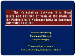
PPT-CT Scan Findings of Head

Author : ceila | Published Date : 2022-06-07

Injury Patients and Cerebral herniations Computed tomography CT has become the diagnostic modality of choice for head trauma All patients underwent CT scan

Presentation Embed Code

Download Presentation

Download Presentation The PPT/PDF document "CT Scan Findings of Head" is the property of its rightful owner. Permission is granted to download and print the materials on this website for personal, non-commercial use only, and to display it on your personal computer provided you do not modify the materials and that you retain all copyright notices contained in the materials. By downloading content from our website, you accept the terms of this agreement.

CT Scan Findings of Head: Transcript


Injury Patients and Cerebral herniations Computed tomography CT has become the diagnostic modality of choice for head trauma All patients underwent CT scan from skull base to the vertex and sequential axial slices of 10 mm were obtained . Plagiocephaly. Presented by: . Amanda Pozarnsky, CO, . BSPO. Board Eligible . Prosthetist. History of Cranial Remolding. How long has cranial remolding existed????. History of Cranial Remolding . For thousands of years, cranial remolding techniques have been used for aesthetic purposes . of . head.txt. Data capture. Data for the head figure was captured by a laser scanner.. The object is mounted on a turntable, and illuminated by a laser stripe. The stripe forms a line image in a camera retina. Because laser projector and camera are not in line, the line image is not straight, but “wiggles” according to object depth.. Luminosity is linked to cross section for a colliding beam accelerator via the equation . For experiments where there is an identifiable reference process (e.g. . Bhabha. scattering . in LEP experiments) that can be accurately calculated, things are reasonably straightforward. For pp scattering there is no equivalent process.. This information is provided by Cleveland Clinic and is not intended to replace the medical advice of your doctor or health care provider. Please consult your health care provider for advice about a s When to Image Based on . Choosing . Wisely. ®. . and ACR Appropriateness . Criteria. ®. What Is R-SCAN?. 2. C. ollaborative activity . for referring clinicians and radiologists to improve patient . Jitsupa Litleangdej . -------------------------------------------------------------------. 5. th . year medical students of . Naresuan university, Phitsanulok , Thailand. . The Correlation between Mild Head Injury and Positive CT Scan of the Brain in the Patients with Moderate Risks at Naresuan University Hospital . Squamous. Cell Carcinoma of the Head and Neck. Yasir. . Rudha. , MD; . Amr. . Aref. , MD; Paul . Chuba. , MD; Kevin O’Brien, MD. Departments of Radiation Oncology, St. John Hospital and Medical Center and St. John Macomb/Oakland Hospital. emergency department They may not be needed as often as you think If you or someone you care for is in the emergency department, you want the medical staff to do everything they can to nd out wh Indication. Current Imaging. Proposed Imaging . Surgical excision of T1/T2N0 without reconstruction or adjuvant radiotherapy. No post treatment imaging . No post treatment imaging . Surgical excision of T3-T4 or N+ with or without adjuvant radiotherapy. EASTERN MEDITERRANEAN UNIVERSITY. SCHOOL OF COMPUTING AND TECHNOLOGY. Disk. Scheduling. Disk Performance Parameters. Normally the disk I/O operation depends on the computer system, the nature of the I/O channel and disk controller hardware.. Structure and Function. The Head. Cranium—8 bones. Frontal (1). Parietal (2). Temporal (. 2). Occipital (1). Ethmoid . (1). Sphenoid (. 1). Question. Is the following statement true or false?. The skull, which is the framework of a head, has two subsections: the cranium and cervical vertebrae.. Motion. Too high of a helical pitch. Oil in the x-ray tube housing. Failure to turn on a beam hardening correction. History:. An adult head is scanned using a helical/spiral mode and the resulting images exhibit hypo and hyper intensities at the brain/bone interface and a blurry appearance throughout the slice. What is causing these artifacts?. supplement 1A: CT scan showed the comminuted fracture at left lateral mass and avulsion fracture at left insertion site of TAL. . Male, 28 years old. A heavy box about 100 kilograms weight fell on his head while he worked as porter. Eccentric axial loading was his main mechanism of injury.. Dr Madeleine Adams. Consultant Paediatric Oncologist. All Wales Paediatric Cancer Predisposition Service. Surveillance for LFS (children). Ultrasound scan of abdomen and pelvis – every 3-4 months. Whole body and brain MRI – annual .

Download Document

Here is the link to download the presentation.
"CT Scan Findings of Head"The content belongs to its owner. You may download and print it for personal use, without modification, and keep all copyright notices. By downloading, you agree to these terms.

Related Documents