/
 Sclerotic Arterial Disorders  Sclerotic Arterial Disorders

Sclerotic Arterial Disorders - PowerPoint Presentation

celsa-spraggs
celsa-spraggs . @celsa-spraggs
Follow
345 views
Uploaded On 2020-04-04

Sclerotic Arterial Disorders - PPT Presentation

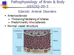
Arterio sclerosis ThickeningHardening of Arteries Predominantly Athero sclerosis Normal vessel structure Pathophysiology of Brain amp Body USSJJQ203 Normal Structure Normal Structure ID: 775371

hdl cholesterol risk atherosclerosis hdl cholesterol risk atherosclerosis fat disease chd ldl factors coronary levels smc amp serv aneurysm

Share:

Link:

Embed:

Download Presentation from below link

Download Presentation The PPT/PDF document " Sclerotic Arterial Disorders" is the property of its rightful owner. Permission is granted to download and print the materials on this web site for personal, non-commercial use only, and to display it on your personal computer provided you do not modify the materials and that you retain all copyright notices contained in the materials. By downloading content from our website, you accept the terms of this agreement.


Presentation Transcript

Slide1

Sclerotic Arterial Disorders

Arterio

sclerosisThickening/Hardening of ArteriesPredominantly AtherosclerosisNormal vessel structure

Pathophysiology of Brain & Body

USSJJQ-20-3

Slide2

Normal Structure

Slide3

Normal Structure

Slide4

Functions of Vessel Cells

Endothelial CellsSemi-PermeableNon-thrombogenicVascular ToneRegulation of SMC GrowthLipid Oxidation (LDL)

Smooth Muscle Cells (SMC)

Constriction/Dilation

Collagen, Elastin Synthesis

Growth Factors/ Cytokines

SMC Proliferation/ Migration

Slide5

Arteriosclerotic Disorders

Narrowed Vessels Organ IschaemiaEndothelial Damage ThrombosisSubsequent Infarction Brain, HeartVessel Wall Weakening Dilatation (Aneurysm) RuptureOccur in Pathogenesis of:AtherosclerosisIschaemic Heart Disease/HypertensionDiabetes

Features

Slide6

Arteriosclerosis - types

AtherosclerosisDominant formIntimal fibrofatty plaque (atheroma)Adds rigidity, leads toIschaemia & aneurysm formationArteriolosclerosisSeen in diabetes and hypertensionThickened media, narrow lumenAffects small arteries and renal arteriolesMönckeberg's Medial Calcific SclerosisVessel wall becomes calcified/rigid (large pulse)No ↓ in lumen size (but can become occluded)

Slide7

Types of Arteries & Associated Diseases

Large/Elastic

(

eg

Aorta)

Composition

: Elastin/Smooth Muscle Cells (SMC)

Disease involvement

:

Atherosclerosis

Medium/Muscular

(

eg

Coronary, Renal)

Composition

: SMC/less elastic tissue

Disease involvement

:

Atherosclerosis

Small/Arterioles

(Parenchymal)

Composition

: SMC

Disease involvement

: Arteriolosclerosis (Hypertension/DM)

Slide8

Atherosclerosis (AS)

WHO: 45% of ‘Western’ deaths linked to AS

Coronary AS

Ischaemic

Heart Disease (IHD)

Acute Myocardial Infarction (AMI)

Congestive Heart Failure (CHF)

Cerebral Vessel AS

Brain Infarct (Stroke)

Aorta (and other major vessel) AS

Aneurysm

Rupture

Ischemia

Necrosis

Gangrene

Slide9

* lipid core

fibrous cap Collagen stained blue

The Culprit

Slide10

Fig. 11.7

AHA Classification of Atherosclerosis

Slide11

Non-denuding, subtle injuryHaemodynamic disturbances/hypercholesterolemiaLDL accumulationActivation/interaction of ECs, SMCs & lymphocytesSMC proliferation, collagen deposition, extracellular matrixFibrofatty atheromaEffects/complications

Development of AS

Slide12

Response to Subtle, Chronic ‘Insult’

Slide13

Risk Sites

Slide14

EC permeability ↑ EC turnover↑ LDL endocytosis↑ leucocyte adhesionHigh shear stress or ‘stagnant’ areas responsible?

Role of Haemodynamics

Slide15

Endothelial Dysfunction

Slide16

Initiation of Fatty Streak

Slide17

Fatty Streak

Slide18

Slide19

Slide20

Slide21

Fibro-fatty Atheroma

Slide22

Slide23

Slide24

Summary of Atherosclerotic Process

Multifactorial process (risk factors)

Initiated by endothelial dysfunction

Up regulation of endothelial and leukocyte adhesion molecules

Macrophage

diapedesis

LDL

transcytosis

LDL oxidation

Foam cells

Recruitment and proliferation of smooth muscle cells (synthesis of connective tissue proteins)

Structural

organisation

of plaque

Formation of arterial thrombi

Slide25

Consequences of plaque formation

Generalised

Narrowing/Occlusion

Rupture

Thrombi/Emboli

Leading to specific problems

Myocardial and cerebral infarcts

Aortic aneurysms

Peripheral

Vascular Disease (PVD)

AS becomes increasingly

complicated

Calcification, fissuring, haemorrhage

Slide26

Plaques

 Complicated Lesions

Slide27

AS Clinical Development

Slide28

Slide29

Slide30

Slide31

Fusiform Aneurysm (Abdominal Aorta)

Slide32

Berry Aneurysm (Circle of Willis)

Slide33

Dissecting Aneurysm (aortic arch)

Slide34

AS Risk Factors: Non-Modifiable

Age

A dominant influence

Atherosclerosis begins in the young, but does not precipitate organ injury until later in life

Gender

Men more prone than women, but by age 60-70 about equal frequency

Family History

Familial cluster of risk factors

Genetic differences

Slide35

AS Risk Factors: Modifiable(potentially controllable)

Hyperlipidaemia

(

Hypercholesterolaemia

)

Hypertension

Cigarette smoking

Diabetes Mellitus

Elevated

Homocysteine

Factors that affect

haemostasis

and thrombosis

Infections:

Herpes virus

Chlamydia/

Chlamydophila

pneumoniae

Obesity

Sedentary lifestyle

Stress

Slide36

Hypercholesterolemia & AS - link

Experimental diets in animals

Plaques (

atheromas

) are mostly cholesterol

Epidemiologic

Cholesterol

Risk

HDL

Risk (seems paradoxical)

LDL

Risk

Genetic/Acquired Disorders

Cholesterol

Familial Hypercholesterolemia

Diabetes Mellitus

Hypothyroidism

Dietary & Pharmacologic

Cholesterol

Atherosclerosis

Slide37

Keys, 1970

Seven Countries Study: CHD Events are

Correlated with Saturated Fat

Slide38

LaRosa

et al, 1990

Six Year CHD Mortality from MRFIT

Desirable

Borderline

High

CHD Mortality is Correlated with Plasma Cholesterol Levels

Slide39

Slide40

Cholesterol and lipoproteins

Hydrophobic lipids (TG, CE) in coreHydrophilic lipids (UC, PL) on surfaceClassification based on size/densityReflects compositionReflects role

Slide41

Lipoprotein Classes

Doi H et al.

Circulation

2000;102:670-676; Colome C et al. Atherosclerosis 2000;149:295-302; Cockerill GW et al. Arterioscler Thromb Vasc Biol 1995;15:1987-1994.

HDL

LDL

Chylomicrons,

VLDL, and

their catabolic remnants

> 30 nm

20–22 nm

9–15 nm

D<1.006 g/ml

D=1.019-1.063g/ml

D=1.063-1.21 g/ml

Lipids Online

Generalisation

!

Exogenous

(dietary)

Endogenous

(by liver)

Transport

(

to

liver)

Slide42

Figure 2 Principal steps in early atherogenesis.

Barter P Eur Heart J Suppl 2005;7:F4-F8

© The European Society of Cardiology 2005. All rights reserved. For Permissions, please e-mail: journals.permissions@oupjournals.org

Slide43

HDL Maturation

(Cholesterol and Atherosclerosis, Grundy)

HDL is secreted in a

discoidal form from the liver and gut.

As it acquires cholesterol from tissues in the circulation, it matures into a spherical form through the action of lecithin:cholesterol acyl transferase

Slide44

HDL Metabolism and Reverse Cholesterol Transport

ABC1 = ATP-binding cassette protein 1; A-I = apolipoprotein A-I; CE = cholesteryl ester; FC = free cholesterol; LCAT = lecithin:cholesterol acyltransferase; SR-BI = scavenger receptor class BI

A-I

Liver

CE

CE

CE

FC

FC

LCAT

FC

Bile

SR-

B

I

A-I

ABC1

Macrophage

Mature

HDL

Nascent

HDL

Slide45

Slide46

HDL is Protective

110

30

21

0

20

40

60

80

100

120

< 35

35–55

> 55

Incidence

per 1,000 (in 6 years)

HDL-C (mg/

dL

)

Assmann G, ed.

Lipid Metabolism Disorders and Coronary Heart Disease.

Munich: MMV Medizin Verlag, 1993

186 events in 4,407 men (aged 40–65 y)

Lipids Online

Slide47

CHD Risk According to Total-C/HDL-C RatioFramingham Study (Men)

Kannel WB.

Am J Cardiol 1987;59:80A–90A

3.0

2.5

2.0

1.5

1.0

0.5

2.0

4.0

6.0

8.0

10.0

12.0

Total-C/HDL-C ratio

CHD risk ratio

3.4

4.4

9.6

Slide48

Effects of Lifestyle Modificationson HDL-C Levels

Weight reduction

For every 3 kg (7

lb

) of weight

loss

, HDL-C levels

increase

1 mg/

dL

.

Smoking cessation

HDL-C levels in smokers are 7–20%

lower

than those in nonsmokers.

HDL-C levels return to normal within 30–60 days after smoking cessation.

Exercise

Aerobic exercise (

eg

, running) increases HDL-C in ‘dose-dependent’ manner.

Slide49

(Continued)

Total fat intake

Low-fat diets lower HDL-C in all patients

But populations with low total fat intake have reduced CHD

How much due to environmental/lifestyle factors?

Alcohol

Alcohol increases HDL-C in a ‘dose-dependent’ manner

Caloric restriction

Caloric restriction

acutely

lowers HDL-C

Effects of Lipid-Modifying Drugs on HDL-C Levels

Niacin

15–35%

Fibrates

10–15%

Estrogens

10–15%

Statins

5–10%

Slide50

Is Atherosclerosis Reversible?

Primate experiments

High fat diet, atherosclerotic lesions regress once diet normal

Humans

Decrease fat and caloric intake (wars, famine, wasting disease),

atheromas

decrease

Angiography after cholesterol lowering, plaque size decreases

What needs to happen for plaques to regress?

LDL lowered

Macrophages ingest lipid

Reverse cholesterol transport, depends on HDL

Slide51

AS - Clinical Prevention

Lifestyle Changes

Smoking, exercise, salt, saturated fats

Trans-FAs to Omega - 3 FAs

Less cake, more oily fish!

 Cholesterol (diet, Rx)

Antiplatelet Drugs

Eg

1 ‘baby’ (75mg) aspirin/day when 40

Slide52

Dietary Components and CHD Risk

Summary of the Nurses’ Health Study

Vit E (Supplement vs no Supplement)

Margarine (<1 tsp/mo vs >4 tsp/d)

Alcohol (1 drink/d vs none)

Nuts (5 servings/wk vs almost never)

Folic Acid (>545 ug/d vs <190 ug/d)

Fibre (23g/d vs 12 g/d)

Whole grains (>1.7 serv vs <0.25 serv)

Eggs (<1/wk vs >1/d)

Saturated Fat (10.7% vs 18.8%)

Total Fat (29.1% vs 46.1%)

Fruit

(3.8 serv vs 0.6 serv)

Vegetables

(6.8

serv

vs

1.5

serv

)

Slide53

Slide54

Coronary Calcium Scanning

Electron Beam CT Coronary Artery Scanning

Very expensive machinesScan is ECG-TriggeredV rapid scan (not mechanical)3mm contiguous scans

Slide55

3D vessel probe of the Left Main and Left Anterior Descending coronary artery.

Curved MultiPlanar Reformatted (MPR) images are automatically rendered and quantify this LAD lesion at 48% diameter stenosis. (Images - Toshiba America Medical Systems and Vital Images SUREPlaque and University of California Irvine, Cardiac CT Center)

CT Angiography

Slide56

Carotid Intimal Medial Thickness (Ultrasound)