/
1 Qs 21 1 Qs 21

1 Qs 21 - PowerPoint Presentation

cheryl-pisano
cheryl-pisano . @cheryl-pisano
Follow
393 views
Uploaded On 2017-03-17

1 Qs 21 - PPT Presentation

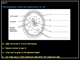
The drawing below shows the ultrastructure of E coli Label structures A to E on the drawing State a function of part D What term is given to this bacterial shape d List three ways in which prokaryotic cells differ from eukaryotic cells ID: 525221

cells cell growth increases cell cells increases growth reference temperature rate table prokaryotic light ans eukaryotic eukaryotes bacterial viruses

Share:

Link:

Embed:

Download Presentation from below link

Download Presentation The PPT/PDF document "1 Qs 21" is the property of its rightful owner. Permission is granted to download and print the materials on this web site for personal, non-commercial use only, and to display it on your personal computer provided you do not modify the materials and that you retain all copyright notices contained in the materials. By downloading content from our website, you accept the terms of this agreement.


Presentation Transcript

Slide1

1

Qs 21The drawing below shows the ultrastructure of E. coli.Label structures A to E on the drawing.State a function of part D.What term is given to this bacterial shape?d) List three ways in which prokaryotic cells differ from eukaryotic cells.Slide2

2

Ans 21A = plasma membrane B = cell wall C = nuclear mass / nucleoid D = mesosome E = ribosomescontain the enzymes for respiration/cell wall synthesisCoccusprokaryotic cells contain no membrane-bound organelles, eukaryotes do prokaryote cells have no nuclear membrane, eukaryotes do; prokaryotes have a nuclear mass / nucleoid eukaryotes have a nucleus / prokaryotes have one long circular chromosome, eukaryotes have many linear chromosomes prokaryotes have no nucleoli, eukaryotes do prokaryotic cell walls contain the structural polysaccharide murein, eukaryotic cell walls (if present) contain cellulose

prokaryotes contain 70S ribosomes, eukaryotes contain 80S ribosomesSlide3

3

Qs 22The diagram shows a cholera bacterium. It has been magnified 50 000 times.Name AName two structures present in an epithelial cell from the small intestine that are not present in a cholera bacterium.Cholera bacteria can be viewed using a transmission electron microscope (TEM) or a scanning electron microscope (SEM).c) Give one advantage of using a TEM rather than a SEM.d) Calculate the actual width of the cholera bacterium between points B and C

Give your answer in micrometres and show your working.Slide4

4

Ans 22(Plasma / cell) membraneNucleus / nuclear envelope / nuclear membrane / nucleolus Mitochondrion; Smooth / rough ER Lysosome; Microvillus / brush border Golgi body Linear / non-circular DNA / chromosome 80S / denser / heavier / larger ribosomesHigher resolution / higher (maximum) magnification / higher detail (of image)OR Allows internal details / structures within (cells) to be seen / cross section to be taken0.42–0.46 µm [divide measured width (µm) by magnification]Slide5

Qs 23

The diagram shows part of an epithelial cell from an insect’s gutThis cell is adapted for the three functions listed below. Use the diagram to explain how this cell is adapted for each of these functions. Use a different feature in the diagram for each of your answers.the active transport of substances from the cell into the bloodb) the synthesis of enzymesrapid diffusion of substances from the lumen of the gut into the cytoplasmSlide6

6

Ans 23a)Mitochondria respireRelease energy/ produce ATPTransport against concentration gradientORInfolding of membraneIncreases (surface) areaMore proteins for active transportb)Ribosomes make proteins / enzymesEnzymes are proteinsORMitochondria respireRelease energy/produce ATP(Energy/ATP) for protein / enzyme synthesisc)

Microvilli increase area / have large areaSlide7

7

Qs 24Filamentous algae are simple photosynthetic organisms that consist of long strands of very similar eukaryotic cells. Each of the cells in the strand is enclosed within a cellulose cell wall. The strand increases in length as the cells divide and elongate. The photographs below show some cells in strands of a filamentous alga, as seen using a light microscope (x 200)Name the process that is involved in the cell division of the filamentous algaCircle the structure that would not be found in a cell from the strand of a filamentous alga Lysosome Mitochondrion Plasmid RibosomeSlide8

8

Qs 24 (continued)An investigation was carried out into the effect of temperature on the rate of growth of a filamentous alga. Several short strands of the alga were placed into culture solutions which were kept at five different temperatures and at a high light intensity.The number of cells in the strands, in Each culture solution, was counted at the Beginning of the time period and again after 18 days. The rate of growth was then calculated. The results of this investigation are shown in the graph Name the independent variable in this investigation.Using the information in the graph, describe and suggest explanations for the effect of temperature on the rate of growth of the filamentous alga.Suggest why it was important that this investigation was carried out at a high light intensitySuggest two abiotic factors, other than light intensity, that would need to be controlled in this investigation..Slide9

9

Ans 24Mitosis b) Plasmid c) Temperature d)1. rate of growth increases as temperature increases {between 13oC and 22oC / up to 22oC} 2. rate of growth decreases {between 22oC and 25oC / above 22oC} 3. use of manipulated data to support abovee.g. increases by {0.7 (a.u.) / 4.5 times}, decreases by 0.1 (a.u.) 4. reference to enzymes involved (in growth) 5. molecules {move about more / have more kinetic energy}, as temperature increases ;6. (therefore) {enzyme and substrate (molecules) collide more / rate of enzyme-substratecomplexes formation increases} as temperature increases 7. correct reference to denaturation of some {enzyme / protein } (molecules) 8. (therefore) rate of {growth / reactions} decreases as fewer enzyme molecules available

e)

1. idea that (each temperature) has same light intensity

2. correct reference to must be above {threshold / compensation point}

3. (below which) no net photosynthesis takes place

4. reference to {so light is not limiting factor / so temperature is the limiting factor}

5. photosynthesis produces {material } needed for growth

f)

1. {wavelength / colour / frequency} of light

2. CO

2

concentration

3. pH (of solution)

4. reference to {mineral Slide10

10

Qs 25Antibiotics are used to treat bacterial infections in eukaryotic organisms. The table below describes some of the structures that are found in cells. Complete the table by writing the name of each of the structures described and stating whether it is found in prokaryotic cells only (P), eukaryotic cells only (E) or both types of cell (B).Vancomycin is an antibiotic that kills bacterial cells by preventing the synthesis of peptidoglycan, a component of bacterial cell walls.State the term used to describe antibiotics, such as vancomycin, that kill bacterial cells.Suggest how bacterial cells are killed by vancomycin.

Explain why doctors have been advised to limit the prescription of antibiotics.Slide11

11

Ans 25a)b) Bactericidalc)1. cell wall {weaker /cannot form properly 2. {cell / cell wall} bursts (easily) 3. during division4. Due to high internal osmotic pressure d)1. reference to antibiotic acting as selective pressure 2. reference to some bacteria resistant (to antibiotic) 3. idea that resistant bacteria survive and {reproduce / pass on resistance / pass ongene 4. idea that antibiotic no longer effective

5. reference to some infections cannot be treated with antibiotics

Mitochondrion /

mitochondria

E / eukaryotic

Flagellum /

flagella

B / both

plasmid

P / prokaryoticSlide12

12

Qs 26Prokaryotes, mitochondria and chloroplasts have many features in common.The diagram shows a mitochondrion. Two of the features labelled are typical of prokaryotes. a) Place a tick (√) in each of the two boxes that correctly identify these features. Answers in diagramb) The table below shows some features of mitochondria. If the feature is also present in chloroplasts, place a tick (√

) in

the box to the right of that feature

and if it is absent, place a cross

(X) in the box.

Answers in table

√Slide13

13

Qs 26Bacteria have prokaryotic cells. The table shows functions of parts of a prokaryotic cell.Complete the table by naming the part of the cell that carries out each functionAntibiotics can be used to treat disease caused by bacteria. Give two reasons why antibiotics cannot be used to treat disease caused by viruses.Ribosome

Plasmid

Capsule

Flagellum

Ans 26

In table

b) Viruses are not cells

Viruses do not have metabolism – e.g. protein synthesis

Viruses do not have cell walls

Viruses do not synthesise proteins / have ribosomes

Viruses are inside cells / antibiotics need to enter cells

Related Contents


Next Show more