/
Epidural anesthesia Epidural anesthesia

Epidural anesthesia - PowerPoint Presentation

coveurit
coveurit . @coveurit
Follow
353 views
Uploaded On 2020-06-26

Epidural anesthesia - PPT Presentation

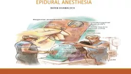
bayan KHawaldeh Introduction Anatomy Epidural space base of skull foramen magnum to the coccyx sacrococcygeal membrane The epidural space surrounds the dura mater posteriorly laterally and ID: 788310

local epidural anesthetic anesthesia epidural local anesthesia anesthetic space spinal anesthetics injection block patient spread volume patients dose nerve

Share:

Link:

Embed:

Download Presentation from below link

Download The PPT/PDF document "Epidural anesthesia" is the property of its rightful owner. Permission is granted to download and print the materials on this web site for personal, non-commercial use only, and to display it on your personal computer provided you do not modify the materials and that you retain all copyright notices contained in the materials. By downloading content from our website, you accept the terms of this agreement.


Presentation Transcript

Slide1

Epidural anesthesia bayan KHawaldeh

Slide2

Introduction

Anatomy–Epidural space – base of skull (foramen magnum) to the coccyx (sacrococcygeal membrane)_The epidural space surrounds the dura mater posteriorly, laterally, andanteriorly. Nerve roots travel in this space as they exit laterally through theforamen and course outward to become peripheral nerves–Distance from skin to epidural space – 4-5 cm–Epidural space contains loose areolar tissue, fat, arterial and venous networks, lymphatics, spinal nerve roots

Slide3

Introduction

Epidural (extradural) anesthesia involves the use of local anesthetics injected into the epidural space (space outside dura) to produce a reversible loss of sensation and motor function. This space extends from the craniocervical junction at C1 to the sacrococcygeal membrane, and anesthesia can theoretically be safely instituted at any level in between. Continuous epidural anesthesia is a neuraxial technique offering a range of applications wider than single-dose spinal anesthesia.

Slide4

Introduction

In practice, an epidural is sited adjacent to the nerve roots that supply the surgical site; that is, the lumbar region is used for pelvic and lower limb surgery and the thoracic region for abdominal surgery. A single injection of local anesthetic can be given but more commonly, a catheter is inserted into the epidural space and either repeated injections or a constant infusion of a local anesthetic drug is used..The majority of the local anesthetic administered is absorbed systemically by the rich venous plexus found within the epidural space. Dura surrounding spinal nerve/nerve roots are a modest barrier to the spread of local anesthetics. A small amount of local anesthetic will be absorbed into epidural fat . What remains will eventually reach its intended site of action, the spinal nerve and nerve roots.

Slide5

Spread of Local Anesthetics within the Epidural Space Local anesthetics administered in the epidural space move in a horizontal and longitudinal direction. Theoretically, if enough local anesthetic is injected, it could spread up to the foramen magnum and down to the sacral foramina . Clinically, the extent of longitudinal spread is volume dependent and cephalad spread is limited. It has been found that an epidural will spread only 4 additional dermatomes when increasing the volume of local anesthetics from 10ml to 30 ml . Horizontal spread occurs through intervertebral foramina, entering the

dural cuff. A small amount of local anesthetic may travel to the anterior epidural space. Diffusion into the CSF occurs at the

dural

cuff through arachnoid granules

Slide6

Distribution, Uptake & Elimination

It takes approximately 6-8 times the amount of local anesthetic in the epidural space to produce the same degree of blockade with a spinal anesthetic. This is due to the following factors:Larger mixed nerves are found in the epidural space.Local anesthetics must penetrate the arachnoid and dura mater.Local anesthetics are lipid soluble and will be absorbed into tissue and epidural fat.Epidural veins absorb a significant amount of local anesthetics. Peak blood concentrations occur 10-30 minutes after a bolus.

Slide7

Factors Affecting Level of Block

Factors affecting the level of epidural anesthesia may not be as predictable aswith spinal anesthesia. In adults, 1 to 2 mL of local anesthetic per segment to beblocked is a generally accepted guideline. For example, to achieve a T4 sensorylevel from an L4–L5 injection would require about 12 to 24 mL. For segmental

or analgesic blocks, less volume is needed

Slide8

Factors Affecting Level of Block

Age : The dose required to achieve the same level of anesthesia decreases with age.Height : patient height affects the extent of cephalad spread. Thus, shorter patients may require only 1 mL of local anesthetic per segment to be blocked, whereas taller patients generally require 2 mL per segment.

Gravity :

Positioning the patient after injection of local anesthetic into the epidural space impacts its spread and height, but not to the degree that it does with spinal anesthesia. For example, positioning the patient in a lateral decubitus position will concentrate local anesthetic and extend block height in the dependent area compared to the non-dependent area

Volume :

The volume of local anesthetic plays a crucial role in the block height ,

For example

, to achieve a T4 sensory level from an L4–L5 injection would require about 12 to 24

mL.

For segmental or analgesic blocks, less volume is needed.

Slide9

Factors Affecting Level of Block

Drug Used :.Additives to the local anesthetic, particularly opioids, tend to have a greatereffect on the quality of epidural anesthesia than on the duration of the block.Epinephrine in concentrations of 5 mcg/mL prolongs the effect of epidurallidocaine, mepivacaine, and

chloroprocaine

more than that of bupivacaine,

levobupivacaine, or ropivacaine. In addition to prolonging the duration and

improving the quality of block, epinephrine delays vascular absorption and

reduces peak systemic blood levels of all epidurally administered local

anesthetics.

Slide10

Failed Epidural Blocks

Unlike spinal anesthesia, in which the procedural endpoint is usually very clear (free-flowing CSF), the onset is very fast, and the technique has a very high success rate, epidural anesthesia is dependent on detection of a more subjective loss of resistance (or hanging drop). Also, the onset of epidural anesthesia is slower, and the

more variable anatomy

of the epidural space and

less predictable spread

of local anesthetic make epidural anesthesia inherently less predictable than spinal anesthesia.

Misplaced injections

of local anesthetic can occur in a number of situations . In some patients, the spinal ligaments are soft, and either good resistance is never appreciated or a false loss of resistance is encountered. Similarly, entry into the paraspinous muscles during an off-center midline approach may cause a false loss of resistance. Other causes of failed epidural anesthesia (such as intrathecal, subdural, and intravenous injection) are discussed in the late section of this chapter on complications.

Slide11

Local Anesthetics used for Epidural Anesthesia

When choosing a local anesthetic for epidural anesthesia, consider the following:local anesthetic potency and duration surgical requirements and duration postoperative analgesia requirementsSeven local anesthetics can be used to produce epidural anesthesia. Only preservative free solutions should be used. Check the label to ensure the solution is “preservative free” and prepared specifically for epidural/caudal anesthesia/analgesia.Short Acting: chloroprocaineIntermediate Acting:lidocainemepivacaine

Long Acting:

Bupivacaine

 etidocaine

 ropivacaine

 levobupivacaine

Slide12

Slide13

Slide14

Slide15

Epidural Technique

Preparation_ Prepare the patient. Discuss options, risks and benefits. Explain what to expect during anepidural anesthetic.Positioning _Proper positioning is essential for a successful block. There are three positions used for the administration of epidural anesthesia: lateral decubitus, sitting , and prone (Used for caudal approach in adults).

Slide16

Patient Positioning

A. Sitting PositionThe anatomic midline is often easier to identify when the patient is sitting thanwhen the patient is in the lateral decubitus position (Figure 45–12). This isparticularly true with obese patients. Patients sit with their elbows resting ontheir thighs or a bedside table, or they can hug a pillow.

Slide17

Slide18

B. Lateral Decubitus

Many clinicians prefer the lateral position for neuraxial blocks (Figure 45–14).Patients lie on their side with their knees flexed and pulled high against theabdomen or chest, assuming a “fetal position.” An assistant can help the patientassume and hold this position.

Slide19

Slide20

Anatomic Approach

A. Midline ApproachFor epidural anesthesia, a sudden loss of resistance (to injection of air or saline) is encountered as the needle passes through the ligamentum flavum andenters the epidural space.

Slide21

Anatomic Approach

B. Paramedian ApproachThe paramedian technique may be selected, particularly if epidural orsubarachnoid block is difficult, particularly in patients who cannot be positionedeasily (eg, severe arthritis, kyphoscoliosis, or prior spine surgery) (Figure 45–15). Many clinicians routinely use the paramedian approach for thoracic epidural

puncture.

Slide22

Slide23

Two techniques make it possible to determine

when the tip of the needle has entered the potential (epidural) space: the “loss ofresistance” and “hanging drop” techniques.

Slide24

Slide25

Slide26

Activating an Epidural

The quantity (volume and concentration) of local anesthetic needed forepidural anesthesia is larger than that needed for spinal anesthesia. Toxic sideeffects are almost guaranteed if a “full epidural dose” is injected intrathecally orintravascularly. Safeguards against toxic epidural side effects include test dosingand incremental dosing. These safeguards apply whether the injection is throughthe needle or through an epidural catheter

Slide27

A test dose

is designed to detect both subarachnoid and intravascularinjection. The classic test dose combines local anesthetic and epinephrine,Unfortunately, epinephrine as a marker of intravenous injection is not ideal.False positives (a uterine contraction causing pain or an increase in heart ratecoincident to test dosing) and false negatives (bradycardia and exaggeratedhypertension in response to epinephrine in patients taking β-blockers) can occur.

Simply aspirating prior to injection is insufficient to avoid inadvertent

intravenous injection; most experienced practitioners have encountered

falsenegative

aspirations through both a needle and a catheter.

Slide28

Incremental dosing

is a very effective method of avoiding seriouscomplications (“each dose is a test dose”).If aspiration is negative, a fraction ofthe total intended local anesthetic dose is injected, typically 5 mL. This doseshould be large enough for mild symptoms (tinnitus or metallic taste) or signs

(slurred speech, altered mentation) of intravascular injection to occur, but small

enough to avoid

seizure or cardiovascular compromise

.

This is particularly important for labor epidurals that are to be used for cesarean delivery

Slide29

If the initial labor epidural bolus was delivered through the needle, and the catheter

was then inserted, it may be erroneously assumed that the catheter is wellpositioned because the patient is still comfortable from the initial bolus. If thecatheter was inserted into a blood vessel, or after initial successful placement,has since migrated intravascularly,

systemic toxicity will likely result if the full

anesthetic dose is injected

. Catheters can migrate intrathecally or intravascularly

from an initially correct epidural position at any time after placement, but

migration is most likely to occur with movement of the patient.

Slide30

Indications

Epidural anesthesia provides excellent operating conditions for surgical procedures below the umbilicus. Procedures include: cesarean section procedures of the uterus, perineum* hernia repairs genitourinary procedures lower extremity orthopedic procedures

Slide31

Indications

Continue…•May be used in–Poor risk patients–Cardiac diseases–Pulmonary diseases–Metabolic disturbances–When GA is contraindicated–When spinal anesthesia is contraindicated–Painful conditions including post-op pain relief

Slide32

Contraindications – similar to spinal

•Severe hemorrhage•Coagulation defects ( hematoma formation )•Previous laminectomy•Uncooperative , Patient refusal•Local inflammation at site

Slide33

Advantages

•Well-defined area of anesthesia•Longer duration•More severe disturbances of spinal anesthesia minimized•Return of gastrointestinal function generally occurs faster than with general anesthesia•The ability to use the epidural catheter for postoperative analgesia•Less respiratory effects•Patent airway•Decreased incidence of deep vein thrombosis and pulmonary emboli formation compared to general anesthesia

Slide34

Disadvantages

•Technically more difficult•Muscle relaxation not complete•Large volume necessary•Danger of dural puncture•Risk of block failure. The rate of failure is slightly higher than with a spinal anesthetic.•Always be prepared to induce general anesthesia if block failure occurs.•Onset is slower than with spinal anesthesia. May not be a good technique if the surgeon is impatient or there is little time to properly perform the procedure.

Slide35