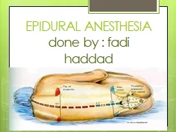
fadi haddad Anatomy Epidural space base of skull foramen magnum to the coccyx sacrococcygeal membrane the peridural space between the ID: 908807
Download Presentation The PPT/PDF document "EPIDURAL ANESTHESIA done by :" is the property of its rightful owner. Permission is granted to download and print the materials on this web site for personal, non-commercial use only, and to display it on your personal computer provided you do not modify the materials and that you retain all copyright notices contained in the materials. By downloading content from our website, you accept the terms of this agreement.
Slide1
EPIDURAL ANESTHESIAdone by : fadi haddad
Slide2Anatomy
Epidural
space
–
base
of
skull
(foramen
magnum)
to
the
coccyx
(
sacrococcygeal
membrane
)
the
peridural
space between the
dura
mater and the
periosteum
lining the vertebral
canal
Distance
from
skin
to
epidural
space
–
4-5
cm
Epidural
space
contains
loose
areolar
tissue,
fat,
arterial
and
venous
networks,
lymphatics
,
spinal
nerve
roots
Slide3Slide4Definition Epidural anesthesia involves the use of local anesthetics injected into the epidural space to produce a reversible loss of sensation and motor
function.
The
injected local
anaesthetic
solution produces analgesia by blocking conduction at the
intradural
spinal nerve roots.
Slide5EPIDURAL ANESTHESIALA deposited
in
epidural
space
Block
spinal
nerve
roots
that
traverse peridural spaceBlocks sympathetic nerves traveling with the anterior rootsApplications range from sensory analgesia, minimal motor block, or dense anesthesia and full motor block – controlled by drug choice, concentration, dosage
Slide6Spread of Local Anesthetics within the Epidural SpaceLocal anesthetics administered in the epidural space move in a horizontal and longitudinal direction. Theoretically, if enough local anesthetic is injected, it could spread up to the foramen magnum and down to the sacral foramina. Clinically, the extent of longitudinal spread is volume dependent and
cephalad
spread is limited. It has been found that an epidural will spread only 4 additional dermatomes when increasing the volume of local anesthetics from 10ml to 30 ml.
Horizontal spread occurs through intervertebral foramina, entering the
dural
cuff. A small amount of local anesthetic may travel to the anterior epidural space. Diffusion into the CSF occurs at the
dural
cuff through arachnoid granules.
Slide7Horizantal
movement
Longtidunal
movement
Slide8Factors Influencing Spread of
Solution
Height
of
patient
Height plays a role in epidural block height. The shorter the patient, the less anesthetic required to achieve the same level of anesthesia as a tall patient. For example, a patient who is 5’3 may require 1 ml per dermatome, whereas a patient who is 6’3” may require 2 ml per dermatome.
Drugs
usedVolumeAge As patients age, less local anesthetic is required to achieve the same level of blockade as their younger counterpart. This is largely due to changes in the size and compliance of the epidural spaceConcentrationLevel of puncture and catheter insertion
Slide9continueGravityPositioning the patient after injection of local anesthetic into the epidural space impacts its spread and height, but not to the degree that it does with spinal anesthesia. For example, positioning the patient in a lateral decubitus position will concentrate local anesthetic and extend block height in the dependent area compared to the non-dependent area. A sitting patient will have more local anesthetic delivered to the lower lumbar and sacral dermatomes.
Slide10Slide11Distribution, Uptake & EliminationIt takes approximately 6-8 times the amount of local anesthetic in the epidural space to produce the same degree of blockade with a spinal anesthetic. This is due to the following factors:Larger mixed nerves are found in the epidural space.Local anesthetics must penetrate the arachnoid and dura
mater.
Local anesthetics are lipid soluble and will be absorbed into tissue and epidural fat.
Epidural veins absorb a significant amount of local anesthetics. Peak blood concentrations occur 10-30 minutes after a bolus.
Slide12Local Anesthetics used for Epidural AnesthesiaWhen choosing a local anesthetic for epidural anesthesia, consider the following:local anesthetic potency and duration surgical requirements and duration postoperative analgesia requirements
Seven local anesthetics can be used to produce epidural anesthesia. Only preservative free solutions should be used. Check the label to ensure the solution is “preservative free” and prepared specifically for epidural/caudal anesthesia/analgesia.
Short Acting
:
2-
chloroprocaine
Intermediate Acting:
lidocaine
mepivacaineLong Acting:Bupivacaine etidocaine ropivacaine levobupivacaine
Slide13Slide14Slide15Types – selective blockade possible because it
can
be performed
at
any
level
of
spine
Cervical
epiduralThoracic epiduralLumbar epiduralCaudal epidural
Slide16Cervical epidural
Slide17Slide18IndicationsEpidural anesthesia provides excellent operating conditions for surgical procedures below the umbilicus. Procedures include: cesarean section procedures of the uterus, perineum* hernia repairs genitourinary procedures lower extremity orthopedic procedures
Slide19continueMay be used
in
Poor
risk
patients
Cardiac
diseases
Pulmonary diseasesMetabolic disturbancesWhen GA is contraindicatedWhen spinal anesthesia is contraindicatedPainful conditions including post-op pain relief
Slide20AdvantagesWell-defined area
of
anesthesia
Longer
duration
More
severe
disturbances
of spinal anesthesia minimizedReturn of gastrointestinal function generally occurs faster than with general anesthesiaThe ability to use the epidural catheter for postoperative analgesiaLess respiratory effectsPatent airwayDecreased incidence of deep vein thrombosis and pulmonary emboli formation compared to general anesthesia
Slide21DisadvantagesTechnically more
difficult
Muscle
relaxation
not
complete
Large
volume
necessaryDanger of dural punctureRisk of block failure. The rate of failure is slightly higher than with a spinal anesthetic.Always be prepared to induce general anesthesia if block failure occurs.Onset is slower than with spinal anesthesia. May not be a good technique if the surgeon is impatient or there is little time to properly perform the procedure.
Slide22complicationLow blood pressure
Loss of bladder control
Itchy skin
Feeling sick
Inadequate pain relief
Headache
Temporary
or permanent nerve
damage
Infection
Slide23Contraindications – similar to spinal
Severe
hemorrhage
Coagulation
defects ( hematoma formation )
Previous
laminectomy
Uncooperative , Patient refusalLocal inflammation at site
Slide24Epidural
anathesia
Spinal
anathesia
Site
of
injection
In
the
epidural
space
Subarachnoid
space
Onset
and
duration
Slow onset and continous duration (use catheter)Rapid
onset and limited durationadvantagesCan be used in analgesiaNot usedNeedle doseCurved,longand blunt (touhy) 10_30mlSmall and sharp 1_4mlspaceAny space usually lumber
lumberQuality of sensory and motor nerve block
lessMore liabletoxicityHypotention gradual total spinal +++ systemic toxicity +++Sudden++
Slide25Differences between Spinal and Epidural Anesthesia
Spinal anaesthesia
Extradural Anaesthesia
Level: below L1/L2, where the spinal cord ends
Level: at any level of the vertebral column.
Injection: subarachnoid space
i.e
punture
of the
dura materInjection: epidural space (between Ligamentum flavum and dura mater) i.e without punture of the dura materIdentification of the subarachnoid space: When CSF appears Identification of the Peridural space: Using the Loss of Resistance technique.Dosis: 2.5- 3.5 ml bupivacaine 0.5% heavyDosis: 15- 20 ml bupivacaine 0.5%Onset of action: rapid (2-5 min)Onset of action: slow (15-20 min)Density of block: more denseDensity of block: less dense
Hypotension: rapid
Hypotension: slow
Headache: is a probably complication
Headache: is
not
a probable.
7 November 2009
25
Slide26Thank youuuuuu doctors