/
Intrapartum Evaluation of Fetal Wellbeing Intrapartum Evaluation of Fetal Wellbeing

Intrapartum Evaluation of Fetal Wellbeing - PowerPoint Presentation

dandy
dandy . @dandy
Follow
69 views
Uploaded On 2023-05-31

Intrapartum Evaluation of Fetal Wellbeing - PPT Presentation

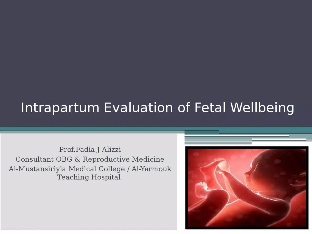
ProfFadia J Alizzi Consultant OBG amp Reproductive Medicine AlMustansiriyia Medical College AlYarmouk Teaching Hospital Objectives At the end of this lecture the students will be able to ID: 1000349

bravado fetal decelerations rate fetal bravado rate decelerations heart ctg amp baseline variability minutes contractions bpm labour reassuring fetus

Share:

Link:

Embed:

Download Presentation from below link

Download Presentation The PPT/PDF document "Intrapartum Evaluation of Fetal Wellbein..." is the property of its rightful owner. Permission is granted to download and print the materials on this web site for personal, non-commercial use only, and to display it on your personal computer provided you do not modify the materials and that you retain all copyright notices contained in the materials. By downloading content from our website, you accept the terms of this agreement.


Presentation Transcript

1. Intrapartum Evaluation of Fetal Wellbeing Prof.Fadia J AlizziConsultant OBG & Reproductive MedicineAl-Mustansiriyia Medical College / Al-Yarmouk Teaching Hospital

2. Objectives At the end of this lecture the students will be able to: How to assess fetal wellbeing during labour ( intrapartum).What are the test available to assess fetal wellbeingIdentify fetus at risk in utero Interpret data of CTG & Fetal blood sampling ( Measurement of pH)Management of fetal distress during labour Case based discussion & slide test

3. Physiology of the fetal heart rate Fetal heart rate is predominantly a product of the intricate balance of the autonomic nervous system, with influences extrinsic to the heart, such as humoral factors.Mechanisms of fetal heart rate control include the SA & AV nodes and cardiac conducting tissue. The fetal parasympathetic nervous system reaches maturity by term, whereas sympathetic nervous system development is still ongoing. As pregnancy progresses, this maturation of vagal innervation manifests as a decline in resting fetal heart rate and an increase in baseline variability. Other factors networked in fetal heart rate control include baroreceptors, chemoreceptors, circulating catecholamines, and other endocrine factors.

4. Relationship of fetal hypoxia & acidosis to FHR Correct functioning of the fetomaternal circulation, uteroplacental exchange, and fetal factors have implications for fetal oxygenation. Any inadequacy of these factors may cause fetal compromise, which will be exaggerated in labour, where with every uterine contraction a period of reduced uteroplacental perfusion ensues.The fetus is conditioned to thrive in a mildly hypoxic and acidic environment. Oxygen exchange between the mother and the fetus is facilitated by this relative gradient. The insufficient supply of oxygen leads to changes in fetal pH- which then subsequently results in signs of distress in the fetus. One such sign is a change in fetal heart rate, caused by the impact of hypoxia and acidosis on the control mechanisms

5. Relationship of fetal hypoxia & acidosis to FHR Correct functioning of the fetomaternal circulation, uteroplacental exchange, and fetal factors have implications for fetal oxygenation. Any inadequacy of these factors may cause fetal compromise, which will be exaggerated in labour, where with every uterine contraction a period of reduced uteroplacental perfusion ensues.The fetus is conditioned to thrive in a mildly hypoxic and acidic environment. Oxygen exchange between the mother and the fetus is facilitated by this relative gradient. The insufficient supply of oxygen leads to changes in fetal pH- which then subsequently results in signs of distress in the fetus. One such sign is a change in fetal heart rate, caused by the impact of hypoxia and acidosis on the control mechanisms HypoxiaMetabolicacidosis

6. Intrapartum Evaluation of fetal wellbeingMethods of evaluation of fetal wellbeing during labour ( Intrapartum assessment ) are :Fetal movement ?CTG Transabdominal ultrasound scanningInternal monitoring : Fetal scalp electrode & Fetal electrocardiography Fetal blood pH & /or Lactate Fetal Pulse OximetryPromising

7. Antenatal vs. Intrapartum Methods of Evaluation Methods of evaluation of fetal wellbeing during pregnancy ( Antenatal assessment ) are :1. Maternal perception of fetal movement ( Subjective test)2. Cardiotocography (CTG) :Non Stress Test (NST)Contraction Stress Test 3. Ultrasound : Biophysical Profile (BPP)4. Doppler US:Maternal :Uterine artery Doppler US Fetal Doppler US : ( Umbilical artery & Middle cerebral artery ) Methods of evaluation of fetal wellbeing during labour ( Intrapartum assessment ) are :Fetal movement ?CTG Transabdominal ultrasound scanningInternal monitoring : Fetal scalp electrode & Fetal electrocardiography Fetal blood pH &/or lactate Fetal Pulse Oximetry

8. Cardiotocography (CTG) Cardiotocography (CTG) is used during pregnancy & labour to monitor fetal heart rate and uterine contractions.An abnormal CTG during labour may indicate the need for fetal blood sampling and potential intervention. It shows 2 important things: Contraction of the uterus Fetal heart rate trace

9. How to read a CTGTo interpret a CTG you need a structured method of assessing its various characteristics. The most popular structure can be remembered using the acronym  DR C BRAVADO:DR: Define riskC: ContractionsBRa: Baseline rateV: VariabilityA: AccelerationsD: DecelerationsO: Overall impressionDR C BRAVADO

10. DR: Define risk Every woman attend labour wither she is high or low risk should have admission CTG:Low risk intrapartum fetal monitoring : use of intermittent auscultation (pinard or hand-held Doppler) at specific intervals ( One minute monitoring / 30minute ) , with the aim of escalating monitoring if concerning features ariseHigh risk intrapartum fetal monitoring: use intermittent (One minute monitoring / 15 minute) or continuous CTG monitoring according to the woman condition . DR C BRAVADO

11. Methods of assessment

12. DR: Define riskHigh risk groups Maternal medical illnessGestational diabetesHypertensionAsthmaObstetric complicationsMultiple gestationPost-date gestationPrevious cesarean sectionIntrauterine growth restrictionPremature rupture of membranesCongenital malformationsOxytocin induction/augmentation of labourPre-eclampsiaOther risk factorsAbsence of prenatal careSmokingDrug abuseDR C BRAVADO

13. ContractionsRecord the number of contractions present in a 10 minute period.Each big square on the CTG chart is equal to one minute, so look at how many contractions occurred within 10 big squares.Individual contractions are seen as peaks (bell-shaped) on the part of the CTG monitoring uterine activity.Assess contractions for the following:NumberDuration: How long do the contractions last?Intensity: How strong are the contractions (assessed using palpation)?DR C BRAVADO

14. Contractions

15. Tachysystole : represents an excessive frequency of contractions and is defined as the occurrence of more than five contractions in 10 minutes, in two successive 10-minute periods or averaged over a 30-minute period. Hyperstimulation : refers to an exaggerated response to uterine stimulants, presenting as an increase in frequency of the contractions, strength of uterine contraction, increased uterine tone between contractions and/or prolonged contractions for over 2 minutes. These may lead to fetal heart rate changes. Therefore, any increased uterine activity (frequency, duration or strength) associated with CTG changes should be considered as uterine hyperstimulation.ContractionsDR C BRAVADO

16. Components of the fetal heart rate trace that are assessed by CTG include:Baseline rateFH variabilityAccelerationsDecelerations Baseline rateDR C BRAVADO

17. 1. Baseline heart rate:The baseline must be for a minimum of 2 minutes in a ten-minute segment. Otherwise, the baseline for that segment is described as indeterminateNormal baseline: 110 and 160 bpm (110-150 bpm)Tachycardia: a baseline value above 160 bpm lasting more than 10 minutes.Bradycardia: a baseline value below 110 bpm lasting more than 10 minutes. Baseline rateDR C BRAVADO

18. Fetal heart rate

19. Fetal heart rate Causes  of feta tachycardia Fetal hypoxiaChorioaminitisHyperthyroidismFetal or maternal anaemiaFetal tachyarrhythmiaCauses  of feta bradycardia Prolonged cord compressionCord prolapseEpidural and spinal anaesthesiaMaternal seizures

20. VariabilityBaseline variability refers to the variation of fetal heart rate from one beat to the next.Variability occurs as a result of the interaction between the nervous system, chemoreceptors, baroreceptors and cardiac responsiveness. It is, therefore, a good indicator of how healthy a fetus is at that particular moment in time.Normal variability indicates an intact neurological system in the fetus.Normal variability is between 5-25 bpm.Persistent absence of variability is considered a pre-terminal feature and carries with it high probability of a hypoxic fetusDR C BRAVADO

21. VariabilityTo calculate variability you need to assess how much the peaks & troughs of the heart rate deviate from the baseline rate (in bpm) or average bandwidth amplitude of the signal in 1-minute segmentsThe tracing shows an amplitude range of ~ 10 BPM (moderate variability ).DR C BRAVADO

22. VariabilityDR C BRAVADO

23. VariabilityGrades of fluctuation are based on amplitude range (peak to trough):Absent variability = Amplitude range undetectableMinimal ( reduced) = < 5 BPMModerate ( normal ) = 6 to 25 BPMMarked (Increased) (Saltatory pattern) = > 25 BPMNormal or not ?

24. VariabilityVariability can be categorized as either reassuring, non-reassuring or abnormal. 1. Reassuring: 5 – 25 bpm2. Non-reassuring:less than 5 bpm for between 30-50 minutesmore than 25 bpm for 15-25 minutes3. Abnormal:less than 5 bpm for more than 50 minutesmore than 25 bpm for more than 25 minutessinusoidalDR C BRAVADO

25. VariabilityReduced variability can be caused by any of the following:Fetal sleeping: this should last no longer than 40 minutes (this is the most common cause)Fetal acidosis (due to hypoxia): more likely if late decelerations are also presentFetal tachycardiaDrugs: opiates, benzodiazepines, methyldopa and magnesium sulphatePrematurity: variability is reduced at earlier gestation (<28 weeks)Congenital heart abnormalitiesDR C BRAVADO

26. VariabilityDR C BRAVADO

27. Variability: Increased Variability (Saltatory Pattern): a bandwidth value exceeding 25 bpm lasting more than 30 minutes. A Saltatory pattern for more than 30 minutes may indicate hypoxia even without decelerationsDR C BRAVADO

28. Variability: A sinusoidal CTG pattern is rare, however, if present it is very concerning as it is associated with high rates of fetal morbidity and mortality.A sinusoidal CTG pattern has the following characteristics:A smooth, regular, wave-like patternFrequency of around 2-5 cycles a minuteamplitude of 5−15 bpm, Stable baseline rate around 120-160bpmNo beat to beat variabilityA sinusoidal pattern usually indicates one or more of the following:Severe fetal hypoxiaSevere fetal anaemiaFetal/maternal haemorrhageDR C BRAVADO

29. Variability: DR C BRAVADO

30. AccelerationsAccelerations: Abrupt& transient (onset to peak in less than 30 seconds) increases in FHR above the baseline, of more than 15 bpm in amplitude, and lasting more than 15 seconds. these tend to be associated with fetal activityAlthough their presence is reassuring, absence in an otherwise normal CTG should not cause concern.DR C BRAVADO

31. AccelerationsDR C BRAVADO

32. Decelerations: Decreases in the FHR below the baseline, of more than 15 bpm in amplitude, and lasting more than 15 seconds. Decelerations are considered to be a reflex response to protect the myocardial workload when a fetus is exposed to a hypoxic or a mechanical stress, to help maintain an aerobic metabolism within the myocardiumTypes of Deceleration:Early DecelerationsLate deceleration Variable decelerationProlonged decelerations DR C BRAVADO

33. Early Decelerations Early Decelerations: Decelerations that are gradual (onset to nadir ≥30s) that return to the baseline. The nadir occurs with the peak of a contraction.Early decelerations are uniform in shape and mirror the contraction. They are likely to be seen in the late first stage and second stage of labour and are believed to be caused by fetal head compression. they should recover with relaxation of the uterusThey do not indicate fetal hypoxia/acidosis.

34. Early Decelerations

35. Early Decelerations

36. Variable Decelerations Abrupt decrease in FHR of > 15 beats per minute measured from the most recently determined baseline rate. The deceleration lasts > 15 seconds and less than 2 minutes . This a rapid drop (onset to nadir in <30s) is followed by a rapid recovery to the baselineThe morphology of one deceleration may vary from the next, thus giving rise to its nomenclature. They can be further classified as uncomplicated (<60 s) or complicated (>60 s).Variable decelerations constitute the majority of decelerations during labour & occurs with umbilical cord compression & are considered a sign of fetal hypoxiaThey are seldom associated with fetal hypoxia/acidosis, unless they evolve to exhibit a U-shaped component( rather than V) with a reduced or an increased variability within the deceleration , and/or their individual duration exceeds 3 minutes .

37. Variable Decelerations

38. Variable Decelerations

39. Late Decelerations When contractions are adequately monitored, late decelerations start more than 30 seconds after the onset of a contraction, have a nadir after the acme, and a return to the baseline after the end of the contraction ( i.e. gradual onset and/or a gradual return to the baselineThis type of deceleration indicates there is insufficient blood flow to the uterus and placenta. As a result, blood flow to the fetus is significantly reduced causing fetal hypoxia and acidosis.Causes of reduced uteroplacental blood flow include:1Maternal hypotensionPre-eclampsiaUterine hyperstimulation

40. Late Decelerations

41. Late Decelerations

42. Prolonged Deceleration Prolonged decelerations: Decelerations lasting more than 3 minutes. It indicate hypoxemia & usually prompts urgent delivery of the fetus Decelerations exceeding 5 minutes, with FHR maintained at less than 80 bpm and reduced variability within the deceleration, are frequently associated with acute fetal hypoxia/acidosis and require urgent intervention.

43. Prolonged Deceleration

44. Over all assessment Once you have assessed all aspects of the CTG you need to determine your overall impression.The overall impression can be described as either reassuring, suspicious or abnormalDR C BRAVADO

45. Classification of CTGs1. Normal antenatal CTG trace (reassuring) : All features are Baseline fetal heart rate (FHR) is between 110-160 bpmVariability of FHR is between 5-25 bpmDecelerations are absent or earlyAccelerations x2 within 20 minutes2. Suspicious (non-reassuring ) : 1 non-reassuring feature & 2 reassuring features3. Abnormal (Pathological): 1 abnormal feature OR 2 non-reassuring features Need for urgent intervention: Acute bradycardia, or a single prolonged deceleration for 3 minutes or more DR C BRAVADO

46. Classification of CTGsNICE - GUIDELINE

47. Classification of CTGsFIGO GUIDELINE

48. Over all assessment DR C BRAVADO

49. Over all assessment DR C BRAVADO

50. Over all assessment DR C BRAVADO

51. Over all assessment DR C BRAVADO

52. Fetal scalp electrode & Fetal electrocardiography The fetal scalp electrode is placed on the fetal scalp. The fetal heart rate is derived from the R-R interval of the fetal electrocardiographic (f ECG) trace picked up by the device. The placement can be difficult, carries the risk of infection, and has the potential to become displacedIt may allow more reliable recording of fetal heart rate where trans-abdominal detection has been substandard.

53. Fetal blood samplingThis involves taking fetal scalp blood into capillary tubes for analysis of fetal pH &/ or lactate. Digital fetal scalp simulation during the procedure can cause fetal heart accelerations, which may be a reassuring signIndication: when uncertainty exists regarding the significance of CTG findings ; To avoid unnecessary operative deliveriesLimitations of this procedure : Acquiring samples Can be time consumingCan be difficultMay fail to yield an adequate sample for analysis ( failure rate 10-20%) It can be falsely reassuring in the presence of chorioamnionitis or thick meconium

54. Fetal blood samplingAn alternative method in FBS is measuring lactate. It requires a smaller amount of bloodThe analysis is done within minutesfailure rate is low (1.3–1.7%) theoretically only metabolic academia is detected

55. Fetal Pulse OximetryContinuous fetal pulse oximetry is an emerging technology for intrapartum fetal assessment. With transcervical placement of an infrared sensor against the fetal cheek, it is possible to monitor fetal oxygen saturation continuously and thus, potentially, identify the truly compromised fetus more accurately.

56. Transabdominal ultrasound scanningIt is now commonplace in the setting of fetal distress in labour. Advantages : visualize fetal cardiac activity rapidlyHelps in decision making before and during an operative vaginal delivery (e.g. forceps, vacuum cup). The use of ultrasound to correctly identify fetal head position and orientation can help select an appropriate and safe technique for assisted delivery.

57. 23 years old woman at her 39 weeks of gestation; she is in the labour room in active labour on Oxytocin infusion. She has more than five contractions /10 minutes; her CTG shows belowWhat is you’re your management : Management:Stop OxytocinAdminister O2 by tight face mask Avoiding supine position Starting IV fluids Administering tocolytics if hyperstimulation persists (terbutaline 0.25 mg SC )Expediting delivery if hypoxia persists despite tocolysis (AVD / CS)Late deceleration

58. 23 years old woman at her 39 weeks of gestation; she is in the labour room in active labour on Oxytocin infusion. She has more than five contractions /10 minutes; her CTG shows belowWhat is you’re your management : Mode of delivery depends on Fetal scalp pH: If late decelerations persist for more than 30 minutes despite the above maneuvers, fetal scalp pH is indicated. Scalp pH > 7.25 is reassuring, pH 7.2-7.25 may be repeated in 30 minutes. Deliver for pH < 7.2  or minimal baseline variability with late or prolonged decelerations and inability to obtain fetal scalp pHLate deceleration

59. Aim is to have a healthy baby Thank you

60. Not a Handicap one Please help me