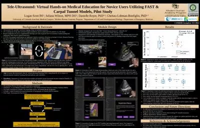
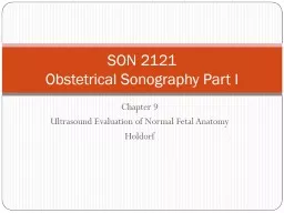
PPT-Lecture 9 Ultrasound Evaluation of Normal Fetal Anatomy
Author : tawny-fly | Published Date : 2018-12-25
Holdorf SON 2121 Obstetrical Sonography Part I Normal Fetal Abdomen and Abdominal Wall Diaphragm The superior aspect of the abdominopelvic cavity is defined by the
Presentation Embed Code
Download Presentation
Download Presentation The PPT/PDF document "Lecture 9 Ultrasound Evaluation of Nor..." is the property of its rightful owner. Permission is granted to download and print the materials on this website for personal, non-commercial use only, and to display it on your personal computer provided you do not modify the materials and that you retain all copyright notices contained in the materials. By downloading content from our website, you accept the terms of this agreement.
Lecture 9 Ultrasound Evaluation of Normal Fetal Anatomy: Transcript
Holdorf SON 2121 Obstetrical Sonography Part I Normal Fetal Abdomen and Abdominal Wall Diaphragm The superior aspect of the abdominopelvic cavity is defined by the diaphragm muscle It appears sonographically as a hypoechoic curved line separating the more echogenic lungs from the liver and stomach. Diagnostic Ultrasound. High frequency sound waves emitted from sound source (transducer). Transducer placed on patient’s body. Sound waves echo off interfaces of internal structures and return to transducer. Conrad R. Chao, MD. Professor of Obstetrics and Gynecology. Chief of Maternal and Fetal Medicine. University of New Mexico. What is FGR. SGA = birthweight below 10. th. percentile. Associated with higher morbidity, mortality, and subsequent adult disease (Barker hypothesis). Holdorf. Lecture. . 5. Fetal Syndromes. Outline. There are hundreds of “syndromes” in the medical literature. It is impossible to remember them all.. The following “syndromes” have appeared on the ARDMS and ARRT registries in the past.. kkk. Kkk . . The objective of this systematic review is to identify, evaluate and synthesise the available evidence for the advantages of using Fetal Renal Artery Doppler, Middle Cerebral Artery Doppler and Ductus Venosus Doppler in those pregnancies complicated by intrauterine growth restriction. . Assistant professor. Dept. of . OBGyn. PESIMSR, KUPPAM. INTRODUCTION. Originally . introduced by . Saling. . in 1967 . AIM. to identify fetuses at risk for severe adverse outcome . to be able to intervene . Endoscopic . ultrasonography. (EUS) employs the technology of endoscopy and internally placed high-frequency ultrasound waves to visualize the gastrointestinal wall and adjacent structures. . EUS has emerged as an important modality for the diagnosis and staging of benign and malignant lesions of the gut wall and surrounding structures of the . Many techniques are available to assess . the. growth and . developmental status . of the . fetus, including . Ultrasound. . Maternal . serum . screening . Amniocentesis. Chorionic . villus . sampling. Focus on neonatal emergencies. 2. . and . 3 . October. 2019. For ST4-ST8 . R. egistrars, AANPS, Senior . F. ellows or Consultants . To book . your. place: . . E,Keshavarz. MD. Assistant Professor of Radiology. SBMU. ISUOG. . (. I. nternational . S. ociety. of . U. ltrasound in . O. bstetrics and . G. ynecology. ). . AIUM. . (. A. merican . I. nstitute of . Fetal Guidelines Dr Mary Moran • National • United Kingdom • International • Implications National Survey 2016 • Prof Keelin O’Donoghue ( Principal Investigator; Consultant Obstetrician & Dr Sarah Bohndiek. Learning outcomes. After . these lectures, . you should be able to:. Explain how ultrasound interacts with tissue. Understand where ultrasound imaging contrast comes from. Describe how ultrasound signals are generated and detected. (HRUS. ): A POSSIBLE RELEVANCE IN CLINICAL . PRACTICE. VALUTAZIONE DEI NERVI CRANICI CON ECOGRAFIA AD ALTA RISOLUZIONE (HRUS): POSSIBILE RILEVANZA NELLA PRATICA CLINICA. Ferdinando Sartucci . 1,2,3. , . Methods. Results. Ultrasound (US): portable, real-time imaging, cheap, no radiation emission.. US is among one of the most utilized imaging modalities used to understand 3D anatomy in a 2D image. 7. .. Public Comment Draft. WG-1/12. . . 4 April 2024. Background. Fetal Cardiac Assessment using Ultrasound has evolved since Sup78 was published in 2010. Practice now includes many more measurements beyond visual assessment, such as: .
Download Document
Here is the link to download the presentation.
"Lecture 9 Ultrasound Evaluation of Normal Fetal Anatomy"The content belongs to its owner. You may download and print it for personal use, without modification, and keep all copyright notices. By downloading, you agree to these terms.
Related Documents