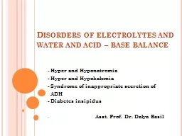
/
Disorders of Water, Electrolytes & Acid–Base Metabolism Disorders of Water, Electrolytes & Acid–Base Metabolism

Disorders of Water, Electrolytes & Acid–Base Metabolism - PowerPoint Presentation

daniella
daniella . @daniella
Follow
344 views
Uploaded On 2022-04-07

Disorders of Water, Electrolytes & Acid–Base Metabolism - PPT Presentation

Lecture 5 Introduction A complex system of chemical buffers together with highly specialized mechanisms of the lungs and kidneys continuously work together to ensure a precise balance of water electrolytes and pH in both the intracellular and extracellular compartments of the human body ID: 910479

acid water plasma base water acid base plasma body hyponatremia electrolytes mmol bicarbonate composition concentration loss alkalosis hypernatremia increased

Share:

Link:

Embed:

Download Presentation from below link

Download Presentation The PPT/PDF document "Disorders of Water, Electrolytes & A..." is the property of its rightful owner. Permission is granted to download and print the materials on this web site for personal, non-commercial use only, and to display it on your personal computer provided you do not modify the materials and that you retain all copyright notices contained in the materials. By downloading content from our website, you accept the terms of this agreement.


Presentation Transcript

Slide1

Disorders of Water, Electrolytes & Acid–Base Metabolism

Lecture 5

Slide2

Introduction

A complex system of chemical buffers together with highly specialized mechanisms of the lungs and kidneys continuously work together to ensure a precise balance of water, electrolytes, and pH in both the intracellular and extracellular compartments of the human body.

Although these systems display impressive flexibility and responsiveness to perturbation by illness or injury, they do have limits, at which point medical evaluation and treatment are required.

2

Slide3

TOTAL BODY WATER: VOLUMEAND DISTRIBUTION

3

Slide4

TOTAL BODY WATER: VOLUMEAND DISTRIBUTION

The minimum daily requirement for water can be estimated from renal & insensible losses:

renal (1200 to 1500 mL in urine) and

“insensible” losses (≈400 to 700 mL)

evaporation from the skin and respiratory tract.

Activity, environmental conditions, and disease all have dramatic effects on daily water (and electrolyte) requirements.

On average, an adult must take in

≈1.5 to 2.0 L

of water daily to maintain fluid balance.

Because primary regulatory mechanisms are designed to

first maintain intracellular hydration status, imbalances in TBW are initially reflected in the ECF compartment.

4

Slide5

TOTAL BODY WATER: VOLUME AND DISTRIBUTIONChanges in Extracellular Fluid Volume

Causes

Clinical Manifestations

Trauma (and other causes of acute blood loss),

“third spacing” of fluid (

eg

,

burns, pancreatitis, peritonitis), vomiting, diarrhea, diuretics, renal or adrenal (

ie

, sodium wasting)

Disease

Thirst, anorexia, nausea,

lightheadedness,

Orthostatic hypotension, syncope, tachycardia, oliguria, decreased skin turgor and “sunken eyes”, shock, coma, deathECF lossHeart failure, cirrhosis, nephrotic syndrome, iatrogenic (intravenous fluidoverload)Weight gain, edema,dyspnea (secondaryto pulmonary edema),tachycardia, jugularvenous distention (HF),portal hypertension(ascites) cirrhosis, esophageal VaricesECF gain

5

Slide6

WATER AND ELECTROLYTES: COMPOSITION OF BODY FLUIDS

The primary cationic (positively charged) electrolytes are:

Sodium (Na

+

), potassium (K

+

), calcium (Ca

2+

), and magnesium (Mg

2+

), Whereas the anions (negatively charged) include:Chloride (Cl-), bicarbonate ( HCO3-

), phosphate (HPO

2

4- , H2PO24- ), sulfate ( SO24- ), organic ions such as lactate, and negatively charged proteins. Na+, K+, Cl-, and HCO3- in the plasma or serum are commonly analyzed in an electrolyte profile because their concentrations provide the most relevant information about the osmotic, hydration, and acid-base status of the body. 6

Slide7

WATER AND ELECTROLYTES: COMPOSITION OF BODY FLUIDS

Any

increase

in the concentration of one anion

is accompanied by a corresponding

decrease

in other

anions

, or by an increase in one or more cations or both because total

electrical neutrality

must be maintained. Similarly, any decrease in the concentration of anions involves a corresponding increase in other anions, a decrease in cations, or both. In the case of polyvalent ions (eg, Ca2+

, Mg

2+

), it is important to distinguish between the substance concentration of the ion itself and the concentration of the ion charge. Thus, although the concentration of total calcium ions in normal plasma is ≈2.5 mmol/L, the concentration of the total calcium ion charge is 5.0 mmol/L (also called 5 milliequivalents per liter [mEq/L]). 7

Slide8

WATER AND ELECTROLYTES: COMPOSITION OF BODY FLUIDSSodium

Disorders of Na

+

homeostasis can occur because of: Excessive loss, gain, or retention of Na

+

, or as

The result of excessive loss, gain, or retention of H

2

O.

It is difficult to separate disorders of Na

+ and H2O balance because of their close relationship in establishing normal osmolality in all body water compartments.Homeostasis within a narrow window is necessary for life, and the body must defend against excessive gains or losses.

8

Slide9

WATER AND ELECTROLYTES: COMPOSITION OF BODY FLUIDSSodium: Hyponatremia

Hyponatremia

is defined as a decreased plasma Na

+

concentration (sodium level goes below 135  mmol/L).

Hyponatremia typically manifests clinically as:

Nausea, generalized weakness,

Mental confusion at values below

120 mmol/L

, &Severe mental confusion plus seizures at less than 105 mmol/L.The

rapidity

of development of hyponatremia influences the Na

+ concentrations at which symptoms develop ie, clinically apparent symptoms may manifest at higher Na+ concentrations [≈125 mmol/L] when hyponatremia develops rapidly. 9

Slide10

WATER AND ELECTROLYTES: COMPOSITION OF BODY FLUIDSSodium: Hyponatremia

Symptoms are due to changes in

osmolality

rather than to the Na

+

concentration by itself.

CNS symptoms are due to movement of H

2

O into cells to maintain osmotic balance and subsequent swelling of CNS cells.

These symptoms can occur more rapidly in children, so there is a need to be particularly aware in the pediatric population.

Hyponatremia can be: hypo-osmotic, hyperosmotic, or iso-osmotic. Thus measurement of plasma osmolality is animportant initial step in the assessment of hyponatremia.

the most common form is hypo-osmotic hyponatremia.

10

Slide11

11

Slide12

12

Slide13

WATER AND ELECTROLYTES: COMPOSITION OF BODY FLUIDSSodium: Hyponatremia

Hyperosmotic Hyponatremia

Hyponatremia in the presence of increased quantities of

other solutes

in the ECF is the result of an extracellular shift of water or an intracellular shift of Na

+

to maintain osmotic balance between ECF and ICF compartments.

The most common cause of this type of hyponatremia is severe

hyperglycemia

.

As a general rule,

Na

+

is decreased by ≈1.6 to 2.0 mmol/L for every 100 mg/dL increase in glucose above 100 mg/dL.Correction of hyperglycemia will restore normal blood Na+. It also may occur when mannitol and glycine, used for irrigation during certain surgical procedures, enter the intravascular fluid compartment. 13

Slide14

WATER AND ELECTROLYTES: COMPOSITION OF BODY FLUIDSSodium: Hyponatremia

Isosmotic Hyponatremia

If the measured

Na

+

concentration in plasma is decreased, but measured plasma osmolality,

glucose, and urea are normal, the most likely explanation is

pseudohyponatremia

caused by the electrolyte exclusion effect.

This occurs when

Na

+

is measured by an indirect ion-selective electrode in patients with severe

hyperlipidemia or hyperproteinemia. Pseudohyponatremia is confirmed if direct ISE value is normal.If direct ISE is not available, simultaneous calculation and measurement of plasma osmolarity is very useful. Measured osmolarity is normal in pseudohyponatremia but calculated osmolarity – based as it is on erroneously low plasma sodium result – is reduced.14

Slide15

WATER AND ELECTROLYTES: COMPOSITION OF BODY FLUIDSSodium: Hyponatremia

Isosmotic Hyponatremia

The volume of total solids (primarily protein and lipid) in an aliquot of plasma is approximately 7%, so that approximately 93% of plasma volume is actually water.

The main electrolytes (Na

+

, K

+

, Cl

-

, HCO

3-) are confined to the water phase. When a fixed volume of total plasma (eg

, 10 µL) is pipetted for dilution before flame photometry or indirect ISE analysis, only 9.3 µL of plasma water that contains the electrolytes is added to the diluent.

Thus a concentration of Na

+ determined by flame photometry or indirect ISE to be 140 mmol/L is the concentration in the total plasma volume, not in the plasma water volume. If the plasma contains 93% water, the concentration of Na+ in plasma water is [140 × (100/93)], or 150 mmol/L. Although it is the electrolyte concentration in plasma water that is physiologic.15

Slide16

16

Predicted influence of water (H

2

0) content on sodium measurements for a 100-mmol/L sodium chloride solution by direct ion-selective electrode versus flame emission photometry or indirect ion-selective electrode.

Red areas

represent nonaqueous volumes, which could consist of lipids, proteins

Indirect ISE

Slide17

WATER AND ELECTROLYTES: COMPOSITION OF BODY FLUIDSSodium: Hyponatremia

Hypo-Osmotic Hyponatremia

Typically, when

plasma Na

+

concentration is

low

, calculated or measured osmolality also

will be low.

This type of hyponatremia can be due to:

Excess loss of Na

+

(depletional hyponatremia) or increased ECF volume (dilutional hyponatremia). Differentiating these initially requires clinical assessment of TBW and ECF volume by history and physical examination. 17

Slide18

WATER AND ELECTROLYTES: COMPOSITION OF BODY FLUIDSSodium: Hyponatremia

Hypo-Osmotic Hyponatremia

Depletional

hyponatremia results from a loss of

Na

+

from the ECF space that exceeds the concomitant loss of water.

The net loss of

Na

+

from the ECF space also stimulates thirst and production of vasopressin, both of which contribute to the maintenance of hyponatremia.

Hypovolemia

is apparent in the physical examination (orthostatic hypotension, tachycardia, decreased skin turgor).

If urine Na+ is low (<10 mmol/L), the kidneys are properly retaining filtered Na+ and the loss is extrarenal, most commonly from the GIT or skin.18

Slide19

WATER AND ELECTROLYTES: COMPOSITION OF BODY FLUIDSSodium: Hyponatremia

Hypo-Osmotic Hyponatremia

Alternatively, if urine

Na

+

is

increased

in this setting (generally

>20 mmol/L

), renal loss of

Na

+

likely. Renal loss of Na+ occurs with: Use of diuretics (which inhibit reabsorption of Cl- and Na+ in the ascending loop), Adrenal insufficiency (no aldosterone), or Salt-wasting nephropathies, as can occur with interstitial nephritis.19

Slide20

WATER AND ELECTROLYTES: COMPOSITION OF BODY FLUIDSSodium: Hyponatremia

Hypo-Osmotic Hyponatremia

Renal loss of Na

+

in excess of H

2

O can also occur in

metabolic alkalosis

from prolonged vomiting, because increased renal

HCO

3

-

excretion

is accompanied by Na+ ions. In this case, urine sodium is increased (>20 mmol/L), but urine chloride remains low.In proximal renal tubular acidosis (RTA) type 2, bicarbonate is lost because of a defect in HCO3- reabsorption, and Na+ is coexcreted to maintain electrical neutrality. As with extrarenal Na+ loss, management is centered around the reversal of underlying cause and restoration of ECF volume. 20

Slide21

WATER AND ELECTROLYTES: COMPOSITION OF BODY FLUIDSSodium: Hyponatremia

Hypo-Osmotic Hyponatremia

Dilutional hyponatremia

is a result of

excess H

2

O retention

and often can be detected during the physical examination as

edema

.

In advanced renal failure, water is retained because of decreased filtration and H

2

O excretion.

When ECF is increased but the circulating blood volume is decreased, as occurs in hepatic cirrhosis and nephrotic syndrome, a vicious cycle is established. The decreased blood volume is sensed by baroreceptors and results in increased aldosterone and vasopressin, even though ECF volume is excessive. The kidneys reabsorb Na+ and H2O in response to increased aldosterone and vasopressin in an attempt to restore the blood volume, resulting in further increases in ECF and further dilution of Na+. 21

Slide22

WATER AND ELECTROLYTES: COMPOSITION OF BODY FLUIDSSodium: Hyponatremia

Hypo-Osmotic Hyponatremia

In hypo-osmotic hyponatremia with a normal or

euvolemic volume

status, the most common causes are the

syndrome of inappropriate antidiuretic hormone

(ADH) (vasopressin) (SIADH), primary polydipsia, and endocrine disorders such as adrenal insufficiency and hypothyroidism.

Hypothyroidism impairs free H

2

O excretion.

Free water restriction is the mainstay of therapy in SIADH.

22

Slide23

WATER AND ELECTROLYTES: COMPOSITION OF BODY FLUIDSSodium: Hyponatremia

Hypo-Osmotic Hyponatremia

However, in severe or symptomatic hyponatremia from any cause, the use of hypertonic saline solutions may be required to correct serum Na

+

concentrations.

In such cases, the hyponatremia must be corrected cautiously because too rapid correction can lead to brain demyelination.

Current recommendations are to increase Na

+

by 0.5 to 2.0 mmol/L per hour and not to exceed a total increase in Na+ greater than 18 to 25 mmol/L over 48 hours.

23

Slide24

WATER AND ELECTROLYTES: COMPOSITION OF BODY FLUIDSSodium: Hyponatremia

Hypo-Osmotic Hyponatremia

Finally, euvolemic hyponatremia also can be found in

primary polydipsia

when water intake is greater than the renal capacity to excrete excess H

2

O.

This can be the result of psychiatric illness, but diseases that cause hypothalamic disorders, such as sarcoidosis, also may cause polydipsia by altering the thirst reflex.

24

Slide25

WATER AND ELECTROLYTES: COMPOSITION OF BODY FLUIDSSodium: Hypernatremia

Hypernatremia is generally defined as a serum sodium level of more than 145 mmol/L.

Symptoms of hypernatremia are primarily

neurologic

(because of neuronal cell loss of H

2

O to the ECF) and include tremors, irritability, ataxia, confusion, and coma.

As with hyponatremia, the rapidity of development of hypernatremia will determine the plasma Na

+

concentration at which symptoms occur.

Acute development may cause symptoms at 160 mmol/L, although in chronic hypernatremia, symptoms may not occur until Na

+

exceeds 175 mmol/L.

In chronic hypernatremia, the intracellular osmolality of CNS cells will increase to protect against intracellular dehydration.25

Slide26

WATER AND ELECTROLYTES: COMPOSITION OF BODY FLUIDSSodium: Hypernatremia

Because of this, rapid correction of hypernatremia can cause dangerous cerebral edema because CNS cells will take up too much water if the ICF is hyperosmotic when

normonatremia

is achieved.

In many cases, the symptoms of hypernatremia may be masked by underlying conditions.

Hypernatremia rarely occurs in an alert patient with a normal thirst response and access to water.

Most cases are observed in patients with

altered mental status or infants, both of whom may not be capable of rehydrating themselves.

26

Slide27

WATER AND ELECTROLYTES: COMPOSITION OF BODY FLUIDSSodium: Hypernatremia

Hypernatremia arises in the setting of:

Hypovolemia (excessive water loss or failure to replace normal water losses),

Hypervolemia (a net Na

+

gain in excess of water gain), or

Normovolemia

.

Again, assessment of TBW status by physical examination and measurement of urine Na

+

and osmolality are important steps in establishing a diagnosis.

27

Slide28

28

Slide29

WATER AND ELECTROLYTES: COMPOSITION OF BODY FLUIDSSodium: Hypernatremia

Hypovolemic Hypernatremia

Hypernatremia in the setting of decreased ECF is caused by renal or extrarenal loss of hypo-osmotic fluid, leading to dehydration.

Thus, once hypovolemia is established by physical examination, measurement of urine Na

+

and osmolality is used to determine the source of fluid loss.

Patients who have large extrarenal losses will have

concentrated urine

(

often >800

mOsmol

/L

) with low urine

Na+ (<20 mmol/L), reflecting a proper renal response to conserve Na+ and water to restore ECF volume.Extrarenal causes include diarrhea, skin losses (burns, fever, or excessive sweating), and respiratory losses coupled with failure to replace the water. 29

Slide30

WATER AND ELECTROLYTES: COMPOSITION OF BODY FLUIDSSodium: Hypernatremia

Hypovolemic Hypernatremia

When gastrointestinal loss is excluded, and the patient has normal mental status and access to H

2

O, a hypothalamic disorder (tumor or granuloma) inducing diabetes insipidus (DI) should be suspected.

In patients with poorly controlled diabetes with glucose values greater than 600 mg/dL, an osmotic diuresis can occur that results in extreme dehydration and hypernatremia.

This condition is referred to as hyperosmolar

hyperglycemic nonketotic syndrome and occurs most commonly in elderly individuals with type 2 diabetes.

30

Slide31

WATER AND ELECTROLYTES: COMPOSITION OF BODY FLUIDSSodium: Hypernatremia

Normovolemic

Hypernatremia

Hypernatremia in the presence of normal ECF volume is often a prelude to hypovolemic hypernatremia.

Insensible losses through the lung or skin must be suspected and are characterized by concentrated

urine as the kidneys conserve water.

Another cause of

normovolemic

hypernatremia is water diuresis, which is manifested by polyuria.

The differential for polyuria (generally defined as >3 L urine output/d) is a water or solute diuresis.

Solute diuresis is exemplified by the osmotic diuresis

of diabetes mellitus and generally is characterized by urine osmolality greater than 300

mOsmol

/L and hyponatremia. Water diuresis, a manifestation of DI, is characterized by dilute urine (osmolality <250 mOsmol/L) and hypernatremia.31

Slide32

WATER AND ELECTROLYTES: COMPOSITION OF BODY FLUIDSSodium: Hypernatremia

Normovolemic

Hypernatremia

DI can be central or nephrogenic.

Central DI is due to decreased or absent vasopressin secretion resulting from head trauma or pituitary tumor

Nephrogenic DI is due to renal resistance to vasopressin as a result of drugs (

eg

, lithium) or electrolyte disorders.

When thirst and access to water are uncompromised, many patients with DI will remain

normonatremic

because their free water losses are compensated by intake.

32

Slide33

WATER AND ELECTROLYTES: COMPOSITION OF BODY FLUIDSSodium: Hypernatremia

Normovolemic

Hypernatremia

Such patients display symptoms of only polyuria and polydipsia.

However, overt hypernatremia can become manifest with progression of underlying causes,

impaired thirst, or restricted access to water.

Administration of vasopressin can be used to treat central DI, although patients with nephrogenic DI may be resistant to it.

Correction of underlying disorders or discontinuation of offending drugs may be required to normalize Na

+

concentrations in

nephrogenic DI.

33

Slide34

WATER AND ELECTROLYTES: COMPOSITION OF BODY FLUIDSSodium: Hypernatremia

Hypervolemic

Hypernatremia

The presence of excess TBW and hypernatremia indicates a net gain of water and Na

+

, with

Na

+

gain in excess of water.

This rare condition is observed most commonly in hospitalized patients receiving hypertonic saline or sodium bicarbonate.

34

Slide35

WATER AND ELECTROLYTES: COMPOSITION OF BODY FLUIDSPotassium

The total body potassium of a 70-kg subject is

≈3.5 mol

(40 to 59 mmol/kg), of which only 1.5 to 2% is present in the ECF.

Nevertheless, plasma K

+

is often a good indicator of total K

+

stores.

Disturbance of K

+ homeostasis has serious consequences. For example, a decrease in extracellular K+ (hypokalemia) is characterized by muscle weakness, irritability, and paralysis.

Plasma K

+

concentrations < 3.0 mmol/L are often associated with marked neuromuscular symptoms andindicate a critical degree of intracellular depletion. At lower concentrations, tachycardia and cardiac conduction defects are apparent on electrocardiogram (ECG) and can lead to cardiac arrest.35

Slide36

WATER AND ELECTROLYTES: COMPOSITION OF BODY FLUIDSPotassium

36

Slide37

WATER AND ELECTROLYTES: COMPOSITION OF BODY FLUIDSPotassium

High extracellular K

+

(

hyperkalemia

) concentrations

may produce symptoms of mental confusion, weakness, and weakness of the respiratory muscles.

Cardiac effects of hyperkalemia include bradycardia and conduction defects.

Prolonged, severe hyperkalemia

>7.0 mmol/L

can lead to peripheral vascular collapse and cardiac arrest. Symptoms or ECG abnormalities are almost always present at K

+

concentrations above

6.5 mmol/L. Concentrations greater than 10.0 mmol/L in most cases are fatal, although fatalities can occur at significantly lower values. 37

Slide38

WATER AND ELECTROLYTES: COMPOSITION OF BODY FLUIDSPotassium: Hypokalemia

Causes of hypokalemia (plasma K

+

<3.5 mmol/L) are classified as:

Redistribution of extracellular K

+

into ICF, or

True K

+

deficits, caused by:

decreased intake or loss of potassium-rich body fluids.

38

Slide39

39

Slide40

WATER AND ELECTROLYTES: COMPOSITION OF BODY FLUIDSPotassium: Hypokalemia

Redistribution

Insulin promotes acute entry of K into skeletal muscle and liver by increasing Na, K-ATPase activity.

In alkalosis, K

+

moves from ECF into cells in exchange with H

+

.

Pseudohypokalemia

can occur in cases of very high white blood cell or platelet counts.when the blood sample is kept at room temp. for a relatively long period.

40

Slide41

WATER AND ELECTROLYTES: COMPOSITION OF BODY FLUIDSPotassium: Hypokalemia

Autosomal dominant channelopathy characterized by muscle weakness or 

paralysis

when there is a fall in potassium levels in the blood

caused most commonly by mutations in the alpha subunit of the skeletal muscle calcium channel gene Cav1.1

Clinically, redistributive hypokalemia is generally a transient phenomenon that is reversed once underlying conditions are corrected.

41

Slide42

WATER AND ELECTROLYTES: COMPOSITION OF BODY FLUIDSPotassium: Hypokalemia

True Potassium Deficit

Hypokalemia reflecting true total body deficits of K

+

as a consequence of potassium loss can be classified into

renal

and

nonrenal losses

, based on daily excretion of K

+

in the urine. If urine excretion of K+ is < 25 mmol/d, it can be concluded that the kidneys are functioning properly and are attempting to reabsorb K

+

. The cause may be:

decreased K+ intake Causes of decreased intake include chronic starvation and postoperative intravenous fluid therapy with K+-poor solutions. extrarenal loss of K+-rich fluidGastrointestinal loss of K+ occurs most commonly with diarrhea and loss of gastric fluid through vomiting. 42

Slide43

WATER AND ELECTROLYTES: COMPOSITION OF BODY FLUIDSPotassium: Hypokalemia

Urine excretion exceeding 25 mmol/d in a hypokalemic setting is inappropriate and indicates that the kidneys are the primary source of K

+

loss.

Renal losses of K

+

may occur:

during the diuretic (recovery) phase of acute tubular necrosis and

Magnesium deficiency also can lead to

increased renal loss of K

+, which is attributable to a reduction in the inhibitory effect of magnesium on luminal potassium channelsDue to metabolic acidosis (renal tubular acidosis)

43

Slide44

WATER AND ELECTROLYTES: COMPOSITION OF BODY FLUIDSPotassium: Hypokalemia

In addition to redistribution of K

+

into cells in an

alkalotic setting

, K

+

can be lost from the kidneys in exchange for reclaimed H

+

ions.

44

Metabolic alkalosis

Slide45

WATER AND ELECTROLYTES: COMPOSITION OF BODY FLUIDSPotassium: Hyperkalemia

Hyperkalemia (plasma K

+

>5.0 mmol/L) is a result of (singly or in combination)

Redistribution,

Increased intake, or

Increased retention.

In addition, preanalytical conditions—such as:

Hemolysis,

Thrombocytosis (>10

6/µL), and Leukocytosis (>10

5

/µL together with delayed sample analysis)—have been known to cause marked

pseudohyperkalemia45

Slide46

46

Slide47

WATER AND ELECTROLYTES: COMPOSITION OF BODY FLUIDS Chloride

In the absence of acid-base disturbances, Cl

-

concentrations in plasma generally will follow those of Na+

.

However, determination of plasma Cl

-

concentration is useful in the differential diagnosis of acid-base disturbances and is essential for calculating the anion gap.

Fluctuations in serum or plasma Cl

-

have little clinical consequence, but do serve as signs of an underlying disturbance in fluid or acid-base homeostasis.The specific replacement of chloride is rarely targeted at chloride deficit independently, but it is a corner stone of management for metabolic alkalosis.

47

Slide48

WATER AND ELECTROLYTES: COMPOSITION OF BODY FLUIDS Chloride: Hypochloremia

Hypochloremia

 is defined as a chloride level less than 95 mmol/L.

In general, causes of hypochloremia parallel causes of hyponatremia.

Persistent gastric secretion and prolonged vomiting result in significant loss of Cl

-

and ultimately in

hypochloremic

alkalosis and depletion of total body Cl

-

with retention of HCO3- . Respiratory acidosis, which is accompanied byincreased HCO3-

, is another common cause of decreased Cl

-

with normal Na+. 48

Slide49

WATER AND ELECTROLYTES: COMPOSITION OF BODY FLUIDS Chloride: Hyperchloremia

Increased plasma Cl

-

concentration, similar to increased Na

+

concentration, occurs with dehydration, prolonged diarrhea with loss of sodium bicarbonate, DI, and overtreatment with normal saline solutions, which have a Cl

-

content of 150 mmol/L.

In fact, mounting evidence suggests that use of saline (NaCl) solution for maintenance, intraoperative, and resuscitative therapy can result in a host of hyperchloremia induced side effects.

A rise in Cl

- concentration also may be seen in respiratory alkalosis because of renal compensation for excreting HCO

3

-

.49

Slide50

Case Studies

Disorders of Water, Electrolytes

50

Slide51

Case 1

A 45‐year‐old man was brought into the A&E department late at night in a comatose state. It was impossible to obtain a history from him, and clinical examination was difficult, but it was noted that he smelt strongly of alcohol.

The following analyses were requested urgently.

51

Slide52

Case 1

Why is his measured osmolality so high?

52

Slide53

Case 1 Comments

The osmolality can be calculated as 291.5, using the formula:

Calculated Osmolality= 2 Na + 2 K + Glucose + Urea 

=(2X137) + (2X4.3) + 4.2 + 4.7 = 291.5

Osmolal

Gap = 465 – 291.5 = 173.5

The difference between this figure and the

value for the directly measured osmolality

(465 mmol/L) could be explained by the presence of another osmotic active substance in plasma.

53

Slide54

Case 1 CommentsFrom the patient’s history, it seemed that ethanol might be contributing significantly to the plasma osmolality, and plasma ethanol was measured the following day, on the residue of the specimen collected at the time of emergency admission.

The result was

170 mmol/L

, very close to the difference between the measured and calculated

osmolalities

.

54

Slide55

Case 2

A 15-year-old male patient with diabetes mellitus (DM) presented to the emergency department (ED) with abdominal pain. Routine chemistry investigations were performed on a plasma sample shortly after arrival with the following results:

55

Slide56

Case 2 Comments

Patients with DM and increased plasma glucose concentration have a high plasma osmolality in the extracellular compartment.

This causes shifting of water from inside the cells into the extracellular compartment and a subsequent dilution of plasma components, including sodium.

Therefore the hyponatremia in this case can be explained solely on the basis of the increased plasma glucose concentration.

56

Slide57

Case 2 Comments

As a general rule, Na

+

is decreased by ≈1.6 to 2.0 mmol/L for every 100 mg/dL

increase in glucose above 100 mg/dL.

In this case, the plasma glucose of 673 mg/dL will depress plasma sodium by approximately 9 mmol/L.

Therefore, using a correction formula, the sodium concentration becomes:

127 + 9 = 136 mmol/L

The hyponatremia may be corrected simply by restoring the plasma glucose concentration to normal.

57

Slide58

Case 3A 60-year-old male was admitted to hospital with persistent cough, disorientation, and confusion. He was a heavy smoker and was not taking any medications. On examination, he

appeared euvolemic. The following results were obtained in blood and urine samples sent to the laboratory:

58

Slide59

59

7-28

Slide60

Case 3 Comments

This patient had severe, symptomatic hyponatremia.

There was no biochemical evidence of renal, adrenal, or pituitary disease.

On examination, the patient did not appear to be volume depleted or overloaded.Based on his laboratory results and clinical history, a diagnosis of the syndrome of inappropriate diuretic hormone secretion (SIADH) was considered.

60

Slide61

Case 3 Comments

The low serum sodium and osmolality values in the context of inappropriately increased urine sodium and osmolality values were consistent with those in SIADH.

The hyponatremia slowly improved in response to fluid restriction.

A chest radiograph and subsequent lung biopsy revealed the presence of small cell lung carcinoma, the source of the inappropriate secretion of arginine vasopressin.

SIADH is common in small cell carcinoma, occurring in up to 10% of patients.

61

Slide62

Case 4A 2-month-old male infant presented with vomiting and severe diarrhea. Biochemistry analysis on a plasma sample collected from the child were as follows:

62

Slide63

Case 4 Comments

The results show hypernatremia and biochemical evidence of dehydration (uremia).

Diarrheal fluid is hypotonic in relation to plasma;

therefore, unless the patient’s water intake is high, severe diarrhea can easily lead to a hypernatremic

state, especially in infants and young children.

Diarrheal fluid from the small bowel is also rich in

bicarbonate; therefore episodes of diarrhea may result in decreased plasma bicarbonate (total CO

2

) levels with concurrent increases in the plasma chloride concentration, as demonstrated in this case.

63

Slide64

Case 4 Comments

In the presence of hypernatremia, water moves out of the intracellular compartment into the extracellular compartment in an attempt to decrease the osmolality of the plasma.

The clinical signs of dehydration in cases of hypernatremia are not always apparent or perhaps as obvious as with cases involving a normal or low plasma sodium concentration.

64

Slide65

ACID-BASE PHYSIOLOGY

The pH of plasma may be considered to be a function of two independent variables:

The

PCO2, which is regulated by the lungs and represents the acid component of the carbonic acid/bicarbonate buffer system, and

The concentration of titratable base (base excess or deficit), which is regulated by the kidneys.

The plasma total CO

2

concentration generally is taken as a measure of the base excess or deficit in plasma and ECF.

65

Slide66

ACID-BASE PHYSIOLOGY Bicarbonate and Dissolved CO

2

Bicarbonate is the second largest fraction (behind Cl

-

) of plasma anions.

Conventionally, it is defined to include:

Plasma bicarbonate ion ( HCO

3

-

),

Carbonate ion ( CO32- ), and CO2 bound in plasma carbamino

compounds (RCNHCOOH).

Actual bicarbonate ion concentration is not measured in clinical laboratories.

The analyte usually measured in plasma is total CO2, which includes bicarbonate and dissolved CO2 (dCO2) but is often referred to as “serumbicarb.” 66

Slide67

ACID-BASE PHYSIOLOGY Bicarbonate and Dissolved CO2

67

Slide68

ACID-BASE PHYSIOLOGYBuffer Systems and Their Role in Regulating the pH of Body Fluids

A buffer is a mixture of a weak acid and a salt of its conjugate base that resists changes in pH when a strong acid or base is added to the solution.

The action of buffers in the regulation of body pH can be demonstrated by using the bicarbonate buffer system as an example.

If a strong acid is added to a solution containing HCO

3

-

and H

2

CO

3

, the H+ will react with HCO3- to form more H2CO3

and subsequently CO

2

and H2O.The hydrogen ions are thereby bound, and the increase in the H+ concentration will be minimal. 68

Slide69

Bicarbonate and Carbonic Acid Buffer SystemPhosphate Buffer System Plasma Protein Buffer System Hemoglobin Buffer System

69

ACID-BASE PHYSIOLOGY

Buffer Systems and Their Role in Regulating the pH of Body Fluids

Slide70

ACID-BASE PHYSIOLOGYRespiratory Mechanism in the Regulation of Acid-Base Balance

central chemoreceptors located on the anterior surface of the medulla oblongata and

by peripheral chemoreceptors located in the carotid arteries and aorta.

70

In a resting state, the respiration rate is normally

12 to 15

breaths/min.

For an average-sized adult with a tidal volume of ~ 0.5 L,

6 to 8 L

of air is moved per minute in either direction.

Involuntary increases in rate and depth of respiration are regulated by the medullary respiratory center in the brainstem, which is stimulated by:

Slide71

ACID-BASE PHYSIOLOGYRespiratory Mechanism in the Regulation of Acid-Base Balance

Peripheral chemoreceptors are stimulated by a fall in

pH

caused by accumulation of

CO

2

or a decrease in

P

O

2

.Central chemoreceptors are stimulated only by a decrease in pH of the CSF.

71

Slide72

ACID-BASE PHYSIOLOGYRespiratory Response to Acid-Base Perturbations

Most metabolic acid-base disorders develop

slowly

, within hours in diabetic ketoacidosis and

months

in chronic renal disease.

The respiratory system responds

immediately

to a change in acid-base status, but

several hours

may be required for the response to become maximal. The maximum response is

not attained

until both central and peripheral chemoreceptors are fully stimulated.

For example, in the early stages of metabolic acidosis, plasma pH decreases, but because H+ ions equilibrate rather slowly across the blood-brain barrier, the pH in CSF remains nearly normal. 72

Slide73

ACID-BASE PHYSIOLOGYRespiratory Response to Acid-Base Perturbations

However, because peripheral chemoreceptors are stimulated by decreased plasma pH,

hyperventilation

occurs, and plasma

P

CO

2

is decreased.

When this occurs, the

P

CO2 of the CSF decreases immediately because CO2 equilibrates rapidly across the blood-brain barrier, leading to a rise in pH of the CSF that inhibits the central chemoreceptors. As plasma bicarbonate gradually falls because of acidosis, bicarbonate concentration, and pH in the CSF will also eventually fall.

At this point, stimulation of respiration becomes maximal from both central and peripheral chemoreceptors.

73

Slide74

ACID-BASE PHYSIOLOGYRenal Mechanisms in the Regulation of Acid-Base Balance

Various functions of the kidneys respond to different alterations in acid-base status.

In the case of acidosis, excretion of acids is increased and that of base is conserved;

In alkalosis, the opposite occurs.

The pH of the urine changes correspondingly and may vary in random specimens from pH 4.5 to 8.0.

The ability to excrete variable amounts of acid or base makes the kidney the final defense mechanism against changes in body

pH.

The various acids produced during metabolic processes are buffered in the ECF at the expense of HCO

3

-

.

74

Slide75

Renal excretion of acid and conservation of HCO3- occur through several mechanisms, including: Na+

-H

+

exchange, Production of ammonia and excretion of NH4+ &Reclamation of HCO

3

-

.

75

ACID-BASE PHYSIOLOGY

Renal Mechanisms in the Regulation of Acid-Base Balance

Slide76

76

Hydrogen ion excretion, sodium hydrogen ion exchange, and ammonia production

in the renal tubules.

Conversion of HPO

2

4-

to H

2

PO

2

-

;

reaction of hydrogen ions with NH

3; excretion of undissociated acids; Na+-H+ exchange; NH3 production; andsynthesis of carbonic acid from CO2.

Slide77

Reclamation of Filtered Bicarbonate

77

ACID-BASE PHYSIOLOGY

Renal Mechanisms in the Regulation of Acid-Base Balance

Slide78

Normally, ≈90% of filtered HCO3- (or ~4500 mmol/d) is reclaimed in the proximal tubule, which parallels Na+

reabsorption.

Thus, for each mmol H

+ secreted into the tubular fluid, 1 mmol Na+ and 1 mmol HCO

3

-

enter the tubular cell and return to the general circulation.

When plasma HCO

3

-

concentration increases above ≈28 mmol/L, the capacity of the proximaland distal tubules to reclaim HCO3- is exceeded and HCO3- is excreted in the urine. RTA type 2 is caused by a decreased ability to reabsorb HCO

3

-

in the proximal tubules, leading toa decrease in blood pH. 78ACID-BASE PHYSIOLOGYRenal Mechanisms in the Regulation of Acid-Base Balance

Slide79

ABNORMAL ACID-BASE STATUS

Abnormalities in acid-base status of the blood are always accompanied by characteristic changes in electrolyte concentrations in the plasma.

H

+

ions cannot accumulate without concomitant accumulation of anions, such as Cl

-

or lactate,

or without exchange for cations, such as K

+

or Na

+.Consequently, the electrolyte composition of blood serum or plasma is often determined along with measurements of blood gases and pH to assess acid-base disturbances.Acid-base disturbances are traditionally classified as: Metabolic acidosis, Metabolic alkalosis,

Respiratory acidosis, or Respiratory alkalosis.

79

Slide80

ABNORMAL ACID-BASE STATUSClassification and Characteristics of Simple Acid-Base Disorders

80

Slide81

ABNORMAL ACID-BASE STATUSClassification and Characteristics of Simple Acid-Base Disorders

A logical approach to the classification of acid-base disorders is to consider that an

acidosis

can occur only as the result of one (or a combination) of three mechanisms:

Increased addition of acid,

decreased elimination of acid, and

increased loss of base.

Similarly, alkalosis occurs only by:

increased addition of base,

decreased elimination of base, and

increased loss of acid.

81

Slide82

ABNORMAL ACID-BASE STATUSMetabolic Acidosis (Primary Bicarbonate Deficit)

Metabolic acidosis is readily detected by decreased plasma bicarbonate (or a negative extracellular base excess)—the primary perturbation in this acid-base disorder.

Causes include the following:

Increased production of organic acids that exceeds the rate of elimination

eg

, production of acetoacetic acid and β-hydroxybutyric acid in diabetic ketoacidosis

Bicarbonate is “lost” in the buffering of excess acid.

Reduced excretion of acids (H

+

) as occurs in renal failure and some RTAs, resulting in an accumulation of acid that consumes bicarbonate.

Excessive loss of bicarbonate secondary to increased renal excretion (decreased tubular reclamation) or excessive loss of duodenal fluid (as in diarrhea).

82

Slide83

ABNORMAL ACID-BASE STATUSMetabolic Acidosis (Primary Bicarbonate Deficit)

Plasma

c

HCO

3

-

falls; the fall is associated with a rise in the concentration of inorganic anions (mostly chloride) or, rarely, a concomitant fall in the sodium concentration.

When any of these conditions exists, the ratio of

c

HCO

3- /CO2 is decreased because of the primary decrease in bicarbonate. The resulting drop in pH stimulates respiratory compensation via hyperventilation, which lowers PCO

2

in order to raise the

pH. 83

Slide84

ABNORMAL ACID-BASE STATUSMetabolic Acidosis (Primary Bicarbonate Deficit)

Metabolic acidosis are classified as those associated with an increased anion gap or a normal anion gap.

All anion gap metabolic acidosis, besides inborn errors of metabolism, can be explained by one (or a combination) of eight underlying mechanisms listed here according to the

common mnemonic device

MUDPILES

84

Slide85

Anion Gap

Body water compartments exist in a state of electroneutrality (anions=cations)

Routine measurements: Na, K, Cl & HCO

3

levels

Anion Gap

is the difference between unmeasured anions and unmeasured cations.

Formula: AG=(Na + K)- (Cl + HCO

3

)The "real" balance is given by the equation:

It has a reference range of 10-20 mmol/L

85


[Na]+ [K] + [other cations] = [Cl] + [HCO3] + [other anions]

([Na]+ [K]) - ([Cl] + [HCO3])= [other anions] - [other cations] =

"Anion Gap“

Slide86

Osmolal

Gap

Osmolal

gap is the difference between the measured osmolality and the calculated one.

Osmolal

Gap= measured osmolality - calculated osmolality

The

osmolal

gap indirectly indicates the presence of osmotically active substances other than sodium, urea or glucose. (ethanol, methanol or β-hydroxybutyrate)

86

Slide87

ABNORMAL ACID-BASE STATUSMetabolic Acidosis (Primary Bicarbonate Deficit)

Increased Anion Gap Acidosis (Organic Acidosis)

87

Slide88

88

Slide89

ABNORMAL ACID-BASE STATUSMetabolic Alkalosis (Primary Bicarbonate Excess)

Alkalosis occurs when:

excess base

is added to the system,

base

elimination

is decreased, or

acid-rich fluids are lost.

The patient will

hypoventilate

to raise PCO

2

, thereby lowering the pH toward normal.

However, hypoxia usually prevents the patient from achieving a PCO2 greater than 55 mm Hg. Above pH 7.55, tetany may develop, even in the presence of a normal serum total calcium conc.Due to a decreased concentration of ionized calcium resulting from increased binding of calcium ions by albumin. 89

Slide90

ABNORMAL ACID-BASE STATUSMetabolic Alkalosis (Primary Bicarbonate Excess)

90

Slide91

ABNORMAL ACID-BASE STATUSMetabolic Alkalosis (Primary Bicarbonate Excess)

Measurement of urine Cl

-

can be helpful because causes of metabolic alkalosis fall into:

Cl

-

responsive,

Cl

-

resistant, and

Exogenous base categories.

91

Slide92

Chloride-Responsive Metabolic AlkalosisMost cases of Cl-

-responsive metabolic alkalosis occur as a result of hypovolemia.

Urine Cl

- will be less than 10 mmol/L because both

the available Cl

-

is reabsorbed with Na

+,

excess bicarbonate is reabsorbed in the absence of sufficient Cl

-

to maintain electrical neutrality.When the ECF is severely depleted, the resulting acid-base disorder is often referred to as

contraction alkalosis

.

Urine Na+ is not useful for classifying metabolic alkalosis, because an obligatory loss of Na+ will occur when filtered HCO3- exceeds reclamation. 92ABNORMAL ACID-BASE STATUSMetabolic Alkalosis (Primary Bicarbonate Excess)

Slide93

ABNORMAL ACID-BASE STATUSMetabolic Alkalosis (Primary Bicarbonate Excess)

Common causes of contraction alkalosis include:

prolonged vomiting or nasogastric suction,

excessive loss of hydrochloric acid

Pyloric or upper duodenal obstruction, and

The use of certain diuretics.

Treatment consists of replacing TBW with water and NaCl tablets or saline infusion.

93

Slide94

Chloride-Resistant Metabolic Alkalosis In these conditions, urine Cl

-

will usually be greater than 20 mmol/L.

This condition is far less common than Cl

-

responsive metabolic alkalosis and is almost always associated with an underlying disease

Primary hyperaldosteronism, Cushing’s syndrome or with excess addition of exogenous base.

K

+

and H

+ are “wasted” by the kidneys as a consequence of increased Na

+

reabsorption

stimulated by increased aldosterone or cortisol. The associated hypokalemia often further contributes to the alkalosis and should be treated with K+ replacement therapy. 94ABNORMAL ACID-BASE STATUSMetabolic Alkalosis (Primary Bicarbonate Excess)

Slide95

Excessive licorice ingestion may cause a form of Cl--resistant alkalosis. Black licorice contains

glycyrrhizic

acid, which inhibits the enzyme 11-

β hydroxysteroid dehydrogenase, which catalyzes the conversion of cortisol to cortisone.

The excess cortisol exerts a mineralocorticoid effect on the distal tubule aldosterone receptors.

Conditions known collectively as

Bartter’s syndrome

, a rare cause of Cl

-

-resistant metabolic alkalosis are several genetic (autosomal recessive)

defects in Cl- reabsorption within the thick ascending limb of the loop of Henle.

95

ABNORMAL ACID-BASE STATUS

Metabolic Alkalosis (Primary Bicarbonate Excess)

Slide96

Mechanisms of action of licorice through inhibition of 11-β-hydroxysteroid dehydrogenase type 2 (11-β-HSD), 5-β-reductase and direct action on the mineralocorticoid receptors (MR).

96

Slide97

ABNORMAL ACID-BASE STATUSMetabolic Alkalosis (Primary Bicarbonate Excess)

Exogenous Base

Examples in this category include:

Citrate toxicity after massive blood transfusion,

Aggressive intravenous therapy with bicarbonate solutions, and

Ingestion of large quantities of antacids

in the treatment of gastritis or peptic ulcer (milk-alkali syndrome).

The latter is far less commonly seen since the

introduction and now widespread use of H

2

-receptor antagonists and proton pump inhibitors.

97

Slide98

ABNORMAL ACID-BASE STATUSRespiratory Acidosis

Any condition that decreases elimination of CO

2

through the lungs results in an increase in PCO

2

(hypercapnia).

Thus respiratory acidosis occurs only through decreased elimination of CO

2

.

98

Slide99

99

Slide100

100

Slide101

ABNORMAL ACID-BASE STATUSRespiratory Alkalosis

A decrease in

P

CO2

(hypocapnia) and the resulting primary deficit in

c

CO

2

(respiratory alkalosis) are caused by an increased rate and/or depth of respiration.

Therefore the basic cause of respiratory alkalosis is

excess elimination of acid by the respiratory route. Excessive elimination of CO2 reduces the PCO2

and causes an increase in the

c

HCO3- / cCO2 ratio. The latter shifts the normal equilibrium of thebicarbonate/carbonic acid buffer system, reducing the hydrogen ion concentration and increasing the pH. 101

Slide102

102

Slide103

Case Studies

Acid Base Balance

103

Slide104

Case 1A 2-year-old baby with a suspected gastrointestinal infection presented to the Emergency Department (ED) with a recent history of diarrhea. Routine chemistry results in blood were as follows:

104

Slide105

Case 1 Comments

These are typical findings observed in severe, acute gastroenteritis.

The diarrhea has resulted in the gastrointestinal loss of

sodium,

potassium

, and

bicarbonate

.

The loss of bicarbonate is compensated by an increase in chloride concentration, resulting in normal anion gap metabolic acidosis (hyperchloremic acidosis).

Although the chloride concentration observed in this patient is within the reference interval, it is actually increased relative to the sodium concentration.

Fluid secreted into the gut lumen contains higher amounts of sodium than chloride.

105

Slide106

Case 2

A 2-month-old male born at 38 weeks of gestation presented to the hospital after a 6-weeks history of weight loss as a result of semi-projectile vomiting. Routine chemistry results on admission bloods were as follows:

106

Slide107

Case 2 Comments

This patient has pyloric stenosis, characterized by gastric outlet obstruction, projectile vomiting, and subsequent loss of sodium, potassium, and hydrochloric acid from the gut.

The loss of hydrochloric acid results in metabolic alkalosis (increased pH; increased bicarbonate) and hypochloremia.

Potassium is also lost via the kidneys, caused by secondary hyperaldosteronism in response to hypovolemia.

107

Slide108

Case 2 Comments

Aldosterone promotes the reabsorption of sodium ions in the distal convoluted tubules in exchange for either potassium or hydrogen ions, which are excreted.

In addition, metabolic alkalosis tends toward hypokalemia, the result in part of transcellular shifting of potassium ions into the intracellular compartment.

In this particular case, the history of vomiting is unusually long, causing severe metabolic alkalosis and hypokalemia.

108

Slide109

Pyloric stenosis

Pyloric stenosis (also called infantile hypertrophic pyloric stenosis) is a type of 

gastric outlet obstruction

, which means a blockage from the stomach to the intestines.Pyloric stenosis affects about 3 out of 1,000 babies in the United States. Runs in families — if a parent had pyloric stenosis, then a baby has up to a 20% risk of developing it.

Most infants who have it develop symptoms 3 to 5 weeks after birth.

109

Slide110

Pyloric stenosis

It's thought that babies who develop pyloric stenosis are not born with it, but have progressive thickening of the pylorus after birth.

A baby will start to show symptoms when the pylorus is so thick that the stomach can't empty properly.

The cause of this thickening isn't clear.

It might be a combination of several things; for example:

use of erythromycin (an antibiotic) in babies in the first 2 weeks of life or

antibiotics given to mothers at the end of pregnancy or during breastfeeding can be associated with pyloric stenosis.

110

Slide111

Case 3

A young woman was admitted in a confused and restless condition. History taking was not easy, but it seemed that she had been becoming progressively unwell over the preceding week or two. Acid–base analysis was performed and results were as follows:

111

Slide112

Case 3

What is her acid–base disorder? What are the most

likely causes, and what investigations could narrow

this down?

112

Slide113

Case 3

She has a metabolic acidosis.

Despite the long list of possible causes of metabolic acidosis, the most common causes are relatively few, and are DKA, renal failure, salicylate overdose and lactic acidosis.

Usually these can be differentiated on the basis of the history; by measuring urea and electrolytes (U&Es) and glucose (and salicylate if indicated);

and performing urinalysis (using a dipstick, and

looking especially for ketones).

Lactate can also be measured if required, but is often not necessary.

This woman was a newly presenting type 1 diabetic.

113