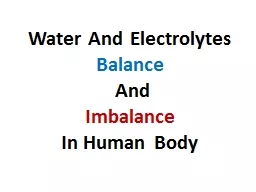
Electrolytes Electrolytes are ions capable of carrying an electricl charge Anions Anode Cations Cathode Major cations of the body Na K Ca 2 amp Mg 2 Major anions of the body ID: 776596
Download Presentation The PPT/PDF document " Part 1 Electrolytes Lecture 14" is the property of its rightful owner. Permission is granted to download and print the materials on this web site for personal, non-commercial use only, and to display it on your personal computer provided you do not modify the materials and that you retain all copyright notices contained in the materials. By downloading content from our website, you accept the terms of this agreement.
Slide1
Part 1
Electrolytes
Lecture 14
Slide2Electrolytes
Electrolytes are ions capable of carrying an electricl chargeAnions: (-) → AnodeCations: (+) → CathodeMajor cations of the bodyNa+, K+, Ca2+ & Mg2+Major anions of the bodyCl-, HCO3-, HPO42- & SO42-
2
Cathode
Anode
Slide3Essential Component in Numerous processes
Volume and osmotic pressure (Na+, K+, Cl-)Myocardial rhythm and contraction (K+, Mg2+, Ca2+)Cofactors in enzyme activation (Mg2+, Ca2+, Zn2+).Regulation of ATPase ion pump (Mg2+)Acid/Base balance (pH) (HCO3-, K+, Cl-)Coagulation (Mg2+, Ca2+)Neuromuscular (K+, Mg2+, Ca2+)The body has complex systems for monitoring and maintaining electrolyte concentrations
3
Slide4Water
Maintenance of water homeostasis is vital to life for all organismsMaintenance of water distribution in various body fluids is a function of electrolytes (Na+, K+, Cl- & HCO3-)
4
Slide5Water
Average water content of human body is 40-75% of total body weight.Solvent for all body processesTransport nutrients to cellsRegulates cell volumeRemoves waste products → urineBody Coolant → sweatingWater is located in intracellular and extracellular compartmentsIntracellular fluid (ICF) is the fluid inside the cellsExtracellular fluid (ECF) and subdivided into theintravascular extracellular fluid (plasma) and the interstitial cell fluid that surrounds the cells in the tissue
5
Slide66
Slide7Water
Normal plasma ~ 93 % H2O, the rest is a mixture of Lipids and proteins.Concentration of ions within the cells and plasma is maintained by:Energy consumption: Active transport Diffusion: Passive transportMaintaining conc. of electrolytes affect distribution of water in compartments.Most membranes freely permeable to water.Conc. of ions on one side affect flow of water across the membrane.
7
Slide8Osmolality
Physical property of a solution is based on the concentration of solutes per kilograms of solvent (mOsm/Kg)Sensation of thirst & arginine vasopressin hormone (AVP) [formerly, Antidiuretic hormone (ADH)] are stimulated by hypothalamus in response to increased blood osmolalityThirst → more water intakeAVP → increase water absorption in kidney
8
Slide9Clinical Significance
Osmolality is the parameter to which hypothalamus responds to maintain fluid intake.The regulation of osmolality also affects the Na+ concentration in plasmaaccount for ~90% of osmotic activity in plasmaAnother process which affects Na+ concentration is regulation of blood volume.
9
Slide10Clinical significance
To maintain normal plasma osmolality (275-295 mOsm/Kg) hypothalamus must respond quickly to small changes 1-2% increase in osmolality: 4 fold increase in AVP secretion.1-2% decrease in osmolality: shuts off AVP secretion.Renal water regulation by AVP and thirst play important roles in regulating plasma osmolality. Renal water excretion is more important in controlling water excess, Whereas thirst is more important in preventing water deficit or dehydration. Consider what happens in several conditions.
10
Slide11Water Load
Excess intake of water lower plasma osmolalityKidney is important in controlling water excessAVP and thirst are suppressedWater is not reabsorbed, causing a large volume of dilute urine to be excreted Hypoosmolality and hyponatremia usually occur in patients with impaired renal excretion of water
11
Slide12Water deficit
As a deficit of water, plasma osmolality begins to increase.Both AVP secretion and thirst are activated. Although AVP contributes by minimizing renal water loss, thirst is the major defense against hyperosmolality and hypernatremia. A concern in infants, unconscious patients, or anyone who is unable to either drink or ask for water.
12
Slide13Regulation of blood volume
Blood volume is essential in maintaining blood pressure and ensure perfusion to all tissues and organs.Regulation of both sodium & water are interrelated in controlling blood volumeRenin-angiotensin-aldosterone: system of hormones that respond to decrease in blood volume and help maintain the correct blood volume.
13
Slide14Regulation of blood volume
Changes in blood volume is detected by receptors in: the cardiopulmonary circulation , carotid sinus, aortic arch and glomerular arterioles
14
They activate effectors that restore volume by:
appropriately varying vascular resistance,
cardiac output,
and renal Na and H
2
O retention.
Slide1515
Angiotensin
converting enzyme (ACE)
Slide1616
Slide17M. Zaharna Clin. Chem. 2009
17
Slide18Regulation of blood volume
Other Factors effecting blood volume: Atrial natriuretic Peptide (ANP) → sodium excretion → blood volumeVolume receptors → release of AVP → conserve water → blood volumeGlomerular filtration rate (GFR): in volume expansion and in volume depletion
18
Slide19Increase in Blood Volume
19
Slide20Determination of Osmolality
Serum or urine sample (plasma not recommended due to the use of anticoagulants)Based on properties of a solution related to the number of molecules of solutes per kilogram of solvent such as:Freezing point ( osmolality freezing point temp.)Vapor pressure ( osmolality Vapor pressure)
20
Slide21Determination of Osmolality
Freezing Point Osmometer:Standardized method using NaCl reference solution.Specimen is supercooled to -7ºC, to determine freezing point osmolality causes depression in the freezing point temp. More solutes present the longer the specimen will take to freeze.
21
Slide22Osmolal
Gap
Osmolal gap is the difference between the measured osmolality and the calculated one.Osmolal Gap= measured osmolality - calculated osmolalityThe osmolal gap indirectly indicates the presence of osmotically active substances other than sodium, urea or glucose. (ethanol, methanol or β-hydroxybutyrate)
22
Slide23Case Study
A sixty-seven year old white male was found pulseless and resuscitated; then brought to the emergency room. He had been reported to be drinking in a bar all afternoon, and had then fallen from a ten foot balcony to snow covered ground. He arrived in the emergency room with a fractured occiput and was unresponsive.Admission Lab. results:
23
Na=143
mEq
/l
(
136-145)
BUN=4 mg/
dL
(6 – 20)
pH=7.30
(7.35 – 7.45)
Cl=105
mEq
/l
(
95-105)
GLU=104 mg/dL
Osmolality=356
mOsm
/kg
(275 – 295)
Slide24Case Study
Cal. Osmo. = (2 X 143) + (104/20) + (4/3) = 286 + 5.2 + 1.33 = 293Osmolal Gap= measured osmolality - calculated osmolality = 356 – 293 = 63An OG value greater than 15 is considered a critical valueThe presence of low blood pH, elevated anion gap and greatly elevated OG is a medical emergency that requires prompt treatment
24
Slide25M. Zaharna Clin. Chem. 2009
25
Na
+
Slide26Sodium
Most abundant extracellular cation- 90%Major function is maintaining the normal water distribution & osmotic pressure of plasmaRole in maintaining acid-base balance (Na+, H+ exchange mechanism)Normal range Serum: 136-145 mmol/LATPase ion pump: the way the body moves sodium and potassium in and out of cells.3 Na+ out of the cell for every 2 K + in and convert ATP to ADP.
26
Slide2727
Slide28Regulation of Sodium Balance
Plasma Na+ concentration depends: on the intake and excretion of water and, on the renal regulation of Na+Three processes are of primary importance: Intake of water in response to thirst ( p. osmolality)The excretion of water (AVP release)The blood volume status, which affects Na+ excretion through aldosterone, angiotensin II, and ANP (atrial natriuretic peptide).
28
Slide29Nephron
29
Slide30Regulation of Sodium Balance
70 % of sodium that is filtered is reabsorbed in proximal tubules.Remainder occurs in the ascending loop of Henle (without water absorption) & DCT under regulation of: AldosteroneRenin-Angiotensin systemAtrial natriuretic Peptide (ANP) → sodium excretion
30
Slide31Hyponatremia
Defined as a serum/plasma level less than 135 mmol/L.One of the most common electrolyte disorders in hospitalized and non-hospitalized patientsLevels below 130 mmol/L are clinically significant.
31
Slide32Causes of Hyponatremia
The principal clinical concern of hyponatremia is the encephalopathy associated with acute-onset severe hyponatremia and consequent cerebral edema.
32
Slide3333
Slide34Hypernatremia
Hypernatremia: increased sodium concentration > 145 mmol/lResult of excess water loss in the presence of sodium excess, or from sodium gain
34
With severe elevations of sodium, seizures and coma may occur.
Slide35Sodium determination
Methods:Flame emission spectrophotometrymeasurement of light emitted when the element is excited by energy in the form of heat.
35
Slide36Atomic absorption spectrophotometry
36
Slide37Ion Selective electrode
37
Reference electrode
selective membrane at the ion selective electrode, allows measured ions to pass, but excludes the passage of the other ions
Slide38Potassium
Major intracellular cation20 X greater concentration in the cell vs. outside.2% of the bodies potassium circulates within the plasma.Function:Regulates neuromuscular excitabilityHydrogen ion concentrationIntracellular fluid volume
38
K
+
Slide39Effects on Cardiac muscle
39
Ratio of K
+
intracellular & extracellular is important determinant of resting membrane potential across cell membrane
Increase plasma potassium; decreasing the resting membrane potential, increase excitability, muscle weakness
decreases the net difference between the cell’s resting potential and threshold (action) potential
Decrease extracellular potassium; decrease excitability
Slide40Slide4141
Slide42Potassium Role in Hydrogen Concentration
In hypokalemia (low serum K+), As K+ is lost from the body, Na+ and H+ move into the cell. The H+ concentration is, therefore, decreased in the ECF, resulting in alkalosis.
42
Slide43Regulation of potassium
The kidneys are important in the regulation of K+ balance.Initially, the proximal tubules reabsorb nearly all the K+. Then, under the influence of aldosterone, K+ is secreted into the urine in exchange for Na+ in both the distal tubules and the collecting ducts. Thus, the distal tubule is the principal determinant of urinary K+ excretion. Most individuals consume far more K+ than needed; the excess is excreted in the urine but may accumulate to toxic levels if renal failure occurs.
43
Slide44Hypokalemia
Decrease of serum potassium below 3.5 mmol/l
Insulin promotes acute entry of K into skeletal muscle and liver by increasing Na, K-ATPase activity;
44
Hypermineralocorticoid-like effects. In the kidney, cortisol activation of mineralocorticoid receptors alters renal tubular exchange of sodium (retained), potassium (excreted)
Slide4545
Slide46Hyperkalemia
Increase potassium serum levels > 5 mmol/lAssociated with diseases such as renal and metabolic acidosis
46
Slide47Potassium determination
Assay method:Ion selective Electrodea valinomycin membrane is used to selectively bind K+
47
Slide48Chloride
Major extracellular anionCl– is involved in maintaining: osmolality, blood volume, and electric neutrality.In most processes, Cl– ions shift secondarily to a movement of Na+ or HCO3–.Cl– ingested in the diet is Completely absorbed by the intestinal tract.
48
Cl
-
Slide49Chloride
Cl– ions are filtered out by the glomerulus and passively reabsorbed, in Conjuction with Na, by the proximal tubules.Excess Cl– is excreted in the urine and sweatExcessive sweating stimulates aldosterone secretion, which acts on the sweat glands to Conserve Na+ and Cl–
49
Slide50Electric Neutrality
Sodium/chloride shift maintains equilibrium within the body.Na reabsorbed with Cl in proximal tubules.Chloride shiftIn this process, carbon dioxide (CO2) generated by cellular metabolism within the tissue diffuses out into both the plasma and the red cell. In the red cell, CO2 forms carbonic acid (H2CO3), which splits into H+ and HCO3- (bicarbonate). Deoxyhemoglobin buffers H+, whereas the HCO3- diffuses out into the plasma and Cl- diffuses into the red cell to maintain the electric balance of the cell
50
Slide51Chloride shift
51
Slide52Hypochloremia
Hypochloremia: < 98 mmol/l
52
Slide53Hypercholremia
Hypercholremia: > 109 mmol/l
53
Slide54Assay
Coulometric titration (ref. method)measure amount of analyte by measuring amount of current and time required to complete reaction use Ag electrode to produce Ag+Ag (s) » Ag+ + e-Ag+ + Cl- » AgCl Ion selective electrode
54