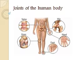
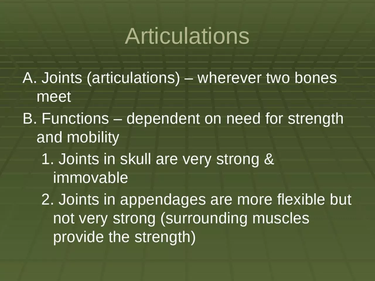
/
Articulations A. Joints (articulations) – wherever two bones meet Articulations A. Joints (articulations) – wherever two bones meet

Articulations A. Joints (articulations) – wherever two bones meet - PowerPoint Presentation

davies
davies . @davies
Follow
64 views
Uploaded On 2024-01-03

Articulations A. Joints (articulations) – wherever two bones meet - PPT Presentation

B Functions dependent on need for strength and mobility 1 J oints in skull are very strong amp immovable 2 Joints in appendages are more flexible but not very strong surrounding muscles provide the strength ID: 1038364

bone joints joint movement joints bone movement joint synovial fibrous bones articular cavity surface cartilage fluid articulationsb examples motion

Share:

Link:

Embed:

Download Presentation from below link

Download Presentation The PPT/PDF document "Articulations A. Joints (articulations) ..." is the property of its rightful owner. Permission is granted to download and print the materials on this web site for personal, non-commercial use only, and to display it on your personal computer provided you do not modify the materials and that you retain all copyright notices contained in the materials. By downloading content from our website, you accept the terms of this agreement.


Presentation Transcript

1. ArticulationsA. Joints (articulations) – wherever two bones meetB. Functions – dependent on need for strength and mobility1. Joints in skull are very strong & immovable2. Joints in appendages are more flexible but not very strong (surrounding muscles provide the strength)

2. ArticulationsC. There are 3 structural classifications:1. Fibrous joints – composed of fibrous tissue with no cavity2. Cartilaginous joints – articulating bones are united by cartilage and no cavity present3. Synovial joints – articular bones are separated by a fluid-filled joint cavity

3. ArticulationsD. There are 3 functional classifications:1. Synarthroses – immovable joints (sternocostal, tibiofibular, sutures)2. Amphiarthroses – slightly movable joints (intervertebral joint and pubic symphysis)3. Diarthroses – freely movable joints (most appendicular joints)

4. ArticulationsE. Fibrous Joints1. Characteristics:A) Bones are joined by fibrous tissueB) No joint cavity is presentC) Most are immovable (synarthrotic) but some are slightly moveable (amphiarthrotic)2. Three types of fibrous joints

5. ArticulationsA) Sutures – contain dense fibrous connective tissue until adulthood when they ossify1) Examples are the skull suturesB) Syndesmoses – bones are connected by a filamentous sheet or cord (ligament or interosseous membrane)1) Movement can range from slight to considerable2) Examples include the tibiofibular & radioulnar (between the shafts)

6. ArticulationsC) Gomphoses – articulation of tooth with alveolar margin1) Possesses a fibrous connection called the periodontal ligamentF. Cartilaginous joints1. Characteristics:A) Articulating surfaces are united by cartilageB) No joint cavity

7. Articulations2. Two main types of cartilaginous joints:A) Synchondroses – hyaline cartilage unites bones; usually temporary joints (sites of bone growth)1) Cartilage is replaced by bone and the joints become synarthrotic2) Examples are the epiphyseal plate and the first rib and the manubrium

8. ArticulationsB) Symphyses – articular surface of bone covered by hyaline cartilage fused to an intervening pad or plate of fibrocartilage1) It is compressible, resilient and functionally amphiarthrotic2) Examples are the pubic symphysis and the intervertebral jointsG. Synovial Joints1. Articular bones are separated by a fluid-filled joint cavity

9. Articulations2. Most common joint in the body3. Diarthrotic4. Five distinct features of synovial jointsA) Articular cartilage – hyaline cartilage forms a glassy smooth surface over the opposing ends of bonesB) Synovial (joint) cavity – small space between the bonesC) Synovial fluid – largely derived from blood; has a viscous, egg-white consistency

10. ArticulationsD) Articular capsule – 2 parts1) Fibrous capsule (external)2) Synovial membrane (internal)E) Reinforcing ligaments 1) Intrinsic (capsular) – parallel bundles of fibers within the fibrous capsule2) Extracapsular – extend bone to bone3) Intracapsular – located inside the cavity

11.

12.

13. Articulations5. Movement of synovial joints:A) Axis of motion1) Non-axial motion – slipping movements (intercarpal & intertarsal)2) Uniaxial motion – movement in one plane (interphalangeal, ulna/humerus)

14. Articulations3) Biaxial motion – movement in two planes (occipital bone/atlas)4) Multiaxial motion – movement in more than two planes (scapula/humerus and coxal bone/femur)

15. ArticulationsB) Types of motion1) Gliding – bones displaced in relation to one another (intercarpal, intertarsal, and intervertebral joints)2) Angular – changing the angle between two bonesa) Flexion – bending or decreasing the joint angle

16. Articulationsb) Extension – stretching or increasing the joint anglec) Abduction – moving away from the midlined) Adduction – moving towards the midlinee) Circumduction – a limb creates a conical (cone) shape in spacef) Rotation – turning movement of a bone around its own axis

17. Articulations6. Special movementsA) Supination of the hand – turning the palm forward or facing upB) Pronation of the hand – turning the palm backward or facing down C) Inversion (supination) of the foot – movement of the sole inwardD) Eversion (pronation) of the foot – movement of the sole outward

18. ArticulationsE) Dorsiflexion – upward movement of foot/toesF) Plantar flexion – downward movement of foot/toesG) Protraction – movement of the mandible forwardH) Retraction – movement of the protracted part back to its start position

19.

20.

21. ArticulationsI) Elevation – lifting a body part superiorlyJ) Depression – moving the elevated part inferiorlyK) Opposition – touching your thumb to the tips of other fingers7. Types of synovial jointsA) Plane joints – articular surface is flat and only allows for short gliding movements (intercarpal and intertarsal)

22. ArticulationsB) Hinge joints – cylindrical projection of one bone fits into a trough-shaped surface on another bone (elbow & knee)C) Pivot joints – rounded end of one bone protrudes into a sleeve or ring composed of bone or ligament (radius to ulna and axis to atlas)D) Condyloid joints – oval articular surface of one bone fits into a complementary depression in another (metacarpophalanges – knuckles)

23. ArticulationsE) Saddle joints – each articular surface has a concave and convex area (carpometacarpal joint of the thumb)F) Ball and socket joints – the spherical end of one bone articulates with a cuplike socket of another bone (shoulder or hip joints)

24. ArticulationsH. Common Joint Injuries1. Sprain – stretching or tearing of a ligament2. Luxation – bones are forced out of their normal position (a.k.a. dislocation)A) Subluxation – partial dislocation3. Bursitis – inflammation of the bursaA) Bursa – flattened sacs that contain a thin film of synovial fluid; located where ligaments, muscles, and tendons rub against bone

25. Articulations4. Tendonitis – inflammation of the tendon5. Arthritis – inflammatory or degenerative disease of the joint where synovial membranes thicken and fluid production decreases resulting in friction and painA) Osteoarthritis – degenerativeB) Rheumatoid arthritis – autoimmune diseaseC) Gouty arthritis – uric acid accumulation6. Synovitis – inflammation of synovial membranes