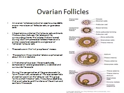
Benign ovarian diseases Ovarian Cysts Prevalence 4 of women are admitted to hospital with an ovarian cyst complication by the age of 65 years 25 of adnexal torsions occur in children ID: 917154
Download Presentation The PPT/PDF document "Ovarian diseases Dr Ismaiel Abu Mahfouz" is the property of its rightful owner. Permission is granted to download and print the materials on this web site for personal, non-commercial use only, and to display it on your personal computer provided you do not modify the materials and that you retain all copyright notices contained in the materials. By downloading content from our website, you accept the terms of this agreement.
Slide1
Ovarian diseases
Dr Ismaiel Abu Mahfouz
Slide2Benign ovarian diseases
Slide3Ovarian Cysts
Prevalence4% of women are admitted to hospital with an
ovarian cyst / complication by the age of 65 years
25%
of adnexal torsions occur in children
90%
of all ovarian
cysts
are benign
Risk
of
Ca
in an ovarian cyst in a woman
of:
Reproductive age:
0.4–0.8/100 000
Age 60–80 years :
60/100 000
Slide4Ovarian cyst events / complications
Rupture
Asymptomatic /
acute abdominal pain
May
follow sexual intercourse or physical activity
Severity
of pain depends on the type of fluid
“Serous
or mucinous/ sebaceous
material/ Blood”
Haemorrhage into a cyst:
Pain of variable degree. Usually mid cycle
Torsion
M
oderate-severe
pain & of sudden onset
Associated
nausea and
vomiting
More pain than tenderness
Infection
Pain, fever, peritoneal irritation ??PID
Slide5Slide6Clinical evaluation
History
History of endometriosis/ PID/ known ovarian cystsBowel
/ urinary symptoms
Anticoagulants
Progesterone only pills: develop recurrent
ovarian
cysts
Pain may be referred down the cutaneous distribution of the Obturator nerve (inner thigh down to the knee)
Examination
+/-
low-grade
fever. BP,PR: usually stable
Abdominal
tenderness
Cervical
excitation on
vaginal
examination
Slide7Investigations
Pregnancy test Urinalysis and cultureFull blood count, urea and electrolytes
? Coagulation screenGenital swabs for infection if PID is
suspected
CA-125 : Not as a routine
Ultrasound examination
Doppler blood
flow of the cyst: Findings
are variable and not
diagnostic
Slide8Ovarian cyst with typical mixed internal echoes suggestive of blood
Slide9Normal ovaries and free
fluid in POD
Slide10Adnexal torsion
Enlarged ovary / adnexal mass
Free fluid if associated with rupture Doppler sonography is not helpful in the diagnosis
Slide11D.Dx
In case of ovarian cyst complication, consider
Ectopic
pregnancy
Pelvic
inflammatory disease
Pelvic
abscess
Fibroid degeneration
Appendicitis
Complications of diverticular
disease
Urinary tract infection
Urinary calculi
Renal
colic
Slide12Management
Expectant Mx: Haemorrhagic cysts and cyst rupture Analgesia
and observationRepeat scan 6 wksSurgery: Laparoscopy /Laparoscopy
if:
H
aemodynamic
compromise
D
iagnostic
uncertainty or likelihood of torsion
No
relief of symptoms within 48 hours of presentation
Consider COCPs
for cyst formers
Slide13Special situation
Ovarian cysts in pregnancy
Most common: Dermoid cysts (50%) then cystadenomas<
5% require
intervention
Conservative management is appropriate
Indications
for intervention:
Symptomatic
relief
Suspicion
of malignancy
Slide14Special situation
Ovarian tumours in children
Ovarian ca represent 1.5% of childhood ca
Most ovarian
tumours
are
benign
Types:
Most common: Epithelial
cysts and
teratoma
Most common ca: Germ
cell
tumours
Most common complication: Torsion (33
% of
cases)
Slide15Malignant disease of the ovary and tubes
Slide16Malignant disease of the ovary
Ca ovary
The second most common gynae ca after uterine
ca
5th most common
ca
in
women
after breast, bowel, lung and uterine
ca
The majority of ovarian
ca
are
epithelial
Slide17Types of Ovarian cysts / tumors
Functional
Follicular
cyst
Corpus luteum cyst
Theca lutein cyst
Inflammatory
Tubo-ovarian
abscess
Benign
tumours/cysts
Endometriotic cyst
Brenner
tumour
Benign teratoma
Fibroma
Malignant
/malignant potential
Epithelia ovarian ca
Malignant teratoma
Endometrioid carcinoma
Dysgerminoma
Secondary ovarian tumor
Cystadenoma, cystadenocarcinoma
Granulosa cell tumor
Arrhenoblastoma
Theca cell tumor
Slide18Classification of ovarian tumours
Ovarian tumors
classified according to their origin, biological behavior or clinical manifestations
WHO
Classification:
Epithelial
Sex cord stromal
Germ cell
Slide19Epithelial tumours (80 - 90%)
Serous Tumors
Benign/Borderline/ ca
Can
be bilateral
Psammoma bodies
BRCA 1 mutations
Mucinous
Tumors
Benign/Borderline/ ca
Pseudomyxoma
peritonei
RT
/ CT resistant
Endometroid
Tumors
Malignant
? Endometriosis
Ass.
with endometrial
ca
Clear
Cell (
Mesonephroid)
Benign/Borderline/ ca
Worst prognosis
Transitional cell (
Brenner)
Usually benign
Mixed
epithelial tumours
Undifferentiated & unclassified
Slide20Germ Cell Tumours (10-15%)
Dysgerminoma
Most commonly
malignant
Abnormal
gonads/Turner
Bilateral
LDH
Chemo/radiosensitive
Endodermal
Sinus Tumors
(
Yolk Sac Tumors)
Young
children < 4yrs
3
rd
decade
Schiller-Duval bodies
AFP
Choriocarcnoma
Malignant
Cyto-
& syncitiotrophoblast
B-HCG
Teratomas
Immature-
can get
malignant
Mature
Solid
Cystic
Monodermal &
highly specialized
Struma
ovarii
Carcinoid
Struma
Ovarii &
Carcinoma
Mixed
forms
GONADOBLASTOMA
Pure
Mixed
with Dysgerminoma or
other Form
Germ Cell
Tumors
Slide21Slide22Sex Cord Stromal Tumours
(5-10%)
Granulosa-Stromal
Cell
Tumors
Granulosa cell
Any
age
Inhibin
A/B or Estradiol
Precocious
puberty
Microscopic
: Call-Exner
bodies
Tumours
in the
Thecoma-fibroma group
Androblastomas
Sertoli-Leydig
Cell Tumors
Well /Intermediate/Poor
differentiated
Secretes
androgen
Fibromas
Associated
with ascites
& hydrothorax
“Meigs syndrome”
Slide23Krukenberg tumour
Secondary Ca of the ovary
Metastasized classically from GIT and breast80%: bilateral ovarian involvement
“ Signet
ring
cells”
Slide24Ovarian tumours
Primary
ovarian ca commonly: 40-60 yrs Teratomas and Sex
Cord:
mostly before puberty
Borderline
malignant:
30-50
yrs
Ovarian ca; a silent killer
Asymptomatic in early stages
75%
diagnosed with
advanced stage
disease
Overall 5-year survival rate:
35%
Most common cause of
death
from gynae ca in UK
Slide25Ovarian ca; risk factors
Ovarian ca
Most cases of EOC are sporadic
The
aetiology
is unknown
Most significant risk factor is genetic
predisposition
Ovarian ca, a challenging disease
Natural history not well understood
No well-defined precursor lesion
Length of time from localised tumor to dissemination
is unknown
No effective screening method for early detection yet
Slide26Risk factors: Heredity
10% of Epithelial ca cases are
familial
Familial
syndromes:
F
amilial
breast-ovarian cancer
syndrome (BRCA I+II)
C
ancer
family syndrome (Lynch
syndrome =
HNPCC)
Account
for 90% of
familial
ovarian
ca
Slide27Additional Risk Factors
Age
Rare <30Peak ≥
60yrs
Reproductive history
Early menarche
Nulliparity
Age
>30 at first
child-bearing
Late
menopause
Fertility drugs
Personal history of breast cancer
Talcum powder
Slide28Protective factors
MultiparityFirst pregnancy before
age of 30Oral contraceptives: 5 years of use decreases
risk
by 50%
Tubal ligation
Hysterectomy
Lactation
Bilateral
oophrectomy
Slide29Diagnostic approach
History
Abdominal bloating, increased girth, pressure
Unusual fatigue
GIT:
nausea, indigestion, gas,
constipation,
diarrhea
Urinary frequency or incontinence
Unexplained weight loss or gain
Shortness of breath
Germ cell tumours
Often
present more
acutely &
at an earlier stage
Typically:
Rapidly
enlarging abdominal/pelvic
mass
Acute
severe lower abdominal pain due to tumour
rupture
, haemorrhage or
torsion
Slide30Diagnostic approach
Examination
Abdominal / pelvic: pelvic masses, ascites, hepatomegaly
Chest:
P
leural effusions, palpable
lymph
nodes
Imaging
TA & TV
scans:
Detection
of
masses and its characters
CT scan (Abdomen / chest):
Assess
spread to LN, pelvic &
abdominal structures
MRI:
Best
to distinguish malignant / benign
tumors
Bloods:
CBC, KFT, LFT, tumour marker
Slide31Diagnostic approachTumour
markers
Serous tumours:
CA
125
Mucinous:
CA
19-9
Granulosa:
Inhibin
Endodermal
sinus:
AFP
Choriocarcinoma:
HCG
Dysgerminoma:
LDH
,
Alkaline phosphatase
Slide32Diagnostic approach
Risk of malignancy index (RMI)
RMI:
Gives
an estimate of the risk of
ovarian ca
for
women
with adnexal
masses
Calculated
using
Ultrasound
findings (U
)
Menopausal status
(M)
CA-125
value (serum levels >30U/ml
abnormal
)
Slide33RMI
RMI = U x M x CA125
Ultrasound findings (U) “Scored
1 point for
each”
Multi-locular
cyst
Evidence
of solid areas
Evidence
of metastases
Presence
of ascites
Bilateral Lesions
U:
U = 0 (U/S score of 0)
U = 1 (U/S score of 1)
U = 3 (U/S score of 2 – 5)
Menopausal
status
Postmenopausal status is graded M = 3
Pre-menopausal status is graded M =
1
Ca-125
Slide34RMI
RISK
RMI
Risk of Cancer
Low
<25
<3
Moderate
25-200
30
High
>200
75
Slide35Ultrasound
Both TA and TV ( TVS has better resolution)
Major limitations
Poor
PPV in asymptomatic women
Inability
to detect
ca
when ovaries are normal size
Allows earlier stage
detection
Slide36Color-flow Doppler
Used in conjunction with TVSMeasures resistance in blood vessels supplying
ovariesMay provide additional information to help distinguish malignant from benign masses
Slide37CA-125
Sustained elevation in 82% of women with advanced ovarian ca
Poor sensitivity
Elevated
in only 50% of women with Stage I
disease
Poor specificity
Elevated
in many gynecologic and non-gynecologic
malignancies
as well as benign
conditions
Slide38CA-125
Malignant conditions Cervical CA
Fallopian tube CA Endometrial CA
Pancreatic CA
Colon CA
Breast CA
Lymphoma
Mesothelioma
Benign conditions
Endometriosis/Menses
Uterine fibroids
PID
Pregnancy
Diverticulitis
Pancreatitis
Liver disease
Renal failure
Appendicitis
IBD
Slide39Benign vs Malignant Tumors
“
Ultrasound & Doppler”
Benign
More likely unilateral
Unilocular
Thin-walled
No papillae
No solid areas
Malignant
More likely bilateral
Multilocular
Thick walls
Papillae present
Mixed echogenicity due to solid areas
Greater Angiogenesis and Blood
Flow
Slide40Benign ovarian cyst
40
Slide41Malignant ovarian mass
Slide42Malignant ovarian mass Doppler with Contrast
Slide43Spread of Ovarian malignancies
Direct seeding:
To peritoneum, omentum,
tubes, ureters
L
ymphatics:
T
o para-aortic
nodes,
umbilicus, diaphragm
B
loodstream:
T
o lower
vagina and in the case of sarcomas and
Teratomas
to the lungs and else
where
D
irect spread:
T
o
any
neighboring
organ or tissue
Slide44Ovarian ca: Staging FIGO ovarian cancer staging: 2014 update
Slide45Stage I
Tumour confined to ovaries
IA: Tumour limited to one ovary, capsule intact, no tumour on surface, negative washings
IB: Tumour involves both ovaries otherwise similar to 1A
IC: Tumour limited to one or both ovaries 1 Surgical spill, 2 Capsule rupture before surgery or tumour on ovarian surface
3
Malignant cells in the ascities or peritoneal washings
.
Stage
II
Tumour involves one or both ovaries with pelvic extension (below the pelvic brim) or primary peritoneal cancer
IIA: Extension and/or implant on uterus and/or fallopian tubes
IIB: Extension to other pelvic intraperitoneal tissues
Stage
III
Tumour involves one or both ovaries with cytologically or histologically confirmed spread to the peritonium outside the pelvis and/or metastasis to the retroperitoneal lymph nodes
IIIA: Positive retroperitoneal lymph nodes and/or microscopic metastasis beyond the pelvis
IIIA1: Positive retroperitoneal lymph nodes only
IIIA1(i): Metastasis ≤10 mm
IIIA1(ii): Metastasis >10 mm
IIIA2: Microscopic, extrapelvic (above the brim) peritoneal involvement ± positive retroperitoneal lymph nodes
IIIB: Macroscopic, extrapelvic, peritoneal metastasis ≤ 2 cm ± positive retroperitoneal lymph nodes
IIIC: Macroscopic, extrapelvic, peritoneal metastasis > 2 cm ± positive retroperitoneal lymph nodes. Includes extension to capsule of liver/spleen without parenchymal involvement of either organ.
Stage
IV
Distant metastasis excluding peritoneal metastasis
IVA: Pleural effusion with positive cytology
IVB: Hepatic and/or splenic parenchymal metastasis, metastasis to extra-abdominal organs (including inguinal lymph nodes and lymph nodes outside of abdominal cavity)
Slide46Ovarian ca: FIGO Grading
Epithelial
tumours of the ovary are
also
sub-classified by
histological grading
Gx
: Grade
cannot be assessed
G1
: Well
differentiated
G2
: Moderately
differentiated
G3
: Poorly
differentiated
Slide47Treatment Options
Surgery
ChemotherapyRadiotherapy
Slide48Standard treatment for ca ovary
Surgery Chemotherapy
Platinum
Taxol
Slide49Surgery
Slide50General surgical principles
Midline incision
Washings of the peritoneal cavity: diaphragm, right and left abdomen, pelvis
Evaluation
of all peritoneal surfaces
Optimal cytoreduction - may improve survival
Infracolic
omentectomy
lymphadenectomy of
pelvic
& para-aortic lymph
nodes
Slide51Types of Surgery
Aim
of surgery
Optimal cytoreduction: maximum residual tumour deposits no more than 1 cm
May consider fertility preserving procedure should that be medically possible
Types of surgery
TAH+BSO
Unilateral
salpingoophrectomy
(if fertility has to be preserved)
Cytoreductive or “debulking”
Peritoneal metastasis reduction
“Second look” laparotomy
Slide52Chemotherapy
Slide53Chemotherapy (CT)
Ovarian ca is a chemo-sensitive
Advanced disease “ has progressed beyond the ovaries,
stage 1c & above; require both surgery and CT
Types of chemotherapy
Adjuvant
:
CT following surgery
Combination
:
Several
agents given simultaneously to enhance their
effectiveness
Neo-adjuvant
:
CT prior to surgery where
Dx
has been established by cytology of ascitic fluid or histology of a tissue
biopsy
Slide54Chemotherapeutic Agents
Alkalyting agents
: Cyclophosphamide
, Cisplatin, Carboplatin,
Melphalan
Plant alkaloids
:
Paclitaxel, Vincristine,
Etoposide
Anticancer antibiotics:
Bleomycin
, doxorubicin
Antimetabolites:
Fluorouracil
, Gemcitabine
Slide55Side effects of chemotherapy
Side effects
Carboplatin
Paclitaxel
Alopecia
No
Yes
Thrombocytopenia
Yes
No
Nausea and vomiting
Yes
Yes
Neurotoxicity
No
Yes
Adverse cardiac effects
No
No
Arthralgia
No
Yes
Myalgia
No
Yes
Hypersensitivity reaction
No
Yes
Diarrhoea
No
No
Nephrotoxicity
Yes
Yes
Neutropenia
Yes
Yes
Slide56Side effects of chemotherapy
Nausea and vomitingF
atigueOral ulcerations
Ototoxicity (cisplatin): hearing loss, tinnitus
P
eripheral
neuritis
N
ephrotoxicity
M
yelosuppression
P
ulmonary
toxicity (bleomycin). Any new-onset cough/shortness of breath should be investigated urgently to exclude pneumonitis or fibrosis.
Slide57Follow up after primary treatment
F
ollow up
Provide
reassurance
Assess for early
recurrence
Follow up
Clinical
CA-125
MRI
Slide58Radiotherapy
Slide59RT; ca ovary
Rarely used as the main Rx for ovarian ca
Can be useful in treating areas where the cancer has spread, either near the main tumor or in a distant
organ, like the brain or spinal cord
Slide60Germ cell tumours (GCTs)
Benign or malignant
15–20% of all ovarian tumoursMalignant Ovarian GCTs are rare; 2–5% of all ovarian ca
Most common ovarian
ca
in the first two decades of life
? Racial prediction: higher incidence in African, South & East Asian & Hispanic patients compared with
Caucasians
Risk
factors:
gonadal dysgenesis
,
abnormal
karyotype
Multimodality Rx,
including surgery &
CT
Most women
have
excellent prognosis
Preservation
of fertility is often
possible
Slide61Radiotherapy for Germ cell tumours
Dysgerminomas are
very radiosensitive No longer forms a part of routine Rx because of
Widespread
use of
chemotherapy
The
long term
toxicities
of radiotherapy
Ovarian failure
Sub-fertility
and
Higher
risk of fatal second
malignancies
Rarely
used in in
a palliative
setting
where
CT refused or
contraindicated
Slide62Standard recommendations
Tumour
Stage
Treatment
Dysgerminoma
Stage IA/IB
Surgery + surveillance
Stage IC
Surgery + postoperative chemotherapy
Non-dysgerminoma
Stage IA, IB of any type or Stage IC immature GCT grade 1/2
Surgery + postoperative surveillance
Stage IC, grade 3 immature
Surgery + surveillance
or
Surgery + postoperative adjuvant chemotherapy
Stage IC or unsuspected stage II
Surgery + chemotherapy
Stage II or greater
Neoadjuvant chemotherapy followed by surgery
Slide63Primary fallopian tube carcinoma (FTC)
0.14% - 1.8% of female genital ca
Only 1200 cases of primary FTC have been reported in the literature Aetiology is unknown but hormonal, reproductive & possibly genetic factors
BRCA-1 and BRCA-2
90% of FTCs
are serous
papillary adenocarcinoma
Slide64Clinical manifestations & Rx
Presentation
40–60 years (median age 55 years)
Symptoms
are vague and non-specific, but are similar to
ovarian ca
Latzko's triad of
symptoms
:
Present in 15% of the cases
Intermittent
profuse serosanguinous vaginal
discharge
Colicky
pain relieved by
discharge
Abdominal
or pelvic mass
0–10% are identified preoperatively
Treatment
As epithelial ovarian ca