/
Brainstorm a list of the three types of muscle and their ch Brainstorm a list of the three types of muscle and their ch

Brainstorm a list of the three types of muscle and their ch - PowerPoint Presentation

debby-jeon
debby-jeon . @debby-jeon
Follow
421 views
Uploaded On 2017-09-24

Brainstorm a list of the three types of muscle and their ch - PPT Presentation

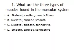
Types of Muscle Tissue cont Muscle Group Major Location Major Function Mode of Control Skeletal Muscle Attached to bones and skin of the face Produces body movements and facial expressions ID: 590360

muscles muscle contraction myosin muscle muscles myosin contraction structure myofibril skeletal amp heads action actin attaches tissue contract bone

Share:

Link:

Embed:

Download Presentation from below link

Download Presentation The PPT/PDF document "Brainstorm a list of the three types of ..." is the property of its rightful owner. Permission is granted to download and print the materials on this web site for personal, non-commercial use only, and to display it on your personal computer provided you do not modify the materials and that you retain all copyright notices contained in the materials. By downloading content from our website, you accept the terms of this agreement.


Presentation Transcript

Slide1

Brainstorm a list of the three types of muscle and their characteristicsSlide2

Types of Muscle Tissue (cont.)

Muscle Group

Major Location

Major

Function

Mode of Control

Skeletal Muscle

Attached to bones and skin of the face

Produces body movements and facial expressions

Voluntary

Smooth Muscle

Walls of hollow organs, blood vessels, and iris

Moves contents through organs; vasoconstriction

Involuntary


Cardiac Muscle

Wall of the heart

Pumps blood through heart

InvoluntarySlide3

Types of Muscle Tissue

Muscle cells

Myocytes called

muscle fibersSarcolemma

– cell membrane

Sarcoplasm

– cytoplasm of cell

Myofibrils

– long structures in

sarcoplasm

Arrangement of filaments in myofibrils produces striationsSlide4

Skeletal Muscle Structure & FunctionSlide5

General Information

prefixes

myo- & mys-

(meaning muscle) & sarco- (meaning flesh) refer to discussion of muscles

Muscles

attach to bones by

tendons

Origin

– where the muscle attaches to the bone that remains relatively stationary

during contraction

Insertion – where the muscle attaches to the bone that moves when the muscles contractSlide6

2. Can categorize muscles by how they work

Synergists – muscles that cause the same action at a joint (biceps &

brachialis; quadriceps group) Slide7

2. Can categorize muscles by how they work

Antagonists

– muscles that produce opposing action (biceps vs/ tricepts; quads vs. hamstrings)Slide8

Functions

produces

movement

maintaining posturestabilize joints b/c tendons go over the joints and help keep bones in place

generating heat

skele

muscle is ~40% of body mass and is most responsible for heat

productionSlide9

Skeletal Muscle

Made of muscle fibers (

myofibril) packed into sheets of muscle

Muscle fibers are cigar-shaped & have several nucleiIndividual muscle fibers are made of smaller pieces… we’ll get to thatSome muscle fibers

are about 1

ft in

length

Have visible stripes

Voluntary

can contract quickly & w/ large force but tires easily and must be rested after use

individual cells are fragile but muscles are strong…why?Slide10

tendon

bone

fascicle

m

uscle

fiber

myofibril

Sarcoplasmic

reticulum

filamentsSlide11

Structure:

Connective Tissue Coverings

Fascia

Covers entire skeletal

muscles

Separates them from

each other

Tendon

A tough, cord-like structure made of fibrous connective tissue

Connects muscles to bonesSlide12

How are tendons made?

All muscles are covered in connective tissue

perimysium

Collagen fiber density inc. near points of attachment  make a

tendon

Attach onto covering around the bones 

periosteumSlide13

Structure:

Connective Tissue Coverings

Aponeurosis

A tough, sheet-like structure made of fibrous connective tissue Attaches muscles to other musclesSlide14

tendon

bone

fascicle

m

uscle

fiber

myofibril

Sarcoplasmic

reticulum

filamentsSlide15

Microscopic anatomy of muscle fibrilsSlide16

1. An action potential reaches the skeletal muscle. The influx of ions causes an electrical impulse to be sent over the muscle. The impulse travels through the

sarcolemms

(cell membrane) and dives into the myofibrils via T-tubules.

Making Muscles ContractSlide17

2. The electrical impulse (change in charge of the muscle’s cytoplasm aka

sarcoplasm) opens voltage-gated calcium in the

sarcoplasmic reticulum channels. Ca2+ flows out of the sarcoplasmic reticulum and bathing the proteins in the myofibrils in Ca2+

Making Muscles ContractSlide18

3. Ca2+ attaches to

actin filaments and untwists the proteins exposing binding sites for the myosin heads. Myosin heads attach to the

actin forming cross bridges.

Making Muscles ContractSlide19

4. ATP supplies

myosin with energy to change shape and pull the

actin

filaments toward the midline of the

sarcomere

. This shortens the

sarcomeres

and thus the entire muscle in a contraction. Another ATP molecule changes the shape of the myosin again. This allows the myosin to release the

actin

and the muscle relaxes.Slide20

How Skeletal Muscles Move

Stimulated by nerve impulses

Axon terminal and the sarcolemma (plasma membrane of muscle cells) are close together with a synaptic cleft in betweenNeuron releases ACh across the cleft  attaches to receptors on the sarcolemma  allows Na+ to enter making cell more + making an action potential  causes contraction of the sarcomeres and thus the muscle

http://highered.mcgraw-hill.com/sites/0072495855/student_view0/chapter10/animation__action_potentials_and_muscle_contraction.htmlSlide21

Question 1

What structure is used to connect actin

and myosin during muscle contraction. Myosin heads AKA “paddles”Slide22

Question 2

Compare/contrast the insertion and origin?Answer

Insertion- usually more distal (limbs)insertion point moves toward the originOrigin- where a muscle attaches to a bone that does not move during muscle movementSlide23

Question 3

Put the following parts of a muscle in order from largest to smallest and explain what each is

MyofibrilFascicleMyofibrilSarcomere

Myosin head AKA “paddles”AnswerFascicle

 myofibril 

sarcomere

 myofibril  myosin headsSlide24

Question 4

After an action potential is sent to the muscle what two things (ions, molecules, etc) a required for muscle contraction? What does each one do?

AnswerATP- allows the myosin heads to pull the Z disc in towards the center of the cell for muscle contraction

Calcium- changes the shape of actin to expose binding sites for the myosin heads Slide25

Question 5

What structure gets pulled in towards the center of the

sarcomere in muscle contraction?Answer: z-lines or z proteinsSlide26

Whisper Down the Lane

You are sitting and you want to stand. Collectively you must describe the progression of events that will occur in order to get the “move” message to your skeletal muscles (focus on just ONE muscle). Tell the person next to you what is going to happen first and they are going to share what you told them plus the next correct, logical step in this process.