/
 Danny Haywood FY1 Arrhythmias  Danny Haywood FY1 Arrhythmias

Danny Haywood FY1 Arrhythmias - PowerPoint Presentation

debby-jeon
debby-jeon . @debby-jeon
Follow
346 views
Uploaded On 2020-04-04

Danny Haywood FY1 Arrhythmias - PPT Presentation

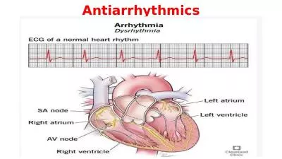
Intro Conduction system of heart Symptomssigns Investigations Tachy vs Brady Bradyarrhythmias Different types Management Tachyarrhythmias Broad vs narrow Types of each Management of each ID: 775332

heart block atrial broad heart block atrial broad type complex disease management mobitz qrs control tachy failure flutter avnrt

Share:

Link:

Embed:

Download Presentation from below link

Download Presentation The PPT/PDF document " Danny Haywood FY1 Arrhythmias" is the property of its rightful owner. Permission is granted to download and print the materials on this web site for personal, non-commercial use only, and to display it on your personal computer provided you do not modify the materials and that you retain all copyright notices contained in the materials. By downloading content from our website, you accept the terms of this agreement.


Presentation Transcript

Slide1

Danny HaywoodFY1

Arrhythmias

Slide2

Intro

Conduction system of heart

Symptoms/signs

Investigations

Tachy

vs

Brady

Bradyarrhythmias

Different types

Management

Tachyarrhythmias

Broad

vs

narrow

Types of each

Management of each

Summary

Some example

ECGs

Slide3

Slide4

Symptoms/signs

Syncope

Dizziness

Palpitations

Heart Failure

Chest pain

Sudden death

No symptoms

Slide5

Investigations

Bedside

ECG

Bloods

TFTs, U+E, FBC, Troponins

Imaging

Echo, CXR

Special tests

Holter monitor

Slide6

ARRhYTHMIAS

Bradyarrhythmias vs Tachyarrhythmias

Brady

HR < 60bpm

Tachy

HR > 100bpm

Slide7

Bradyarrhythmias

Type I heart block

1

st

degree heart block

Prolonged PR interval > 0.2 seconds

Type II heart block

Mobitz

type 1 –

Wenckebach

Gradually increased PR intervals until missed QRS

Mobitz

type 2

Intermittently P wave not followed by QRS

May be pattern

eg

2:1, 3:1 ratio of P waves to QRS complexes – no increase in PR interval

Type III heart block

Complete heart block

No correlation between

P

waves and QRS complexes

Slide8

Management

Acute (eg. Secondary to MI)

If symptomatic/clinical deterioration

IV atropine

External (transcutaneous) pacing

Chronic

Mobitz type II or complete AV block

Permanent pacemaker

Slide9

Tachyarrhythmias

Narrow complex (Supraventricular) vs Broad complex (Ventricular)

Narrow

QRS <0.12 seconds

Broad

QRS >0.12 seconds

Slide10

Narrow complex

Sinus tachycardia

Atrial Fibrillation (AF)

Atrial Flutter

Atrioventricular

nodal re-entry tachycardia (AVNRT)

Atrioventricular

reciprocating tachycardia (AVRT)

Slide11

AF

Continuous, rapid activation of atria – due to rapidly depolarising foci within the atriaOften located by pulmonary veinsNo coordinated mechanical action

Slide12

AF – Causes

ATRIAL PhIB

A – Alcohol

T – Thyroid disease

R – Rheumatic heart disease

I – Ischaemic heart disease

A – Atrial myxoma

L – Lung pathology (pneumonia, PE)

Ph – Pheochromocytoma

I – Idiopathic

B – Blood pressure (hypertension)

Slide13

AF - management

Conservative

Alcohol cessation

Lifestyle factors (diet/exercise/smoking)

Medical

Treat underlying cause

Rate control

vs

rhythm control

Interventional

Catheter ablation

Slide14

Rate control

Older age, permanent AF

Bisoprolol/verapamil and Warfarin (CHADSVASc)

Slide15

Chadsvasc

 C  Congestive heart failure (or Left ventricular systolic dysfunction)1 H Hypertension: blood pressure consistently above 140/90 mmHg (or treated hypertension on medication)1 A2 Age ≥75 years2 D Diabetes Mellitus1 S2 Prior Stroke or TIA or thromboembolism2 V Vascular disease (e.g. peripheral artery disease, myocardial infarction, aortic plaque)1 A Age 65–74 years1 Sc Sex category (i.e. female gender)1

ScoreRiskAnticoagulation Therapy0LowNo antithrombotic therapy (or Aspirin)1ModerateOral anticoagulant (or Aspirin)2 or greaterHighOral anticoagulant

Slide16

Rhythm control

Cardioversion

Pharmacological vs DC

younger, symptomatic, physically active patients

Congestive heart failure

Paroxysmal AF

failure of rate control

< 48 hours

Cardioversion + heparin

> 48hrs – TOE/anti-coagulation (3 weeks)

risk of failure?

High – 4 weeks sotalol/amiodarone then electrical.

Low - electrical

Slide17

Rhythm control

Pharmacological

No structural heart disease

1

st

- Flecainide

2

nd

– Sotalol

3

rd

– Amiodarone

Structural heart disease

Amiodarone

Interventional

Pulmonary vein isolation - catheter ablation

Slide18

Atrial Flutter

Organised atrial rhythm, coming from ectopic focus in atria (usually left)Usually 300bpmVentricular rate depends on degree of AV block eg 2:1 = 150bpmSaw tooth pattern

Slide19

Atrial Flutter

Management

Conservative

Vagal manoeuvres

Medical – similar to AF

Acute

DC cardioversion or IV adenosine (<48 hours)

> 48 hours - 3 weeks anticoag then cardiovert

Chronic

Pill in pocket

Regular anti-arrhythmics

Interventional

Radiofrequency catheter ablation

Slide20

AVNRT

2 pathways within the AV node

1) short refractory period + slow conduction

2) long refractory period + fast conduction

Normally conducts through fast pathway

If premature atrial beat, fast pathway still refractory (long refractory period) therefore travels down slow pathway and back up the fast pathway.

Slide21

avnrt

Slide22

AVRT

Accessory pathway (Bundle of Kent most common)Pre-excitation (delta wave) on ECGWolff-Parkinson-White syndrome

Slide23

Management of SVTs

Haemodynaically unstable

Electrical cardioversion

Conservative

Vagal manoeuvres

Valsalva, carotid massage, cold water

Medical

Adenosine (acute)

Anti-arrhythmics (regular and pill-in-pocket)

Interventional

Catheter ablation

Slide24

Broad complex tachysVT vs VF

VTUnstable electrical cardioversionStable 1st – Class I Anti-arrhythmics (lidocaine)2nd – Amiodarone3rd – DC cardioversion

Slide25

Broad complex tachysVT vs VF

VFCardiac arrestRapid, irregular activity – no cardiac outputUsually provoked by ventricular ectopic beatManagementElectrical defibrillation

Slide26

Broad complex tachys

Something to be aware ofSVT with concomitant bundle branch block = broad complex tachy

Slide27

Summary

Brady vs tachy

Brady

Sinus Brady

1

st

degree heart block

Mobitz I & II

Complete

Tachy

Narrow

Sinus tachy, AF, Flutter, AVNRT, AVRT

Broad

VT, VF,

Remember causes of AF

Slide28

ECGs – test yourself

Slide29

A

Slide30

B

Slide31

C

Slide32

D

Slide33

E

Slide34

F

Slide35

G

Slide36

H

Slide37

I

Slide38

J

Slide39

K

Slide40

L

Slide41

Answers

Sinus rhythm

AF

Atrial Flutter

VT

VF

1

st

degree heart block

Complete heart block

Mobitz

type II

AVRT

Mobitz

type I

AVNRT

Right bundle branch block

Slide42

References

All images and ECGs borrowed gratefully from google images

Kumar & Clarke: Clinical Medicine 7

th

Ed

NICE guidelines: AF (CG36)