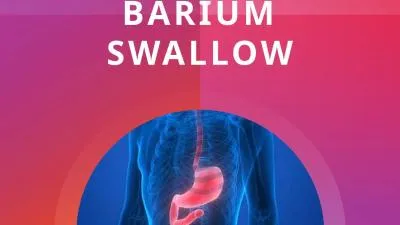
ASST PROF DR PURNACHANDRA LAMGHARE PROFESSOR Fluoroscopic imaging of the esophagus Evaluation of swallowing Also imaging of the oropharynx and of stomach to some extent Using Barium sulphate preparations ID: 912295
Download Presentation The PPT/PDF document "BARIUM SWALLOW DR. NILESH PANDIT" is the property of its rightful owner. Permission is granted to download and print the materials on this web site for personal, non-commercial use only, and to display it on your personal computer provided you do not modify the materials and that you retain all copyright notices contained in the materials. By downloading content from our website, you accept the terms of this agreement.
Slide1
BARIUM SWALLOW
DR. NILESH PANDIT
ASST PROF
DR PURNACHANDRA LAMGHARE
PROFESSOR
Slide2Fluoroscopic imaging of the esophagus.
Evaluation of swallowing.
Also imaging of the oropharynx and of stomach to some extent.
Using Barium
sulphate preparations.
Slide3SWALLOWING
Deglutition.
Passage of bolus from oral cavity to stomach via pharynx and esophagus.
Protection of airway.
Inhibition of air entry into the stomach.
Three phases – oral, pharyngeal, esophageal.
Esophageal phase – primary, secondary, tertiary waves.
Slide4TECHNIQUE
Slide5INDICATIONS
Slide6CONTRAINDICATIONS
Slide7MODIFICATIONS
Slide8ESOPHAGUS
25-40
cm long with a diameter of 1-2
cm
.
Four layers.
Three parts –
Cervical.
Thoracic.
Abdominal.
Two sphincters –
Upper – cricopharyngeus.
Lower – vestibule.
Three normal constrictions –
Cervical – cricoid cartilage.
Thoracic – aortic arch.
Abdominal
– diaphragmatic hiatus
.
Slide9Slide10Slide11SPHINCTERS
Upper esophageal –
Cricopharyngeal
muscle.
C5-C6 level
Relaxes with bolus
Lower esophageal –
Distal 2-4 cm high pressure zone – vestibule.
Prevents GER.
Can be mistaken for hiatal hernia.
Slide12Slide13Slide14Slide15Slide16Slide17Slide18PERISTALSIS
Primary –
Propels bolus.
Secondary –
Propels remaining food.
Tertiary –
Non propulsive.
Abnormal.
Diffuse esophageal spasm –
Intermittent contraction of mid & distal part.
Cork screw appearance.
Slide19Slide20Slide21Slide22ACHALASIA
Impaired relaxation of LES.
Gradually progressive dysphagia for both solid & liquids.
Chest pain, regurgitation.
Abnormal opacity with air fluid levels in mediastinum.
Bird beak sign, smooth tapering.
Pooling/stasis of barium in esophagus.
Slide23Slide24REFLUX ESOPHAGITIS
Most common inflammatory disease.
Reflux of gastric content due to dysfunction of LES.
h/o of episodic heartburn & regurgitation, worsening on lying down.
Double contrast esophagography.
Abnormal motility, mucosal nodularity, ulceration, thickened folds, scarring/ stricture.
Slide25Slide26WEB –
Hypopharynx & proximal esophagus.
Majority from anterior wall.
Symptomatic if lumen obstruction by 50%.
Plummer-Vinson syndrome.
Slide27Slide28SCHATZKI RINGS –
Lower esophageal ring.
Stenotic ring that causes dysphagia.
C.C. – episodes of severe chest pain, uncomfortable sensation in lower chest.
Ba swallow – thin, 2- 4 mm in height, luminal opening of less than 13 mm diameter.
Slide29DIVERTICULUM
Pulsion
– Mucosa & submucosa herniating through muscular layer.
Traction – All three layers.
Slide30Slide31Slide32ZENKER’S DIVERTICULUM
Posterior
hypopharyngeal
diverticulum.
Pulsion
type.
Through area of weakness between horizontal and oblique components of cricopharyngeus – Killian’s dehiscence.
Dysphagia, regurgitation of undigested food, hoarseness, halitosis, neck mass.
Air – fluid level in neck.
Posterior, midline bulge above cricopharyngeus.
Slide33Slide34Slide35KILLIAN-JAMIESON DIVERTICULUM –
Pulsion
type.
Below the
cricophyaryngeus
.
Lateral.
EPIPHRENIC DIVERTICULUM –
Pulsion
type.
AORTOPULMONARY WINDOW DIVERTICULUM –
Traction diverticulum.
Granulomatous disease.
Slide36Slide37Slide38HIATUS HERNIA
Sliding –
GE junction >2cm above hiatus.
>95 %
Paraesophageal
–
Fundus herniates through hiatus.
GE junction normal.
Mixed –
Slide39Slide40Slide41CANDIDA
Double contrast study
Longitudinally oriented Small plaques.
Fine granular appearance secondary to mucosal edema & inflammation.
HERPES –
Severe substernal chest pain during swallowing.
Discrete, superficial ulcers in
midesophagus
without plaques.
CMV
Giant ulcers.
ESOPHAGITIS
Slide42Slide43Slide44TBDirect extension from adjacent lesions.
Sinus
tracts.
Ulcers
.
EOSINOPHILIC
h/o allergies.
Dysphagia.
Diffuse narrowing, strictures, corrugated margins.
Slide45Slide46Slide47STRICTURES
Reflux esophagitis.
Barrett’s esophagus.
Corrosive poisoning.
Esophagitis.
Radiotherapy.
Benign pemphigoid.
GVHD.
Slide48Slide49BOERHAAVE SYNDROME
Rupture of wall – distal left posterolateral 1-4 cm, transmural/ full thickness tear.
Water soluble contrast.
Excessive vomiting in eating disorder, alcoholic binge, excessive coughing, trauma.
Pneumomediastinum.
Left pleural effusion.
Left pneumonthorax.
Extravasation of contrast material.
Slide50Slide51MALLORY-WEISS TEAR
Sudden increase in intra esophageal pressure.
Prolonged forceful vomiting.
Linear mucosal laceration.
Partial thickness tear of mucous membrane.
Hematemesis.
Diagnosis always by endoscopy.
Linear 1-4 cm longitudinal collection in distal esophagus.
Slide52Slide53Slide54SCLERODERMA
Collagen vascular disease.
Diffuse fibrosis.
75-85 % with esophageal involvement.
Aperistalsis due to disruption of smooth muscles including LES.
Dilatation with dysmotility of distal esophagus.
Apparent shortening due to fibrosis.
GER due to dysfunction of sphincter.
Slide55Slide56Slide57LEIOMYOMA
Most common benign esophageal neoplasm (50%).
Slowly progressive, often large but nonobstructive, calcified.
Distal third – 60 %
Asymptomatic, intermittent dysphagia, substernal pain.
Soft tissue mass in posterior mediastinum, punctate areas of calcification.
Smooth submucosal masses, round or ovoid filling defects, obtuse angle with esophageal wall.
At GE junction, can involve cardia & fundus, causes severe dysphagia.
Slide58A calcified esophageal mass
Is almost always
leiomyoma
Slide59Slide60Slide61Slide62CARCINOMA
Tobacco, alcohol – major risk factors.
Majority are squamous type.
Adeno CA – lower part, longer segments.
Symptomatic after 50-75% lumen obstructed.
Dysphagia, more to solids.
Widening of mediastinum, retrocardiac mass, widened azygoesophageal recess, tracheal deviation.
Infiltrating, irregular, polypoidal, ulcerative or varicoid mass.
Narrowing of lumen, shouldering.
Rat tail appearance.
Slide63Slide64Slide65VARICES
Slide66Slide67ABERRANT RIGHT SUBCLAVIAN ARTERY
Slide68Slide69DOUBLE AORTIC ARCH
P
ersistence
of R and L IV branchial arches
Passes on both sides of trachea
Joins posteriorly behind esophagus
Right arch is larger and higher
Left arch is smaller and lower
Barium swallow shows bilateral impressions on frontal
view and Posterior
impression on lateral view
Slide70Slide71COARCTATION OF AORTA
Slide72THANK YOU..!!!