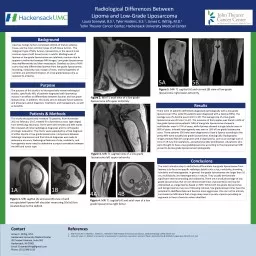
PDF-Figure 3 MRI T1W LS spine sagittal view Arrows show owing osteo

Author : delilah | Published Date : 2022-09-05

367 Figure 4 MRI T2W LS spine axial view Arrows show narrowing of neural foramen on right side and calci cation Forestier and Rotes described it as a disease of

Presentation Embed Code

Download Presentation

Download Presentation The PPT/PDF document "Figure 3 MRI T1W LS spine sagittal view ..." is the property of its rightful owner. Permission is granted to download and print the materials on this website for personal, non-commercial use only, and to display it on your personal computer provided you do not modify the materials and that you retain all copyright notices contained in the materials. By downloading content from our website, you accept the terms of this agreement.

Figure 3 MRI T1W LS spine sagittal view Arrows show owing osteo: Transcript


367 Figure 4 MRI T2W LS spine axial view Arrows show narrowing of neural foramen on right side and calci cation Forestier and Rotes described it as a disease of the spine developing in old peopl. SPINE TRAUMA . CERVICAL SPINE INJURY. THORACO-LUMBAR SPINE INJURY. CERVICAL SPINE INJURY. COMMON MECHANISMS OF INJURY. . HYPERFLEXION- . MVA, CAR COMES TO SUDDEN STOP. HYPEREXTENSION- MVA, CAR STRUCK FROM BEHIND. Note what you are looking at (e.g. AP view of the R shoulder girdle). Differentiate air, fat, soft tissue & fluid, and bone.. Obtain a copy of the radiology report & integrate it with the info from your PT examination in order to formulate your PT . in Ghana . through July. , 2014. If this . surgery had been done . in the USA, it would . have cost . $50 million dollars! . Our costs . in Ghana . are significantly less than. . 10% of that.. The Department of Pathology receives biopsies and reviews cases from Dr. . Sagittal alignment of the spine is changing! Evolution theories Life cycles Sagittal alignment P lanning of the 3D reconstruction of the spine N ormative data about the sagittal plane Segmental Sagittal Spinopelvic Parameters of Children with Early Onset Scoliosis Ron El-Hawary, Peter Sturm , Jason Howard, Krystyna Cowan Patrick Cahill, Amer Samdani, Michael Vitale, Peter Gabos, Nathan Bodin, Charles Cyrus . Wong, M.D.. Department of Neurological Surgery. Vanderbilt University Medical Center. Plain . Xray. Historically used to diagnose all types of pathology related to the spine. It relies on ionizing radiation to provide images. Elana. B. Smith, MD. Kimberly Gardner, MD. Kiran. S. . Talekar. , MD. Adam E. Flanders, MD. Adam C. Zoga, MD. Correspondence can be addressed to:. Elana. B. Smith, MD. Thomas Jefferson University Hospital. Rev. 6/13 72090 – X - Ray trunk spine scoliosis study 72100 – X - Ray lower spine 2 or 3 view 72110 – X - Ray scapula 72202 – X - Ray sacroiliac joints 3 or more views 72220 – X - Ra Cervical Exercise: The Backbone of Spine How important is it?What can be done? Important: If you have had an accident that started your neck pain or if you have pain, numbness, tingling or weakness i Volume 1 , Issue 1 SYNOSTOSIS Andrew Reisner , M.D., William R. B oydston, M.D., Ph.D., Barun Brahma, M.D., Joshua Chern, M.D., Ph.D., David Wrubel, M.D. Craniosynostosis, an early closure of t Brain Necrosis 1 2 Brain Necrosis 4 Brain Necrosis Brain Necrosis retrosplenial cortex (white arrow). Selective vulnerability of brain regions to the effects of toxic compounds may imitate the lesions Tăbăran F, Sándor A, Marinov M, Cătoi C, Mihalca A. Alaria alata Infection in European Mink. Emerg Infect Dis. 2013;19(9):1547-1549. https://doi.org/10.3201/eid1909.130081. Bonthius DJ, Stanek N, Grose C. Subacute Sclerosing Panencephalitis, a Measles Complication, in an Internationally Adopted Child. Emerg Infect Dis. 2000;6(4):377-381. https://doi.org/10.3201/eid0604.000409. Maheshwari A, Fischer M, Gambetti P, Parker A, Ram A, Soto C, et al. Recent US Case of Variant Creutzfeldt-Jakob Disease—Global Implications. Emerg Infect Dis. 2015;21(5):750-759. https://doi.org/10.3201/eid2105.142017.

Download Document

Here is the link to download the presentation.
"Figure 3 MRI T1W LS spine sagittal view Arrows show owing osteo"The content belongs to its owner. You may download and print it for personal use, without modification, and keep all copyright notices. By downloading, you agree to these terms.

Related Documents