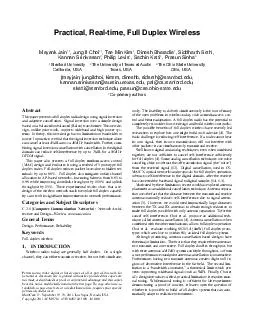
/
Seungyeon   Choi a ,  Sunghoon Seungyeon   Choi a ,  Sunghoon

Seungyeon Choi a , Sunghoon - PowerPoint Presentation

eatfuzzy
eatfuzzy . @eatfuzzy
Follow
342 views
Uploaded On 2020-08-26

Seungyeon Choi a , Sunghoon - PPT Presentation

Choi b Donghoon Lee a Young Wook Choi c and HeeJoung Kim a b a Department of Radiation Convergence Engineering College of Health Science Yonsei University ID: 802527

amp observer model iworid observer amp iworid model obs breast nps rois angular images study results digital signal tomosynthesis

Share:

Link:

Embed:

Download Presentation from below link

Download The PPT/PDF document "Seungyeon Choi a , Sunghoon" is the property of its rightful owner. Permission is granted to download and print the materials on this web site for personal, non-commercial use only, and to display it on your personal computer provided you do not modify the materials and that you retain all copyright notices contained in the materials. By downloading content from our website, you accept the terms of this agreement.


Presentation Transcript

Slide1

Seungyeon

Choia, Sunghoon Choib, Donghoon Leea, Young-Wook Choic,and Hee-Joung Kima, b*a Department of Radiation Convergence Engineering, College of Health Science, Yonsei University, Wonju, Koreab Department of Radiological Science, College of Health Science, Yonsei University, Wonju, Koreac Korea Electrotechnology Research Institute (KERI), Ansan, 15588, Korea

Investigation of model observer and human observer performance of a prototype digital breast tomosynthesis

IWORID 2019

(

Medical Physics & Imaging Lab.)

The 21

st

International Workshop on Radiation Imaging Detectors07-12 July 2019 in Kolympari, Greece

(

Yonsei University)

Slide2

Introduction

IWORID

2019DM is a widely installed device in the hospital (85%), which has a high recall rate due to anatomical superposition.DBT equipment: introduced since the early 2000s, received attention since it began receiving FDA approvalThe process of reading and analyzing many images requires training → time and money consumingITN, Finding Value in Digital Breast Tomosynthesis, 2018

DM

(Digital mammography) & DBT (Digital breast tomosyntheis)Currently three FDA approved DBT

For a fixed radiation dose, optimize:

Projection arcNumber of projectionsReconstruction filtersSame Task, Different Designs

Stephen Glick, ‘Digital Breast Tomosynthesis, VCT Research at the FDA’, U.S. FDA, 2016 AAPM

Vedantham et al., ‘Digital Breast Tomosynthesis

: State of the Art’, Radiology, 2015 No standard DBT system (different detectors, acquisition geometries etc.).

Hologic

GE

Siemens

FDA approved system

Hologic

Selenia

Dimensions

GE

Senoclaire

Siemens

Mammomat

Inspiration

Year of approval

2011

2014

2015Total angle (arc)152450# of projection views15925

Slide3

IWORID

2019IntroductionNon-clinical, physics based phantom testingMTF, NPS, DQE, SNR or CNRUse simple unrealistic phantoms, might not apply for non-linear reconstruction, don’t evaluate objective task performanceEvaluation of Breast image qualityClinical TrialMultiple breast radiologists reading large number of clinical casesRigorous multi-reader, multi-case ROC analysisExpensive, time-consuming, and radiation riskVirtual Clinical TrialImplement before clinical adaptation and regularization approvalTo investigate model observer and human observer performance according to angular range in

a prototype digital breast tomosynthesis

Slide4

Spheroidal masses (Breast carcinoma)

# of tasks

3Sizes of tasks3.9, 4.7. 6.3 mmIWORID 2019

Materials & Methods

Two tissue-equivalent materials mimicked

100

% adipose and 100% gland tissues “swirled” together in an approximate 50/50 ratio by weightTarget

Inhomogeneous breast phantom

Slide5

IWORID

2019Materials & MethodsImage acquisitionImages acquired with 5 different scanning conditions. Acq. Parameter #1#2#3#4

#5

Angular range

#

of

proj

.

15

15

15

15

15


Acq

.

Parameter

#1

#2

#3

#4

#5

Angular

range

#

of

proj

.

15

15

15

15

15

Region of interest extraction

Same ROIs were used for various scanning schemes

ROI size of 12*12

3

signal present ROIs

9

signal absent

ROIs


Used for training data

Signal absent ROIs

Signal present ROIs

Prototype DBT system developed by

Korea

Electrotechnology

Research Institute

(

KERI)

was used with FBP reconstruction.

Slide6

IWORID

2019Materials & MethodsNon-prewhitening (NPW) model observers3D detectabilityThe observer is assumed to fully perceive the volumetric image informationSlice detectabilityTo adapt coronal slice reading in a tomosynthesis, d’ was derived in a form pertaining to a single 2D slice extracted from the volume.


Weighted sum for all Fourier direction

Extract slice information first, then apply weighted sum for in-plane Fourier direction


Coronal slice that want to observe

Slide7

IWORID

2019Materials & MethodsTask function (WTask)Imaging tasks were modeled according to a simple binary hypothesis-testing.“Signal-present (H1)” images were taken from the central coronal slice through each object and ROIs were selected such that signals were at the center.

“Noise-only/signal-absent (H

2

)”

images were taken from the same or neighboring slices without the signal.

,

if

=

(sphere detection)

(sphere detection

)


Object function

(

)


Intensity of mass

and background

Mass ROI image

Non-

prewhitening

(NPW) model observers

Slide8

IWORID

2019Materials & MethodsQuantum & electronic noise (SQ + SE) – local NPSConventional NPS measurements frequently involve computing the NPS over multiple ROIs (or VOIs) within an image and averaging the outcome. Noise was characterized by the local NPS within a ROI at a fixed location, i.e., instead of marching the ROI through the image, NPS was measured at the same location in a large ensemble of reconstructions and then averaged.

⋮⋱

Conventional NPS

Marching multiple sub-images

local area

Multiple acquisitions for exactly same position b/w scans

(subtraction)

Leaving only Q+E noise

local

NPS

Non-

prewhitening

(NPW) model observers

In this work, noise measurements were performed under the assumption of

local

stationarity

(i.e.,

within the ROI itself

, but not necessarily throughout the image).

This method invokes

stationarity

assumptions both

locally within the ROI

and globally over the ensemble of ROIs contributing to the NPS estimate.

Slide9

IWORID

2019Materials & MethodsTransfer function (T) – local MTFLocal impulse response was calculated, by injecting an impulse of 𝜕 into the voxel of lesion located ROI. No noise was added to the simulated projection following the forward model.Inject an impulse of magnitude into voxel j Divide by

and DFT of differenced image


Forward projection

and reconstruction


Non-

prewhitening (NPW) model observers


Slide10

IWORID

2019Materials & MethodsHuman observer study4-AFC study facilitated with an in-house made tool7 observers who have radiological background were participatedA session consisted of 4 training and 24 4-AFC testsObserver performance represented by the average percentage correct Prior to each test, observers were trained using images acquired at the same experimental conditions as the test data, requiring few minutes to gain familiarity with the task.Constant viewing distance of

~50 cm was encouraged.The complete study required

(3 tasks) × (~1 min) (training) + (3 tasks) × (4 schemes) × (~5s) (test)

Slide11

IWORID

2019Results & DiscussionsTest images randomly positioned

Slide12

IWORID

2019Results & DiscussionsAnswer for the test images (6.3 mm, 4.7 mm and 3.9 mm spheroidal masses),randomly positioned

Slide13

IWORID

2019Results & Discussions600400200300200100

120

80

40

200150

50

100

30

20

10

25

15

5

35

25

15

40

30

20

0.3

0.2

0.1

0.35

0.25

0.15

0.05

0.35

0.25

0.15

0.05

0.35

0.25

0.15

0.05


Task function

l

ocal MTF

l

ocal NPS

Spatial frequency (

)


Model observer results

Slide14

IWORID

2019Results & Discussions Std=

Obs. 1

0.92

0.26

Obs. 2

0.71

0.45

Obs. 3

0.89

0.31

0.86

0.35

Obs. 5

0.92

0.26

Obs. 6

0.89

0.31

Obs. 7

0.92

0.26

Average

0.87


Obs. 1

0.92

0.26

Obs. 2

0.71

0.45

Obs. 3

0.89

0.31

0.86

0.35

Obs. 5

0.92

0.26

Obs. 6

0.89

0.31

Obs. 7

0.92

0.26

Average

0.87

Recommended

value range:

0.7 ~ 0.8

In

the human observer study, the average PC from seven observers were

0.87, ranging PC values from 0.71 to 0.92.

Reproducibility

estimated by the standard deviation of the least variable observer


Human observer results

Slide15

IWORID

2019Results & DiscussionsThe performance of the theoretical model observer values resulted in similar trend to the human observers’ PC results.The resulted patterns of PC decreased with increasing the angular ranges from ±10.5° to ±24.5° with different size of tasks.(A)(B)Detectability index (d’) results from modeling observer (A), results from 4AFC (B) with different angular distribution. 

Slide16

IWORID

2019ConclusionIn this study, we focused on the 4-AFC human observer study and NPWE model observer study for detecting mass in the prototype breast tomosynthesis system.For evaluation of angular distribution, projection images were acquired with angular range of ±10.5°, ±14°, ±21°, ±24.5°, with equally 15 projection views.Our results showed that the NPWE model could reasonably predict mass detectability from small to large sizes for different angular ranges. The correlation between theoretical and measured performance is necessary for better description of task-based model observer performance for future study.Incorrect conclusions could be drawn if the observer results are inconsistent under similar/identical conditions: the need of bootstrap sample study.

Slide17

M

edical

Physics & Imaging LaboratoryThanks for your attention

Presenter’s e-mail address :

sychoi324

@yonsei.ac.kr