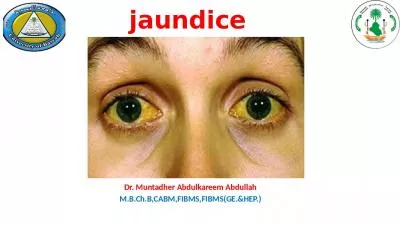
/
Case Report 3 year old male child presented with jaundice Asince 1 mon Case Report 3 year old male child presented with jaundice Asince 1 mon

Case Report 3 year old male child presented with jaundice Asince 1 mon - PDF document

edolie
edolie . @edolie
Follow
342 views
Uploaded On 2022-10-27

Case Report 3 year old male child presented with jaundice Asince 1 mon - PPT Presentation

Abdominal examination revealed abdominal distension with hepatomegaly2 cm with a span of 7 soft splenomegaly5 cm Cardiovascular system examination revealed a pansystolic murmur more prominent al ID: 961032

paucity bile ags syndrome bile paucity syndrome ags drug liver cholestasis revealed patients syndromic duct normal disease ducts alagille

Share:

Link:

Embed:

Download Presentation from below link

Download Pdf The PPT/PDF document "Case Report 3 year old male child presen..." is the property of its rightful owner. Permission is granted to download and print the materials on this web site for personal, non-commercial use only, and to display it on your personal computer provided you do not modify the materials and that you retain all copyright notices contained in the materials. By downloading content from our website, you accept the terms of this agreement.


Presentation Transcript

Case Report 3 year old male child presented with jaundice Asince 1 month of age, abdominal distension since 1 year & pruritus. The child had global developmental delay. His antenatal and family history was non-contributory.General examination revealed mild pallor, icterus, clubbing with dystrophic nails. He had facial dysmorphism with deep set eyes, hypertelorism and chin. Cutaneous examination revealed multiple dark maculopapular lesions inolving both upper and lower limbs. On admission his weight, height and head circumference were below the 3rd centile for his age on WHO growth charts indicating growth retardation.Fig. 1: Characteristic facial features. Deep set eyes, hypertelorism, pointed chin. Abdominal examination revealed abdominal distension with hepatomegaly(2 cm with a span of 7 soft) splenomegaly(5 cm). Cardiovascular system examination revealed a pansystolic murmur, more prominent along the lower left sternal border.Table 1: InvestigationsInvestigations revealed anaemia, direct hyperbilirubinaemia, elevated serum transaminases, normal bleeding and clotting time. Serum cholesterol and triglycerides were mildly elevated. The renal parameters and blood sugars were within normal limits. The viral markers for hepatitis, urine for Bombay Hospital Journal, Vol. 57, No. 2, 2015 190 *Paediatrics, Dept. of Paediatrics, **Gastroenterology, Dept. of Gastroenterology, TNMC and BYL Nair Charitable Hospital, Mumbai. AbstractAlagille syndrome (AGS) is a rare genetic disorder with autosomal dominant inheritance associated with abnormalities of liver, heart, eye, skeleton, and characteristic facial appearance. In the fully expressed syndrome, affected subjects have intrahepatic bile duct paucity and cholestasis, with cardiac malformations (peripheral pulmonary stenosis), ophthalmological abnormalities (posterior embryotoxon being the most common), skeletal anomalies (most commonly butterfly vertebrae), and characteristic facial appearance. AGS, syndromic bile duct paucity or arteriohepatic dysplasia initially described by Alagille et al in 1969. The prevalence of this syndrome has been reported as 1 in 100 000 live births. Alagille Syndrome: Syndromic Paucity of Bile Ducts, a Fully Expressed Syndrome Sujay K. Earan*, Radha Gulati Ghildiyal*, Punam Sankhe*, Pravin M Rathi**, Jane David* Investigations Result Normal rangeHaemoglobin 8 gm/dl10.5-14 gm/dlTotal bilirubin8 mg%0-1mg%Direct bilirubin4.1 mg%0-0.3mg%Alkaline phosphatase429 U/L0-280U/LSGOT196 U/L0-40 U/LSGPT118 U/L0-40U/LProthrombin time12 sec12 secProthrombin index91.6 %100% INR1.21-1.2Serum cholesterol175 mg%150-250 mg%Triglycerides195 mg%Upto 170 mg%Urine reducing substancesNegative Plasma aminoacidogramNormal Urine aminoacidogramNormal reducing substances were negative. His anti Liver Kidney Microsomal 1 antibodies, anti smooth muscle antibodies and anti nuclear antibodies w

ere negative. Alpha 1 antitrypsin levels were also normal.Ultrasonography of the abdomen suggested a normal echotexture with hepatosplenomegaly, abdominal Doppler and oesophagoduodenoscopy were normal. Magnetic resonance cholangiopancreatography was done to rule out extra hepatic biliary obstruction, was normal. Echocardiography showed a peripheral pulmonary artery stenosis. Ophthalmological examination revealed bilateral posterior embryotoxon. Skeletal radiographs revealed absent coccyx and spina bifida involving L5 vertebra. Punch biopsy of the cutaneous papular lesions showed hyperkeratotic stratum corneum with plugging of follicular infundibulum consisting of parakeratotic plugs, suggestive of phrynoderma.Fig. 2: X ray spine lateral view showing absent coccyx.Fig. 3: X ray spine AP view showing spina bifida at L5Liver biopsy revealed, paucity of bile ducts and near total absence of interlobular bile ductules. Intrahepatic cholestasis and xanthomatous changes especially hepatocytes close to interlobular septae were seen. The paucity of bile ducts was confirmed on Masson's Trichrome staining.Fig. 4: Histopathology of liver biopsyHME 10x Portal area with paucity of inter lobular ducts. Swollen hepatocytes with mild cholestasis. Hepatocytes with xanthomatous changeDiscussionAlagille syndrome (AGS) is a rare genetic disorder with autosomal dominant inheritance associated with abnormalities of liver, heart, eye, skeleton, and characteristic facial appearance. AGS, syndromic bile duct paucity or arteriohepatic dysplasia initially described by Alagille et al in 1969. The prevalence of this syndrome has been reported as 1 in 1100000 live births. The diagnostic criteria of AGS include a histological paucity in the bile duct, in association with 3 of the following 5 major clinical features: (1) chronic cholestasis; (2) congenital cardiac disease; (3) skeletal malformation (butterfly vertebrae); (4) ocular posterior embryotoxon; and (5) characteristic facies. Other features include mental and growth retardation, renal disease, intracranial bleeding, endocrine abnormalities, anddermatologic manifestations.The mutant gene has been localised to chromosome 20p. AGS is caused by mutation or deletion of gene encoding Bombay Hospital Journal, Vol. 57, No. 2, 2015 191 Jagged 1(JAG 1), a ligand involved in the Notch signalling pathway.The majority of symptomatic patients present in infancy with manifestations of hepatic disease ranging from mild cholestasis and pruritus to progressive liver failure. Cholestasis manifests by pruritus, raised serum bile acid concentrations, xanthomas, and growth failure. Liver biopsy classically shows intrahepatic bile duct paucity. Depending the timing of the biopsy, there may be portal fibrosis and rarely, bile duct proliferation. In a Bangladesh study in cholestatic jaundice in infancy, 22 of the 62 infants had ne

onatal hepatitis, of the 8 suspected neonatal hepatitis patients biopsied one patient had paucity of intrahepatic bile ducts without any 2external features.Congenital heart disease has been reported in up to 90% of patients with AGS. The most common feature being peripheral pulmonary artery stenosis. The most common complex cardiac malformation associated with pulmonary involvement in these patients is Tetralogy 3,4of Fallot (7-10%). Other cardiac defects seen in AGS, in order of decreasing frequency, include ventricular septal defects, atrial septal defects, aortic stenosis, and coarctation of aorta. Ophthalmological findings in AGS include defects of the anterior chamber (posterior embryotoxon, Axenfeld's anomaly, or Rieger anomaly), and retinal pigmentary 3,4changes. Posterior embryotoxon has been reported in up to 89% of patients. The most common skeletal abnormality in AGS is butterfly vertebrae. Other skeletal anomalies include narrowing of interpeduncular spaces in the lumbar spine (50%), pointed anterior process of C1, spina bifida occulta, hemivertebrae, bony connections between ribs, and short 1,3,4,5fingers.In 1990 Nigale et al reported a case of AGS in a 5 year old female with disseminated skin lesions and persistent neonatal jaundice without any characteristic ophthalmic or bony 6lesions. In 2002 Harshalee Shengde et al reported a case of partial alagille syndrome; the child had three of five major 7features. A Abdel Moniem Deghady et al in their study in Alexandria found that, the incidence of non-syndromic bile duct paucity was 2.8%, while syndromic bile 8duct paucity (AGS) was not found. In a report by Yaccha et al based on the pooled data from eight medical centres in India, among 1008 analysed cases of neonatal cholestasis syndrome, ductal paucity was in 3%, and ductal paucity was due to 9non syndromic variety in 83%. Thus emphasising the rarity of our case in the community.The treatment is essentially symptomatic with substitution of fat soluble vitamins, medium chain triglycerides for failure to thrive and treatment of pruritus. The treatment is aimed at increasing the flow of the bile from the liver using ursodeoxycholic acid. Partial External biliary diversion procedure can be done to provide relief from refractory pruritus. Orthotopic Liver Transplantation should be considered for severe forms, in particular intractable pruritus or severe growth retardation. Bombay Hospital Journal, Vol. 57, No. 2, 2015 192 Patients with significant intracardiac disease require subacute bacterial endocarditis (SBE) prophylaxis.Estimated 20-year survival rates are 80% for those not requiring liver transplantation and 60% for those requiring transplantation. The 20-year predicted survival rate via Kaplan-Meier plots for individuals with significant intracardiac lesions is 40%; those individuals without significant intracardiac

lesions, the survival rate is 80%.References1.Danks DM, Campbell PE, Jack I, etal: Studies of the aetiology of neonatal hepatitis and biliary atresia. Arch Dis Child 1977;52:360-7.2.A S M Bazlul Karim, M Kamal: Cholestatic jaundice during infancy: experience at a tertiary-care center in Bangladesh. Indian J Gastroenterology 2005; 24:52-54.3.Alagille D, Estrada A, Hadchouel M, et al: Syndromic paucity of interlobular bile ducts. J Pediatr 1987;110:195-200.4.Deprettere A, Portman B, Mowat A: Syndromic paucity of the intrahepatic bile ducts: diagnostic difficulty; severe morbidity throughout early childhood. J Pediatr Gastroenterol Nutr 1987; 6: 865-71.5.Rosenfield NS, Kelley MJ, Jensen PS, et al: Arteriohepatic dysplasia: radiologic features of a new syndrome. AJR 1980;135: 1217-236.Nigale V, Tarsi SS, Khopkar US et al: Alagille syndrome a case report. Acta Derm Venerol 1990; 70(6):521-37.Harshalee Shengde, Milind S.Tullu, Asha Shenoy et al: Alagille syndrome. Indian J Pediatr 2002; 69(9): 825-8278.A. Abdel Moniem Deghady, M. Abdel-Fattah, M. Abdel-Kader et al: Diagnostic evaluation of cholestasis in infants and young children in Alexandria. The Internet Journal of Pediatrics and Neonatology. 2006;6(1)9.Yaccha et al: Consensus report on Neonatal cholestasis syndrome. Pediatric Gastroenterology subspeciality chapter of Indian Academy of Pediatrics. Indian Pediatr 2003; 37:845-51 Bombay Hospital Journal, Vol. 57, No. 2, 2015 193 Guidelines, polypharmacy, multimorbidity, and drug-drug interactions A cascade of failureOne common consequence of polypharmacy is the high rate of adverse drug reactions, mainly from drug-drug interactions (the ability of a drug to modify the action or effect of another drug administered successively or simultaneously).Recent guidelines for chronic illness such as chronic obstructive pulmonary disease (COPD), heart failure, and diabetes mellitus have attempted to account for likely comorbidities. Unfortunately, these guidelines consider only one comorbid disease at a time and provide few specific recommendations about how to manage people with multiple comorbidities.Older patients are particularly vulnerable. Prevalence of multimorbidity increases with age, along with the prevalence of polypharmacy.Drugs can also interact with genes, and we have barely touched on the potential for an additive effect between drug-drug interactions and genetic factors. Clopidogrel is a good example of a drug with a genetically variable therapeutic effect. If a genetically poor metaboliser (with lower bioavailability of active drug) is co-prescribed a proton pump inhibitor, which further reduces the therapeutic effect of clopidogrel, vascular risk increases in an unpredictable way.More research is needed to make guidelines effective and safe for patients with multimorbidity.Alessandra Marengoni, Graziano Onder, BMJ, 2015, Vol 350,