/
Surgical Jaundice By Dr. Ahmed Surgical Jaundice By Dr. Ahmed

Surgical Jaundice By Dr. Ahmed - PowerPoint Presentation

elise
elise . @elise
Follow
64 views
Uploaded On 2024-01-29

Surgical Jaundice By Dr. Ahmed - PPT Presentation

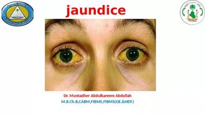
Rashidy Lecturer of General and Pediatric Surgery Jaundice Yellowish discoloration of body tissues amp fluids except the brain CSF tears saliva amp milk due to excess bilirubin in the blood ID: 1043191

bile jaundice bilirubin biliary jaundice bile biliary bilirubin obstructive cbd liver amp stone due duct intrahepatic haemolytic blood ducts

Share:

Link:

Embed:

Download Presentation from below link

Download Presentation The PPT/PDF document "Surgical Jaundice By Dr. Ahmed" is the property of its rightful owner. Permission is granted to download and print the materials on this web site for personal, non-commercial use only, and to display it on your personal computer provided you do not modify the materials and that you retain all copyright notices contained in the materials. By downloading content from our website, you accept the terms of this agreement.


Presentation Transcript

1. Surgical JaundiceByDr. Ahmed RashidyLecturer of General and Pediatric Surgery

2. JaundiceYellowish discoloration of body tissues & fluids (except the brain, CSF, tears, saliva & milk) due to excess bilirubin in the blood.Normal level of bilirubin 0.2 - 0.7 mg %.Becomes manifest when > 2.5 mg %.Biliruin metabolism:1- RBCs destruction old red blood corpuscles are phagocytosed by the reticulo-endothelial cells all over the body but particularly in the bone marrow and spleen.2- Break down of hemoglobin the liberated Hb undergoes breakdown into globin and haeme3- - Globin enters into the amono acid pool - heme is transformed into biliverdin which is reduced to bilirubin the latter being water insoluble, is transported in plasma bound to albumin and constitutes unconjugated bilirubin.

3. 4- Bilirubin is conjugated with glucuronic acid to form bilirubin diglucuronide (conjugated bilirubin) which is water soluble. - Conjugation of bilirubin in the liver is catalyzed by the bilirubin glucuronyl transferase enzyme.5-Conjugated bilirubin is excreted via the biliary passages to the intestine.6- Conjugated bilirubin is converted by bacterial enzymes to stercobilinogen In the terminal ileum and colon which is oxidized to stercobilin responsible for the normal colour of stool. - Up to 20% of stercobilinogen is reabsorbed and 90% of this is promptly reexcreted by the liver, with much of the remainder appearing in urine as urobilinogen.

4.

5. Types and EtiologyAccording to the etiology of excess bilirubin, three types of jaundice are recognized.1- Haemolytic or (prehepatic jaundice) is due to excessive destruction of red blood corpuscles which occurs in the various types of hemolysis. Unconjugated bilirubin is elevated. Fecal stercobilinogen and urinary urobilinogen are increased. Bile salts don’t accumulate in the serum. Causes of hemolytic anaemia:1- Hemolytic anemia.2- Erythroblastosis fetalis.3- RBCs destruction by :- Poison e.g. snake venom - lnfections e.g. malaria & septicemia

6. 2- Hepatocellular or (hepatic jaundice)It is due to liver dysfunction and inability to transform unconjugated bilirubin to the conjugated variety. In addition, due to the presence of intrahepatic cholestasis, some conjugated bilirubin is reabsorbed to the blood. Blood levels of both conjugated and unconjugated bilirubin are elevated. Bile salts levels may also rise. Causes of hepatocellular jaundice includeA- Viral hepatitis B- Decompensated liver cirrhosis. Bacterial infections as septicaemia and pyaemia.C- Drugs including phenothiazines compounds. Certain diuretics, testesterone derivatives, oral contraceptives, oral antidiabetic agents, halothane and arsenicals. - are known to injure the cellular excretion mechanism and cause intrahepatic cholestasis.

7. 3- Obstuctive or post-hepatic jaundice is due to obstruction somewhere in the biliary pathway. Conjugated bilirubin will be reabsorbed to the blood and will be excreted in urine. Bile salts will accumulate in the blood. Faecal stercobilinogen and urinary urobilinogen levels are low. Causes of obstructive jaundice include:A- Causes in the lumen of the bile ducts:(1) Calcular obstructive jaundice :- Stone comes from the gall bladder (15% of patients with calcular cholecystitis have stones in the CBD)- Rarely primary in the CBD due to stricture.(2) Parasites as ascaris or fasciola

8. B- Causes in the wall of the bile ducts (stricture) which include1- Congenital biliary atresia, choledochal cyst2- Inflammatory stricure as with - Sclerosing cholangitis - Secondary to an impacted stone which has been present for along time3-Traumatic stricture is usually iatrogenic following cholecystectomy, choledocholithotomy or instrumental procedure on the common bile duct.4- Malignant stricure (cholangiocarcinoma)C- Causes outside bile ducts e.g., - Carcinoma of the head of pancreas or periampullary carcinoma obstructing the lower part of the common bile duct - Mass of metastatic lymph nodes at the porta hepatis obstructing the hepatic ducts.

9. Clinical features:HistoryThe age and sex of the patient may give a hint to the diagnosis. - Jaundice in the neonatal period may be due to physiological jaundice of the newborn, Rh incompatibility, neonatal hepatitis, congenital syphilis or congenital biliary atresia. - In children haemolytic anaemia should be suspected. - Calcular jaundice is more common in middle age females while malignant obstructive jaundice is commonly seen in elderly males.History of drug intake, recent injection and blood transfusion.Colour of urine and stools. - In haemolytic jaundice urine colour is normal while stool is dark. - In obstructive and hepatocellular jaundice, urine is dark while stool is pale.Pruritis is absent in haemolytic jaundice

10. Recurrent attacks of haemolytic crisis suggest haemolytic jaundiceDepth of jaundice. - Haemolytic jaundice is usually of a mild degree - Hepatocellular and obstructive jaundice may be of a severe degree.- Calcular jaundice is usually fluctuant while malignant jaundice is progressive.Pain, - In haemolytic jaundice abdominal pain may be present during a haemolytic crisis. - Calcular obstructive jaundice is usually accompanied by upper abdominal pain. - Malignant obstructive jaundice is usually painless but may be accompanied by dull aching epigastric pain referred to the back

11. ExaminationGeneral examination focuses on pallor, pyrexia, the degree of jaundice, virchow’s lymph nodes, cachexia, stigmata of liver insufficiency, bleeding tendency and edema of the lower limb.Abdominal examination. - Patients with haemolytic anaemia usually have hepatomegaly and splenomegaly. - In calcular obstructive jaundice abdominal examination is usually negative. - In malignant obstructive jaundice the liver and gall bladder are commonly palpable. Ascitis may be present in advanced cirrhosis or in metastatic malignant lesions.

12. Investigations(A) Laboratory1- Blood picture, in haemolytic jaundice there is anaemia. Special testes for various haemolytic anaemias may be required.2- Serum bilirubin jaundice easily recognizable when the conjugated bilirubin concentation in the serum reaches 2-3 mg/ 100ml or the unconjugated bilirubin level is 3-4 mg/100ml. - In haemolytic jaundice the unconjugated bilirubin level is increased- In obstructive jaundice the conjugated biliribin is high- In hepatocellular jaundice both types of bilirubin are increased (biphasic).3- SGOT(AST) and SGPT(ALT) - In haemolytic jaundice the level of transaminases will be normal. - In acute hepatitis the level will be very high, while in cirrhosis there will be moderate increase - In the early stages of obstructive jaundice the level will be normal but with prolonged obstruction, some degree of parenchymatous damage occurs and the level of theses enzymes will increase.

13. 4- Alkaline phosphatase, - In haemolytic jaundice the level is normal- In hepatocellular jaundice there may be slight increase, - In obstructive jaundice there is moderate increase in the level of this enzyme particularly in malignant obstruction.5- Prothrombin time and concentration (PT&PC) - Are normal in haemolytic jaundice. There is prolongation of prothrombin time and diminished prothrombin concentration in both hepatocellular and obstructive jaundice. To differentiate both conditions, IV vitamin k is prescribed for few days. In obstructive jaundice the prothrombin parameters will improve but not in hepatocellular jaundice.6- Serum albumin is low in patients with cirrhosis or after prolonged malignant cachexia.7- Fecal stercobilinogen is high in haemolytic jaundice, but is low in hepatocellular and obstructive jaundice.

14. (B) Imaging1- Abdominal ultrasound, this is the first investigation. Advantages: Easy Simple. Inexpensive, non invasiveValue:Detection of gall bladder stones in a 95% of cases. less accurate in detection of CBD stoneDetection of the thickness of the wall of the gall bladder.Visualization of intrahepatic and extrahepatic biliary radicles.Estimation of the diameter of CBD.Detection of masses in the porta hepatis or the head of pancreas.In obstructive jaundice- Dilatation of intrahepatic biliary radicles- Increased CBD diameter (normally 4-7 mm)- Presence of stone in CBD, mass of head of pancreas- Chronic inflamed GB with stones

15. 2- CT scan & MRI is particularly indicated in patients suspected having abdominal malignancy e.g., carcinoma of the head of the pancreas. - It will clearly demonstrate the site and extent of the tumour, invasion of adjacent strucures, metastatic lymph nodes or liver metastases.3- ERCP is particulrly indicated in patients with obstructive jaundice suspected of having a lesion involving the lower end of the common bile duct, e.g., ampullary carcinoma.4- PTC is particularly advised in patients with obstructive jaundice suspected of having a lesion involving the upper end of the common bile duct, e.g., postoperative stricture or carcinoma of the hepatic ducts.

16. 2- CT scan & MRI is particularly indicated in patients suspected having abdominal malignancy e.g., carcinoma of the head of the pancreas. - It will clearly demonstrate the site and extent of the tumour, invasion of adjacent strucures, metastatic lymph nodes or liver metastases.3- ERCP (Endoscopic retrograde cholangiopancreatography) Value: Diagnostic and therapeutic role. Diagnostic:Lesion involving the CBD.Detection of calculi in the CBD.Detection of operative injuries of the biliary system.Visualization of pancreatic duct.

17. Therapeutic:Sphinctrotomy (drainage).Extraction of stone.Dilatation & Stenting.Biopsy.4- MRCP (magnetic resonant cholangiopancreatography):This is non invasive procedure produce the same diagnostic information as can be obtained with ERCP but without important risk of complication. MRCP is now becoming the standard technique for investigation of the biliary tree.Disadvantage: Expensive, no therapeutic role

18. 4- PTC (percutaneous transhepatic cholangiography): is particularly advised in patients with obstructive jaundice suspected of having a lesion involving the upper end of the common bile duct, e.g., postoperative stricture or carcinoma of the hepatic ducts. Prerequisites:1- PT and concentration should be done before the procedure if prolonged vitamin K is given I.V for a few days before the procedure.2- Dilated intrahepatic biliary radicles as seen on U/S.Complications:1- Liver injury and hemorrhage  hemobilia or hemoperitoneum.2- Sepsis.3- Biliary peritonitis.

19. Method:1. Under local anaesthesia and U/S intrahepatic biliary passage is punctured in the midaxillary line through 8th intercostal space by Chiba needle Catheter is passed (better to be done on dilated intra hepatic biliary ducts).2. lnjection of radio-opaque material followed by X-Ray films.3. The procedure is done under floroscopic guidance and mild sedation. Antibiotic coverage is recommended.Diagnostic value:1- Particularly indicated to diagnose high obstruction of bile ducts.2- Visualization of the extrahepatic & intrahepatic biliary system.3- It is done if the ERCP fails the to give enough data about obstructing agent.

20. Calcular obstructive jaundicePathology:Formation:1. ln most cases stone in the CBD is originally formed in the gall bladder and has passed through the cystic duct into the common bile duct2. Primary stones in the common bile duct are rare and occur when there is prolonged stasis and infectionSequlea:1- No effect if the stone remains floating in CBD without obstruction2- Passage to the duodenum. A small stone, usually less than 3mm, can pass spontaneously through the sphincter of Oddi. During its passage it may produce acute pancreatitis

21. 3- Obstruction of CBD- Obstructive jaundice- Dilatation of the bile duct and intrahepatic biliary radicles.ln the presence of prolonged obstruction and high pressure in the bile ducts, bile secretion by the liver stops. The bile ducts become full of mucous (white bile).Complication:Bleeding tendency with failure of absorption of fat-soluble vitamins including vitamin KCholangitis due to infection by Gram-negative bacilliSepticaemia and hepatorenalfailureAcute pancreatitis if the stone impacts in the lower part at the common channel of pancreatic duct and CBDBiliary cirrhosis is uncommon in prolonged cases where obstruction is intermittent or incomplete.

22. Clinical pictureType of patient: Common in female (Forty or Fifty, Fatty, Fertile, Flatulent)Symptoms It may be silent1) Jaundice- Onset: slowly progressive- Course: Intermittent, usually does not reach severe degree.- Duration: variable2) UrineDark excess »» direct billirubinFrothy »» excess bile salts  surface tension.3) Stool Bulky offensive clay colored stool.

23. 4) Pain (biliary colic)- Rt.Hypochondrial and epigastric dull aching pain , Radiating to Rt. Shoulder or back, Increased by fatty meals and decreased by antispasmodics- Associated symptoms: there may be nausea & vomiting.5) Fever : Intermittent with rigors (ascending cholangitis).6) Itching due to raised bile salts7) Bleeding tendency Malabsorption of Vit. KCharcot's triads: indicates ascending cholangitis : Pain --- Jaundice --- Fever and rigors.Reynold's pentad Pain --- Jaundice --- Fever and rigors + altered mental state & shock

24. Signs:General(1) Vital signs- Temperature: high if associated with cholangitis.- Pulse: bradycardia (bile salts suppresses SAN & myocardium).- Blood pressure: Hypotension (Bile salts causes vasodilatation)(2) Jaundice (3) Purpura and ecchymosis (signs of bleeding tendency).Abdominal(1) GB: usually not palpable (chronic inflammation & fibrosis) Courvoisier's law(2) Liver may be enlarged due to hydrohepatosis (retention of bile)(3) Spleen usually non palpable except in pigment stone due to hemolytic anaemia.

25. Courvoisier's law- In obstructive jaundice if gall bladder is palpable it is most probably non calcular with exceptionsa. Metabolic stone (healthy GB)b. Associated stone in cystic ductc. Big stone in Hartmann's pouch obstructing CBD from outside and causing mucocele of G.B.- In obstructive jaundice if GB is palpable it is most probably non-calcular (malignant) with exceptions:a. Associated chronic cholecystitisb. Malignant obstruction above cystic duct (Klatskin's tumor) or LN at the portahepatisc. Previous cholecystectomy.

26. Investigations:(A) Laboratory:1- Bilirubin: serum bilirubin mainly direct fraction. It usually does not exceed 10 mg/dl and its level may fluctuate.2- Alkaline phosphatase is increased [normally (3-13) king Armstrong]: >30 KA units, other causes of level should be excluded.3- SGOT & SGPT: usually not increased (unless in cholangitis)4- Gamma GT and 5-nucleotidase: Both are elevated5- PT & PC: Impaired, improved by IV vitamin K6- Serum creatinine:  in hepatorenal failure7- CBC: to exclude hemolytic anemia, increase TLC if there is cholangitis

27. (B) Radiological:1- Abdominal U/S: (U/S is less sensitive to lower part of CBD) & the first to be done- CBD stones are suggested by CBD diamater > 8 cm on U/S.- Dilated intrahepatic biliary radicals.- Chronically inflamed GB with stones.2- ERCP;(indicated in OJ with suspected lesion in lower end of the CBD)- Both common bile ducts and pancreatic duct will be visualized.- Stone will appear as a filling defect.- Detect lesion at ampulla of vater and a biopsy can be takenShoudn’t be done during an attack of cholangitis unless a therapeutic procedure to drain bile is planned because the rise of pressure during injection of the contrast material may lead to severe systemic sepsis

28. (3) PTC (Percutaneous transhepatic cholangiography): (indicated in obstructive jaundice with suspected lesion in upper end of CBD)- Visualize extra and intrahepatic biliary system.lndications:a. lmpacted stone.b. Arrest of dye due to stricture.c. Failure of cannulation of duodenal papilla).(4) MRCP : (good diagnostic value but not therapeutic)

29. Differential diagnosis:(1) From other causes of obstructive jaundice  malignant obstructive jaundice.(2) From other causes of jaundice:a. Hemolytic jaundice (lemon yellow jaundice - dark stool -  indirect bilirubin)b. Hepatocellular jaundice (Biphasic hyperbillirubinemia).(3) Yellow discoloration of sclera:a. Dark races.b. Carotinemia.

30. Treatment:(A) Preoperative Preparations:1- Correct clotting dysfunction: vitamin K injection and fresh blood tr.2- Guard against liver cell failure:- High intake of glucose- Broad spectrum antibiotics (e.g., cephalosporins) if there is evidence of cholangitis3. Guard against renal failure:- Adequate hydration by IV fluids.- IV mannitol. - Oral bile salts.

31. (B) Definitive Treatment1. Aim:- To relieve biliary obstruction by removal of CBD stones (1st priority)- To remove the GB, that is usually the source of CBD stones (2nd priority).2- Methods:(a) If ERCP is available : It is the favoured option of management by endoscopic extraction of calculi followed by cholecystectomy (open or laparoscopic)- Sphincterotomy by diathermy (at 11o'clock to avoid injury to the blood supply).- Removal of stone(s) by dormia basket or ballon catheter.- A large stone can be fragmented before removal either by mechanical, electrohydrolic or laser lithotripsy.- Later on, cholecystectomy.

32. Complications of ERCP:1. Bleeding in 2-9% may be due to coagulation disorder. The PT& PC should be checked and corrected by IV vitamin K.2. Acute cholangitis in 1-3%. It may progress to septicemia and death especially if failed bile duct clearance after sphincterotomy.If endoscopic drainage is failed rapid open surgery is mandatoryto achieve biliary drainage.3. Pancreatitis in 1-4%.

33. B- If failed ERCP or not available:- Cholecystectomy & choledocholithotomy (exploration of CBD and removal of the stones).Procedure:(1) The supraduodenal part of CBD is exposed then opened between 2 stay sutures and all calculi are extracted by stone forceps(2) Bake’s dilator is passed through the ampulla to check that there is no stenosis or residual stones.

34. (3) Flushing of the duct with saline, Some surgeons insert a choledochoscope to check that there is no retained calculi.(4) A T tube is inserted in CBD which is closed around it. The long limb of the tube is brought outside. The aim of the tube is drainage of bile in the early post-operative period.

35. (5) A T tube cholangiogram (intra-operative) can be performed after closure of CBD to check absence of filling defects.(6) Cholecystectomy is then performed.(7) Ten (10) days after surgery T tube cholangiogram is performed. If there is no residual stones with free flow of the contrast to the duodenum  The tube is removed.

36. Additional procedures to prevent future obstruction:1- Impacted stone in the lower end of CBD2- Stricture of CBD.3- Residual Inaccessible stones e.g., intrahepatic stone.Procedures1- If CBD > 2 cm) choledocho-duodenostomy (end to-side) or (side-to-side)2- If CBD < 2cm) - Transduodenal sphincteroplasty is rarely done nowadays and replaced by endoscopic sphincterotomy (ERCP) - Choledocho-jejunostomy (Roux en Y anastomosis)

37.

38. Malignant obstructive JaundiceClinical Picture:Type of patient: Usually elderly maleSymptoms:1) Jaundice- Onset: gradual- Course: rapidly progressive in cancer head of pancreas & intermittent in periampullary carcinoma (due to sloughing of the tumor).- Duration: Short2) Urine - Dark: direct billirubin- Frothy:  bile salts

39. 3) Stool: Bulky offensive clay colored stool.4) Pain:- Epigastric boring pain, radiating to the back- Increased by lying down and decreased by leaning forward5) Fever: If associated with Cholangitis6) Itching 7) Marked weight loss due to malignant cachexia8) Bleeding tendency.

40. Signs:(A)General:(1) General look: weight loss (malignant cachexia)(2) Vital signs: - Temperature: usually normal except if chorangitis develops.- Pulse: bradycardia.- BP: hypotension(3) Jaundice : may be severe (olive green)(4) Itching marks.(5) Signs of bleeding tendency (Purpura and ecchymosis).(6) LNs: enlarged supraclavicular LNs (Virchow's) in advanced malignancy.

41. Signs:(B) Local:1- GB: commonly distended (courvoirsir law)2- Liver:- Enlarged non tender due to hydrohepatosis.- Enlarged, tender, nodular & hard due to metastasis.3- Spleen: enlarged if obstruction is due to lymphoma.4- Ascites:Due to infiltration of portal vein or liver metastasis (malignant ascites).

42. Investigations:(A) Laboratory(1) Liver function tests :- Bilirubin: serum bilirubin specially direct fraction.- Alkaline phosphatase: > 30 KA units- SGOT & SGPT: No rise unless cholangiohepatitis occurs.- Gamma GT and 5-nucleotidase: Both are elevated- PT : Prolonged.(2) Stool:- Clay colored, bulky & offensive.- Contains excess undigested fat.- No or  stercobilinogen.- Occult blood in periamupullary carcinoma.

43. (3) Urine:- Dark colored. - Frothy- No or  urobilinogens.- Bilirubin: direct > 10 mg%.(4) Serum creatinine:  in hepatorenal failure.(5) CBC: anaemia and TLC in cholangitis(6) Tumour markers: CEA, CA19-9(B) Radiological(1) Abdominal U/S:- Dilated intrahepatic biliary radicals & Dilated CBD.- A mass of head of the pancreas may be detected.

44. (2) CT scan (indicated in suspected malignancy) should be triphasic, spiral CT with thin cuts.- Mass in the pancreas, Liver deposits, Metastatic LNs.(3) ERCP:- Both common bile ducts and pancreatic duct will be visualized.- Detects any lesion of the ampulla and biopsy may be taken.- Visualize extra and intrahepatic biliary system- Placing of a stent in the bile duct to allow drainage of bile to relieve jaundice either as a preparation for surgery or permanently in very ill inoperable cases.(4) MRCP(5) Barium meal:- Widening of the C-curve of the duodenum (Cancer head of the pancreas)- Inverted 3-shape (Peri-ampullary carcinoma).

45. Treatment(1) Preoperative preparations: as in calcular OJ(2) Definitive ttt: According to the general conditions of the patientsIf the pt is unfit for surgery (marked bilirubin, very ill pt)- ERCP and endoscopic stentingIf the pt is fit for surgery1-Operable patients with cancer head of pancreas or lesions in the lower end of CBD  Pancreatico-duodenectomy (whipple operation).

46.

47. 2- Inoperable patients  treated by cholecysto-jejunostomy- Lesions at the hilum of the liver (Klatskin tumour) are rarely operable  anastomosis of any dilated biliary segment above the lesion to a loop of jejunum or percutaneous tran-hepatic biliary drainage (PTD).

48. Biliary Atresia (Neonatal obstructive jaundice)Incidence: 1 /10,000 live births.Etiology: Unknown, may be the result of inflammatory processTypes: 1- Correctable type (10%): there is a patent portion of the extrahepatic duct which communicate with intrahepatic ducts2- Non correctable type (90%): the extrahepatic portion is occluded

49. Clinical picture:(1) Jaundice is present since births but is not marked until after several weeks (Olive green jaundice).(2) Dark urine & clay colored stools.(3) Distended abdomen by:- Hepatomegaly, ascites and splenomegaly (late).Complications:Biliary cirrhosis in untreated neglected cases.

50. Differential diagnosis: from other causes of neonatal jaundiceA. Physiological jaundice ( indirect bilirubin)B. Hemolytic diseases e.g., thalasemia , hereditary spherocytosisC. Obstructive jaundice: e.g. inspissated bile syndrome, choledochal cyst, alpha -1 antitrypsin deficiencyD. Hepatocellular jaundice:- Neonatal hepatitis.- Viral infections.- Metabolic defects.

51. Investigations:(A) Laboratory:1- Liver functions tests- total bilirubin (mainly direct)-  alkaline phosphatase(B) Radiological 1- U/S: hepatomegaly (may be ascitis, splenomegaly).2- HIDA scans: - Technetium 99m labeled derivatives of the iminodiacetic acid (HIDA) are excreted in the bile and are used to visualize the biliary tree. - non-visualization of the duodenum (no passage of bile and isotope to the duodenum).3- MRCP(C) Liver biopsy: - In intrahepatic atresia  no bile ducts.- In extrahepatic atresia  dilated intrahepatic bile ducts.

52. Treatment:Should be done before the age of 2 months or around it(1) Intrahepatic  Liver transplantation.(2) Extrahepatic 1- lf there is a patent segment of the proximal bile duct (10% of cases): Roux-en-Y operation (hepatico-jejunostomy).2- lf no patent segment (90% of cases): Kasai operations (porto-enterostomy )in which excision of bile ducts up to liver capsule is performed and Roux-en-Y loops of jejunum is anastomosed to expose area of liver capsule, unsuccessful after 3 months of age.

53. Choledochal CystDefinition: Congenital cystic dilatation of intra and or extrahepatic biliary systemIncidence: More common in female.Etiology:(Unknown)Clinical Picture:The majority of cases are diagnosed in childhood before10 years of age- Triad (Jaundice, pain, right hypochondrial mass)

54. Complications:1- Cholangitis (jaundice, pain, fever)2- Rupture (biliary peritonitis)3- Cholangiocarcinoma4- Biliary cirrhosisInvestigations: as in biliary atresiaTreatment:Surgical excision of the cystic dilatation + choledocho or hepatico-jejunostomy (Roux-en-Y anastomosis).

55. Post-operative jaundiceA. Hemolytic:1- Drugs: e,g. penicillin.2- Incompatible blood transfusion.3- Blood accumulating in the peritoneum.4- Pulmonary infarction.B. Hepatocellular:1- Drug toxicity e.g., Halothane2- Infections e.g., viral hepatitis, septicemia3- Liver damage by - Hypoxia - Operative stress upon liver diseaseC- Obstructive:1- Missed stone in CBD2- Ligation or operative injury to CHD OR CBD3- Biliary leakage into the peritoneal cavity