/
Adrenal  gland Noori    M. Adrenal  gland Noori    M.

Adrenal gland Noori M. - PowerPoint Presentation

eliza
eliza . @eliza
Follow
342 views
Uploaded On 2022-06-15

Adrenal gland Noori M. - PPT Presentation

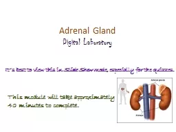
Luaibi Adrenocortical Hormones The two adrenal glands each of which weighs about 4 grams lie at the superior poles of the two kidneys As shown in Figure 771 each gland is composed of two distinct parts the ID: 918630

aldosterone cortisol secretion adrenal cortisol aldosterone adrenal secretion cells hormones acth effects sodium mineralocorticoid activity adrenocortical plasma cortex concentration

Share:

Link:

Embed:

Download Presentation from below link

Download Presentation The PPT/PDF document "Adrenal gland Noori M." is the property of its rightful owner. Permission is granted to download and print the materials on this web site for personal, non-commercial use only, and to display it on your personal computer provided you do not modify the materials and that you retain all copyright notices contained in the materials. By downloading content from our website, you accept the terms of this agreement.


Presentation Transcript

Slide1

Adrenal

gland

Noori

M.

Luaibi

Slide2

Adrenocortical Hormones

The two

adrenal glands

,

each of which weighs about

4

grams, lie at the superior poles of the two kidneys. As shown in Figure 77–1, each gland is composed of two distinct parts, the

adrenal medulla

and the

adrenal cortex

.

The adrenal medulla, the central 20 per cent of the gland, is functionally related to the sympathetic nervous system; it secretes the hormones

epinephrine

and

norepinephrine

in response to sympathetic stimulation. In turn, these hormones cause almost the same effects as direct stimulation of the sympathetic nerves in all parts of the body.

The adrenal cortex secretes an entirely different group of hormones, called

corticosteroids

.

These

hormones are all synthesized from the steroid cholesterol, and they all have similar chemical formulas. However, slight differences in their molecular structures give them several different but very important functions.

Slide3

Slide4

Corticosteroids Mineralocorticoids, Glucocorticoids, and Androgens.

Two major types of adrenocortical hormones, the

mineralocorticoids

and the

glucocorticoids

,

are secreted by the adrenal cortex. In addition to these, small amounts of sex hormones are secreted, especially

androgenic hormones

,

which exhibit about the same effects in the body as the male sex hormone testosterone.

They are normally of only slight importance, although in certain abnormalities of the adrenal cortices, extreme quantities can be secreted

and

can result in masculinizing effects.

The

mineralocorticoids

have gained this name because they especially affect the electrolytes (the “minerals”) of the extracellular fluids-sodium and potassium, in particular.

The

glucocorticoids

have gained their name because they exhibit important effects that increase blood glucose concentration.

They have additional effects on both protein and fat metabolism that are equally as important to body function as their effects on carbohydrate metabolism.

More than 30 steroids have been isolated from the adrenal cortex, but two are of exceptional importance to the normal endocrine function of the human body:

aldosterone,

which is the principal

mineralocorticoid, and cortisol

,

which is the

principal glucocorticoid.

Slide5

Synthesis and Secretion of Adrenocortical Hormones The Adrenal Cortex Has Three Distinct Layers.

the adrenal cortex is composed of three relatively distinct layers:

1.

The

zona

glomerulosa

,

a thin layer of cells that lies just underneath the capsule, constitutes about

15

per cent of the adrenal cortex. These cells are the only ones in the adrenal gland capable of secreting significant amounts of

aldosterone

because they contain the enzyme

aldosterone synthase

,

which is necessary for synthesis of aldosterone.

The secretion of these cells is controlled mainly by the extracellular fluid concentrations of

angiotensin II

and

potassium

,

both of which stimulate aldosterone secretion.

2.

The

zona

fasciculata

,

the middle and widest layer, constitutes about

75

per cent of the adrenal cortex and secretes the glucocorticoids

cortisol

and

corticosterone

,

as well as small amounts of

adrenal androgens

and

estrogens.

The secretion of these cells is controlled in large part by the hypothalamic-pituitary axis via

adrenocorticotropic hormone (ACTH).

3.

The

zona

reticularis

,

the deep layer of the cortex, secretes the adrenal androgens

dehydroepiandrosterone

(DHEA)

and

androstenedione

,

as well as small amounts of estrogens and some glucocorticoids .

Slide6

ACTH

also regulates secretion of these cells, although other factors such as

cortical androgen-stimulating hormone

,

released from the pituitary, may also be involved. The mechanisms for controlling adrenal androgen production, however, are not nearly as well understood as those for glucocorticoids and mineralocorticoids.

Aldosterone

and

cortisol

secretion are regulated by independent

mechanisms

. Factors such as

angiotensin II

that specifically increase the output of aldosterone and cause hypertrophy of the

zona

glomerulosa

have no effect on the other two zones. Similarly, factors such as

ACTH

that increase secretion of cortisol and adrenal androgens and cause hypertrophy of the

zona

fasciculata

and

zona

reticularis

have little or no effect on the

zona

glomerulosa

.

Slide7

Adrenocortical Hormones Are Steroids Derived from Cholesterol.

All human steroid hormones, including those produced by the adrenal cortex, are synthesized from cholesterol. Although the cells of the adrenal cortex can synthesize de novo small amounts of cholesterol from acetate, approximately

80

per cent of the cholesterol used for steroid synthesis is provided by low-density lipoproteins

(LDL)

in the circulating plasma

. The

LDL’s

,

which have high concentrations of cholesterol, diffuse from the plasma into the interstitial fluid and attach to specific receptors contained in structures called

coated pits

on the adrenocortical cell membranes. The coated pits are then internalized by

endocytosis

,

forming vesicles that eventually fuse with cell lysosomes and release cholesterol that can be used to synthesize adrenal steroid hormones.

Slide8

Transport of cholesterol into the adrenal cells is regulated by feedback mechanisms that can markedly alter the amount available for steroid synthesis. For example,

ACTH

, which stimulates adrenal steroid synthesis, increases the number of adrenocortical cell receptors for

LDL

, as well as the activity of enzymes that liberate cholesterol from

LDL

.

Once the cholesterol enters the cell, it is delivered to the mitochondria, where it is cleaved by the enzyme

cholesterol

desmolase

to form

pregnenolone

;

this is the rate-limiting step in the eventual formation of adrenal steroids. In all three zones of the adrenal cortex, this initial step in steroid synthesis is stimulated by the different factors that control secretion of the major hormone products aldosterone and cortisol.

For example, both

ACTH

, which stimulates cortisol secretion, and angiotensin II, which stimulates aldosterone secretion, increase the conversion of

cholesterol

to

pregnenolone

.

Slide9

Synthetic Pathways for Adrenal Steroids.

Figure 77-2 gives, the principal steps in the formation of the important steroid products of the adrenal cortex: aldosterone, cortisol, and the androgens. Essentially all these steps occur in two of the organelles of the cell, the

mitochondria

and the

endoplasmic reticulum

,

some steps occurring in one of these organelles and some in the other.

Each

step is catalyzed by a specific enzyme system.

A change in even a single enzyme in the schema can cause vastly different types and relative proportions of hormones to be formed. For example, very large quantities of masculinizing sex hormones or other steroid compounds not normally present in the blood can occur with altered activity of only one of the enzymes in this pathway.

The chemical formulas of

aldosterone

and

cortisol

, which are the most important mineralocorticoid and glucocorticoid hormones, respectively,

Cortisol has a

keto

-oxygen

on

carbon number

3

and is

hydroxylated

at carbon numbers 11 and

21

. The mineralocorticoid aldosterone has an

oxygen atom

bound at the

number

18

carbon

. In addition to

aldosterone

and

cortisol

, other steroids having glucocorticoid or mineralocorticoid activities, or both, are normally secreted in small amounts by the adrenal cortex.

And several additional potent steroid hormones not normally formed in the adrenal glands have been synthesized and are used in various forms of therapy.

Some of the more important of the corticosteroid hormones, including the synthetic ones.

Slide10

Slide11

Mineralocorticoids

Aldosterone

(very potent, accounts for about

90

per cent of all mineralocorticoid

activity

)

Desoxycorticosterone

(

1/30

as potent as aldosterone, but very small quantities

secreted

)

Corticosterone

(slight mineralocorticoid activity)

9a-Fluorocortisol

(synthetic, slightly more potent than aldosterone)

Cortisol

(very slight mineralocorticoid activity, but large quantity secreted)

Cortisone

(synthetic, slight mineralocorticoid activity)

Glucocorticoids

Cortisol

(very potent, accounts for about

95

per cent of all glucocorticoid activity)

Corticosterone

(provides about

4

per cent of total glucocorticoid activity, but

much

less potent than cortisol)

Cortisone

(synthetic, almost as potent as cortisol)

Prednisone

(synthetic,

four

times as potent as cortisol)

Methylprednisone

(synthetic,

five

times as potent as cortisol)

Dexamethasone

(synthetic,

30

times as potent as cortisol)

Slide12

It is clear from this list that some of these hormones have both glucocorticoid and mineralocorticoid activities

.

It

is especially significant that cortisol has a small amount of

mineralocorticoid

activity, because some syndromes of excess cortisol secretion can cause significant

mineralocorticoid

effects, along with its much more potent

glucocorticoid

effects.

The intense

glucocorticoid

activity of the synthetic hormone dexamethasone, which has almost zero

mineralocorticoid

activity, makes this an especially important drug for stimulating specific

glucocorticoid

activity .

Slide13

Adrenocortical Hormones Are Bound to Plasma Proteins.

Approximately

90

to

95

per cent of the cortisol in the plasma binds to plasma proteins, especially a globulin called

cortisol-binding globulin

or

transcortin

and, to a lesser extent, to

albumin.This

high degree of binding to plasma proteins slows the elimination of cortisol from the plasma; therefore, cortisol has a relatively long half life of

60

to

90

minutes. Only about

60

per cent of circulating aldosterone combines with the plasma proteins, so that about

40

per cent is in the free form; as a result, aldosterone has a relatively short half-life of about

20

minutes. In both the combined and free forms, the hormones are transported throughout the extracellular fluid compartment.

Binding of adrenal steroids to the plasma proteins may serve as a reservoir to lessen rapid fluctuations in free hormone concentrations, as would occur, for example, with cortisol during brief periods of stress and episodic secretion of

ACTH

. This reservoir function may also help to ensure a relatively uniform distribution of the adrenal hormones to the tissues.

Slide14

Adrenocortical

Hormones Are Metabolized in the Liver.

The adrenal steroids are degraded mainly in the liver and conjugated especially to

glucuronic

acid

and, to a lesser extent, sulfates. These substances are inactive and do not have

mineralocorticoid

or

glucocorticoid

activity.

About

25

per cent of these conjugates are excreted in the bile and then in the

feces.The

remaining conjugates formed by the liver enter the circulation but are not bound to plasma proteins, are highly soluble in the plasma, and are therefore filtered readily by the kidneys and excreted in the urine. Diseases of the liver markedly depress the rate of inactivation of adrenocortical hormones, and kidney diseases reduce the excretion of the inactive conjugates.

The normal concentration of aldosterone in blood is about

6

nanograms

(

6

billionths of a gram

) per

100 ml

, and the average secretory rate is approximately

150

μg

/day

(

0.15

mg/day

).

The concentration of cortisol in the blood averages

12

μg

/

100

ml

, and the secretory rate averages

15

to

20

mg/day

.

Slide15

Functions

of the Mineralocorticoids- Aldosterone

*

Mineralocorticoid Deficiency Causes Severe Renal Sodium Chloride Wasting and Hyperkalemia.

*

Aldosterone Is the Major Mineralocorticoid Secreted by the Adrenals.

Aldosterone exerts nearly

90

per cent of the mineralocorticoid activity of the adrenocortical secretions, but cortisol, the major glucocorticoid secreted by the adrenal cortex, also provides a significant amount of mineralocorticoid activity. Aldosterone’s mineralocorticoid activity is about

3000

times greater than that of cortisol, but the plasma concentration of cortisol is nearly

2000

times that of aldosterone.

Cortisol can also bind to mineralocorticoid with high affinity , However, the renal epithelial cells also contain the enzyme

11β-

hydroxysteroid

dyhydrogenase

type 2

. which converts

cortisol

to

cortisone

. because cortisone dose not avidly bind mineralocorticoid receptors, cortisol dose not normally exert significant mineralocorticoid effects . However , in patients with genetic deficiency

of

11β-

hydroxysteroid

dyhydrogenase

type 2

activity cortisol may have substantial mineralocorticoid effects .

This concision is called

apparent mineralocorticoid excess syndrome

(AME)

because the patient has essentially the same pathophysiological change as patient with excess aldosterone secretion except that plasma aldosterone levels are very low. Ingestion of large amounts of Licorice , which contains

glycyrrhetinic

acid

, may also cause

AME

due to its ability to block

11β-

hydroxysteroid

dyhydrogenase

type 2

activity .

Slide16

*Renal and Circulatory Effects of

Aldosterone

1.

Aldosterone

Increases Renal Tubular Reabsorption of

Sodium

and Secretion of Potassium.

2.

Excess

Aldosterone Increases Extracellular Fluid Volume

and

Arterial Pressure but Has Only a Small Effect on Plasma

Sodium

Concentration.

3.

Excess

Aldosterone Causes Hypokalemia and Muscle

Weakness

; Too Little Aldosterone Causes Hyperkalemia

and

Cardiac Toxicity.

4

.

Excess

Aldosterone Increases Tubular Hydrogen

Ion

Secretion, and Causes Mild Alkalosis.

Slide17

*

Aldosterone Stimulates Sodium and Potassium Transport in Sweat Glands, Salivary Glands, and Intestinal Epithelial Cells.

Cellular Mechanism of Aldosterone Action

Although for many years we have known the overall effects of mineralocorticoids on the body, the basic action of aldosterone on the tubular cells to increase transport of sodium is still not fully understood. However, the cellular sequence of events that leads to increased sodium reabsorption seems to be the following.

First

,

because of its lipid solubility in the cellular membranes, aldosterone diffuses readily to the interior of the tubular epithelial cells.

Second

,

in the cytoplasm of the tubular cells, aldosterone combines with a highly specific cytoplasmic

receptor protein (MR)

a protein that has a

stereomolecular

configuration that allows only aldosterone or very similar compounds to combine with it.

Third

,

the aldosterone-receptor complex or a product of this complex diffuses into the nucleus, where it may undergo further alterations, finally inducing one or more specific portions of the

DNA

to form one or more types of messenger

RNA

related to the process of sodium and potassium transport.

Fourth

,

the messenger

RNA

diffuses back into the cytoplasm, where, operating in conjunction with the ribosomes, it causes protein formation. The proteins formed are a mixture of

(1)

one or more enzymes and

(2)

membrane transport proteins that, all acting together, are required for sodium, potassium, and hydrogen transport through the cell membrane.

Slide18

One of the enzymes especially increased is

sodiumpotassium

adenosine

triphosphatase

,

which serves as the principal part of the pump for sodium and potassium exchange at the

basolateral

membranes

of the renal tubular cells.

Additional proteins, perhaps equally important, are epithelial sodium channel proteins inserted into the

luminal membrane

of the same tubular cells that allows rapid diffusion of sodium ions from the tubular lumen into the cell; then the sodium is pumped the rest of the way by the

sodium-potassium pump

located in the

basolateral

membranes of the cell.

Thus, aldosterone does not have an immediate effect on sodium transport; rather, this effect must await the sequence of events that leads to the formation of the specific intracellular substances required for sodium transport. About

30

minutes is required before new

RNA

appears in the cells, and about

45

minutes is required before the rate of sodium transport begins to increase; the effect reaches maximum only after several hours.

Slide19

Regulation of Aldosterone Secretion

The regulation of aldosterone secretion is so deeply intertwined with the regulation of extracellular fluid electrolyte concentrations, extracellular fluid

volume,blood

volume, arterial pressure, and many special aspects of renal function .

The regulation of aldosterone secretion by the

zona

glomerulosa

cells is almost entirely independent of the regulation of cortisol and androgens by the

zona

fasciculate

and

zona

reticularis

.

Four factors are known to play essential roles in the regulation of aldosterone. In the probable order of their importance, they are as follows:

1.

Increased potassium ion concentration in the extracellular fluid

greatly increases

aldosterone secretion.

2.

Increased activity of the renin-angiotensin system (increased levels of angiotensin II) also

greatly increases

aldosterone secretion.

3.

Increased sodium ion concentration in the extracellular fluid

very slightly decreases

aldosterone secretion.

4.

ACTH

from the anterior pituitary gland is necessary for aldosterone secretion but has little effect in controlling the rate of secretion in most physiological conditions .

Slide20

Of

these factors,

potassium ion

concentration

and

the

renin-angiotensin system

are by far the most potent in regulating aldosterone secretion. A small percentage increase in potassium concentration can cause a

several fold

increase in aldosterone secretion.

Likewise, activation of the

renin-angiotensin system

, usually in response to diminished blood flow to the kidneys or to sodium loss, can cause a

several fold

increase in aldosterone secretion. In turn, the aldosterone acts on the kidneys

(1)

to

help them excrete the excess potassium ions and

(2

)

to increase the blood volume and arterial pressure

,

thus returning

the

renin angiotensin

system

toward its normal level of ac

tivity

. These feedback control mechanisms are essential for maintaining life.

With an

angiotensin –converting

enzyme

inhibitor

after several weeks of a low-sodium diet that increases plasma aldosterone concentration .

Note

that blocking angiotensin II formation markedly decreases plasma aldosterone concentration without significantly changing cortisol concentration; this indicates the important role of angiotensin II in stimulating aldosterone secretion when sodium intake and extracellular fluid volume are reduced.

By contrast, the effects of sodium ion concentration percentage

and

of

ACTH

in controlling aldosterone secretion are usually minor. Nevertheless, a

10

to

20

per cent decrease in extracellular fluid sodium ion concentration, which occurs on rare occasions, can perhaps double aldosterone secretion. In the case of

ACTH

, if there is even a small amount of

ACTH

secreted by the anterior pituitary gland, it is usually enough to permit the adrenal glands to secrete whatever amount of aldosterone is required, but total absence of

ACTH

can significantly reduce aldosterone secretion.

Slide21

Functions of the Glucocorticoids

1. Effects of Cortisol on Carbohydrate Metabolism

A.

Stimulation of Gluconeogenesis.

1

.

Cortisol increases the enzymes required to convert amino acids into glucose in

the

liver cells.

2

.

Cortisol causes mobilization of amino acids from the

extra hepatic

tissues

mainly

from muscle.

B .

Decreased Glucose Utilization by Cells.

C.

Elevated

Blood Glucose Concentration and “Adrenal Diabetes.”

2.

Effects of Cortisol on Protein Metabolism

A.

Reduction in Cellular Protein.

B.

Cortisol Increases Liver and Plasma Proteins.

C.

Increased Blood Amino Acids, Diminished Transport of Amino Acids into

Extra hepatic

Cells, and Enhanced Transport into Hepatic Cells.

3.

Effects of Cortisol on Fat Metabolism

A.

Mobilization of Fatty Acids.

B.

Obesity Caused by Excess Cortisol.


Slide22

4.

Cortisol is Important in Resisting Stress and Inflammation

Some

of the different types of stress that increase cortisol release are the

following

:

1.

Trauma of almost any type

2.

Infection

3.

Intense heat or cold

4.

Injection of norepinephrine and other sympathomimetic drugs

5.

Surgery

6.

Injection of necrotizing substances beneath the skin

7.

Restraining an animal so that it cannot move

8.

Almost any debilitating disease

5.

Anti-inflammatory Effects of High Levels of Cortisol

When

tissues are damaged by trauma, by infection with bacteria, or in other ways, they almost always become “

inflamed

.” In some conditions, such as in rheumatoid arthritis, the inflammation is more damaging than the trauma or disease itself. The administration of large amounts of cortisol can usually block this inflammation or even reverse many of its effects once it has begun. Before attempting to explain the way in which cortisol functions to block inflammation .


Slide23

There are five main stages of inflammation:

1.

release

from the damaged tissue cells of chemical substances that activate the

inflammation process chemicals

such as

histamine

,

bradykinin

,

proteolytic

enzymes

,

prostaglandins

,

and

leukotrienes

;

2.

an

increase in blood flow in the inflamed area caused by some of the

released

products from the tissues, an effect called

erythema

;

3.

leakage

of large quantities of almost pure plasma out of the capillaries into the

damaged

areas because of increased capillary permeability, followed by clotting

of

the tissue fluid, thus causing a

non-pitting

type of edema

;

4.

infiltration

of the area by leukocytes;

5.

after

days or weeks, ingrowth of fibrous tissue that often helps in the

healing

process.

When large amounts of cortisol are secreted or injected into a person, the cortisol has two basic

anti-inflammatory effects

:

it

can block the early stages of the inflammation process before inflammation

even

begins, or

if

inflammation has already begun, it causes rapid resolution of the

inflammation and

increased rapidity of healing.

Slide24

These effects are explained further as

follows:

A.

Cortisol Prevents the Development of Inflammation by

Stabilizing

Lysosomes and by Other Effects

.

Cortisol has the

following

effects in preventing inflammation:

1.

Cortisol stabilizes the

lysosomal

membranes

2.

Cortisol decreases the permeability of the capillaries,

3

.

Cortisol

decreases both migration of white blood cells into the

inflamed

area and phagocytosis of the damaged cells.

4.

Cortisol suppresses the immune system, causing lymphocyte

reproduction

to decrease markedly.

5.

Cortisol attenuates fever mainly because it reduces the release of

interleukin-1 from the white blood cells,

B.

Cortisol Causes Resolution of Inflammation.

6.

Other Effects of Cortisol

A

.

Cortisol Blocks the Inflammatory Response to Allergic Reactions.

B.

Effect on Blood Cells and on Immunity in Infectious Diseases

Slide25

Regulation of Cortisol Secretion by Adrenocorticotropic Hormone from the Pituitary Gland ACTH Stimulates Cortisol Secretion

.

Unlike aldosterone secretion by

the

zona

glomerulosa

, which is controlled mainly by potassium and angiotensin acting directly on the adrenocortical cells, almost no stimuli have direct control effects on the adrenal cells that secrete cortisol. Instead, secretion of cortisol is controlled almost entirely by

ACTH

secreted by the anterior pituitary gland.

This hormone, also called

corticotropin

or

adrenocorticotropin

,

also enhances the production of adrenal androgens.

Slide26

ACTH Secretion Is Controlled by

Corticotropin

-Releasing Factor from the Hypothalamus.

In the same way that other pituitary hormones are controlled by releasing factors from the hypothalamus, an important releasing factor also controls

ACTH

secretion. This is called

corticotropin

- releasing factor

(CRF

).

It is secreted into the primary capillary plexus of the

hypophysial

portal system in the median eminence of the hypothalamus and then carried to the anterior pituitary gland, where it induces

ACTH

secretion. CRF is a peptide composed of

41

amino acids

. The

cell bodies of the neurons that secrete

CRF

are located mainly in the

paraventricular

nucleus of the hypothalamus. This nucleus in turn receives many nervous connections from the limbic system and lower brain stem.

The anterior pituitary gland can secrete only minute quantities of

ACTH

in the absence of

CRF

. Instead, most conditions that cause high

ACTH

secretory rates initiate this secretion by signals that begin in the basal

regions of the brain, including the hypothalamus, and are then transmitted by

CRF

to the anterior pituitary gland.

Slide27

ACTH Activates Adrenocortical Cells to Produce Steroids by Increasing Cyclic Adenosine Monophosphate (

cAMP

).

The principal effect of

ACTH

on the adrenocortical cells is to activate

adenylyl

cyclase

in the cell membrane.

This then induces the formation of

cAMP

in the cell cytoplasm, reaching its maximal effect in about

3

minutes. The

cAMP

in turn activates the intracellular enzymes that cause formation of the adrenocortical hormones. This is another example of

cAMP

as a

second messenger

signal system.

The most important of all the

ACTH

-stimulated steps for controlling adrenocortical secretion is activation of the enzyme

protein kinase A

,

which

causes

initial conversion of cholesterol to

pregnenolone

.

This initial conversion is the “rate-limiting” step for all the adrenocortical hormones, which explains why

ACTH

normally is necessary for any adrenocortical hormones to be formed.

Long-term stimulation of the adrenal cortex by

ACTH

not only increases secretory activity but also causes hypertrophy and proliferation of the adrenocortical cells, especially in the

zona

fasciculate

and

zona

reticularis

, where cortisol and the androgens are secreted.

Slide28

Physiologic Stress Increases ACTH and Adrenocortical Secretion

As

pointed out earlier in the chapter, almost any type of physical or mental stress can lead within minutes to greatly enhanced secretion of

ACTH

and consequently cortisol as well, often increasing cortisol secretion as much as

20

-fold. This effect was demonstrated by the rapid and strong adrenocortical secretory responses after trauma

Pain stimuli caused by physical stress or tissue damage are transmitted first upward through the brain stem and eventually to the median eminence of the

hypothalamus.

CRF

is secreted into the

hypophysial

portal system.

Within

minutes

the entire control sequence leads to large quantities of cortisol in the blood.

Mental stress can cause an equally rapid increase in

ACTH

secretion. This is believed to result from increased activity in the limbic system, especially in the region of the

amygdala

and

hippocampus

, both of which then transmit signals to the posterior medial hypothalamus.

Slide29

Inhibitory Effect of Cortisol on the Hypothalamus and on the Anterior Pituitary to Decrease ACTH Secretion.

Cortisol has direct negative feedback effects on

the hypothalamus to decrease the formation of

CRF

and

the

anterior pituitary gland to decrease the formation of

ACTH

.

Both of these feedbacks help regulate the plasma concentration of cortisol.

That is, whenever the cortisol concentration becomes too great, the feedbacks automatically reduce the

ACTH

toward a normal control level.

Slide30

Adrenal Androgens

Several moderately active male sex hormones called

adrenal androgens

(the most important of which is

dehydroepiandrosterone

) are continually secreted by the adrenal cortex, especially during fetal life, Also

progesterone

and

estrogens

, which are female sex hormones, are secreted in minute quantities.

Normally, the

adrenal androgens

have only weak effects in humans. It is possible that part of the early development of the male sex organs results from childhood secretion of adrenal androgens. The

adrenal androgens

also exert mild effects in the female, not only before puberty but also throughout life. Much of the growth of the pubic and axillary hair in the female results from the action of these hormones.

In extra-adrenal tissues, some of the

adrenal androgens

are converted to testosterone, the primary male sex hormone, which probably accounts for much of their androgenic activity.