/
Diseases of adrenal gland-1 Diseases of adrenal gland-1

Diseases of adrenal gland-1 - PowerPoint Presentation

willow
willow . @willow
Follow
65 views
Uploaded On 2023-11-19

Diseases of adrenal gland-1 - PPT Presentation

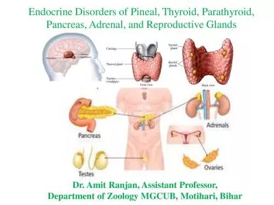
Dr Tariq Aladily tnaladilyjuedujo Department of Pathology The University of Jordan Second semester 20212022 Hypercortisolism AKA Cushing syndrome Can be exogenous iatrogenic or endogenous less common ID: 1033325

cells adrenal results common adrenal cells common results mutation hyperplasia syndrome bilateral deficiency cushing gland acth chronic disease glucose

Share:

Link:

Embed:

Download Presentation from below link

Download Presentation The PPT/PDF document "Diseases of adrenal gland-1" is the property of its rightful owner. Permission is granted to download and print the materials on this web site for personal, non-commercial use only, and to display it on your personal computer provided you do not modify the materials and that you retain all copyright notices contained in the materials. By downloading content from our website, you accept the terms of this agreement.


Presentation Transcript

1. Diseases of adrenal gland-1Dr. Tariq Aladilytnaladily@ju.edu.joDepartment of PathologyThe University of JordanSecond semester 2021/2022

2. Hypercortisolism AKA Cushing syndromeCan be exogenous (iatrogenic) or endogenous (less common)Endogenous causes are divided into ACTH-dependent and indepedendentACTH-secreting PA is the most common cause of endogenous hypercorticolism (60%), AKA Cushing Disease, more common in women, young adults, functional microadenomaIn rare cases, corticotroph hyperplasia (excessive CRH secretion from hypothalamic tumor)Adrenal glands show bilateral nodular hyperplasia

3. hypercortisolismEctopic ACTH production: 5-10% of endogenous Cushing syndrome casesMore common in men, middle ageSmall cell carcinoma of lung, carcinoid tumor, medullary carcinoma of thyroid, pancreatic neuroendocrine tumorsIn some cases, ectopic production of CRHAgain, bilateral adrenal nodular hyperplasiaPathologic changes is less prominent than pituitary cause, secondary to poor prognosis of accompanied cancer

4. HypercortisolismPrimary adrenal adenoma10-20% of ACTH-independent casesLow ACTH level (negative feedback on pituitary)PRKAR1A genetic mutation The other adrenal gland is atrophic

5. hypercortisolismAdrenal carcinoma5-7% of ACTH-independent casesVery large size of adrenal glandsProduces very high level of cortisolGenetic mutations in: activation of beta-catenin (CTNNB1), inactivation of TP53, MEN1 and PRKAR1A The other adrenal gland is atrophic

6. hypercortisolismPrimary adrenal hyperplasia:Independent of ACTHRareShows bilateral adrenal cortical hyperplasia, nodules are larger than 1 cmFamilial disease: inherited mutation in the tumor suppressor gene: armadillo repeat containing 5 (ARMC5)Sporadic disease: 50% show ARNC5 mutation, others show ectopic production of G-protein coupled hormone receptors (similar action of ACTH)Syndromic disease: McCune Albright syndrome, germline activating mutation in GNAS, produces excessive cAMP  multisystemic disease

7. HypercortisolismMicronodular bilateral adrenal hyperplasiaACTH-independentSmall nodules (<1 cm)Two variants: primary pigmented nodular adrenocortical disease or Carney complex (multisystemic disease of endocrine and non-endocrine neoplasms)Both variants harbor mutation in cAMP-dependent protein kinase (PRKAR1A gene), producing excessive cAMP

8. MorphologyPituitary gland shows Crooke hyaline change (homogenous pale color of corticotroph cells instead of normal granular basophilic cytoplasm), seen in all cases of Cushing syndromeAdrenal atrophy (fasciculata and reticularis) is seen in exogenous cause of Cushing syndromeACTH-dependent hyperplasia shows bilateral diffuse enlargement of adrenal glands, variable nodularity and yellow color (grossly), zona reticularis is expanded, eosinophilic and is “lipid poor”, surrounded by outer vacuolated “lipid-rich” zona fasciculata. Nodules can be seen and contain “lipid-rich” cellsMacronodular hyperplasia: multiple large nodules (mixed lipid-rich and poor cells), separated by small nodules Micronodular hyperplasia: small dark nodules (black or brown), the pigment is lipofuscin

9.

10. morphologyAdrenal adenoma can be functional (Cushing syndrome) or non-functional (incidental), both have the same appearanceAdrenocortical adenoma is surrounded by a capsule, weighs <30g, yellow, microscopically is similar to zona fasciculata cellsAdrenocortical carcinoma is large (commonly >200 g), not capsulated, anaplastic cellsBoth diseases are more common in women, middle age, show contralateral adrenal atrophy (when functioning)

11. Clinical symptoms of Cushing diseaseHypertensionCentral obesity, moon face, buffalo humpProximal muscle weakness (atrophy)Hyperglycemia, glucoseurea, polyurea, polydipsiaBone resorption (osteoporosis)Collagen degradation (thin skin, easy bruise, poor wound healing, striationHisrutismMenstrual abnormalitiesImmune suppressionMental and psychotic disturbances

12. Diseases of adrenal gland-2Dr. Tariq Aladilytnaladily@ju.edu.joDepartment of PathologyThe University of Jordam

13. Primary hyperaldosteronismChronic excessive production of aldosteronePatients develop hypertension, hypokalemia, suppression of renin-angiotensin system and decreased renin activityPrimary hyperaldosteronism is caused by one of three diseases:(1) Bilateral idiopathic hyperaldosteronism (60%):Bilateral nodular hyperplasia of zona glomerulosa cellsMost commonly sporadic, old patients, mild hypertensionGermline mutation in KCNJ5Morphology: diffuse enlargement of the adrenal gland, sometime subtle and not obvious

14. Primary hyperaldosteronism(2) Adrenocortical neoplasm (35%):Functional adenoma or carcinomaConn syndrome: adrenal adenoma that secretes aldosterone only, more common in middle-age women50% harbor KCNJ5 mutation, which encodes potassium channel on zona granulosa cells (called GIRK4 protein), mutant protein allows influx of sodium and activation of aldosterone synthase enzyme, other similar mutations affects CACNA1H and ATP1A1 genesMorphology: adenoma is small, more common on left adrenal, buried within the gland (difficult to be seen in radiology), yellow in color and resemble fasciculata cells. Spironolactone bodies: intracellular eosinophilic material following treatment with antihypertensive drugsThe other adrenal gland is NOT atrophic

15. Primary hyperaldosteronism(3) Familial hyperaldosteronism (5%)Four subtypesFH-1 is the most common (AKA glucocorticoid-remediable aldosteronism), mutation in CYP11B2 (encoding aldosterone synthase), becomes sensitive to ACTHThe other four subtypes are rare

16. Secondary hyperaldosteronismActivation of renin-angiotensin systemIncreased level of plasma renin, occurs in:Decreased renal perfusion (renal artery stenosis or arteriolar nephrosclerosis)Arterial hypovolemia and edema (congestive heart failure, cirrhosis, nephrotic syndrome)Pregnancy (estrogen-induced)

17. Adrenogenital syndromesAdrenal secretes dehydroepiandrosterone and androstenedione which converts to testosteroneSecretion is ACTH-dependentAdrenocortical neoplasm associated with virilization: carcinoma is more common than adenoma, can be pure or mixed with hypercortisolism

18. Congenital adrenal hyperplasiaGroup of autosomal recessive disorders Deficiency in enzymes responsible for synthesizing cortisol Steroid precursors accumulate and shifts to synthesis of androgens resulting in virilizationMaybe associated with deficiency in aldosterone synthesis, too21-hydroxylase deficiency: the most common deficiency (90%), mutation in CYP21A1 gene, variable degree of deficiency, results in either:Salt wasting syndrome: associated with deficiency in aldosterone, cortisol and catecholeamine synthesis, appears in utero or shortly after birth (hyponatremia, hypokalemia, hypotension, cardiovascular collapse, virilization in females)Simple virilization: no salt wasting, genital ambiguityLate-onset adrenal virilism: common, partial enzyme deficiency: hirsutism, acne, irregular menses Morphology: bilateral nodular hyperplasia of adrenals, brown, hyperplasia of pituitary corticotroph cells

19.

20. Adrenocortical insufficiencyPrimary: adrenal failure (acute or chronic)Secondary: ACTH deficiencyPrimary acute adrenocortical insufficiency: sudden withdrawal of exogenous steroids, crisis in chronic insufficiency, massive adrenal hemorrhage (newborns, difficult delivery and hypoxia), coagulopathyWaterhouse-Friderichsen syndrome: overwhelming bacterial infection (classically occurs in Neisseria meningitidis), direct injury to adrenal vessels resulting in hemorrhage and damage to drenals

21. Primary chronic adrenocortical insufficiencyCalled Addison diseaseUncommonProgressive destruction of adrenal cortexSymptoms appear after 90% damage to adrenocortical tissueCauses: autoimmune inflammation, infections (HIV, TB)

22. pheochromocytomaTumor of adrenal medulla (chromaffin cells)Secretes catecholamines, results in hypertension10% bilateral, 10% biologically malignant, 10% not associated with hypertension, 10% arises in extra-adrenal sites (carotid body, called paraganglioma), 10% familial (early-life onset)Genetic mutations in growth-factor receptor pathway genes (RET, NF1) or increased activity of hypoxia-induced transcription factors (HIF-1α, HIF-2α)Morphology: variable size, may show necrosis, bleeding, incubation with potassium dichromate produces dark brown color (chromaffin)Histology: small nests of cells separated by supporting sutentacular cells (zellballen)Malignancy is determined by the presence of metastasis, not histology

23. Endocrine diseases of pancreasDr. Tariq Aladilytnaladily@ju.edu.joDepartment of PathologyThe University of Jordan

24. Diabetes mellitusGroup of metabolic disorders results in chronic hyperglycemiaMulti-organ damage (kidneys, retina, cardiovascular)Very commonIncreased morbidity and mortalityRisk factors: family history, life-style, obesity

25. Type-1 diabetes5% of all DM casesAutoimmune destruction of pancreatic β-cells  deficiency in insulinCommonly affects children and adolescentsSymptoms tend to appear rapidly, although the cell damage takes timeAssociated with HLA-DR3, DR-4, DQ8Polymorphism in CTLA4 and PTPN22 genes (similar to autoimmune thyroiditis)Environmental factor: virus epitopes, only 50% of monozygotic twins share DM-1Failure of self-tolerance in T-cells for islet antigensOther less common causes DM: chronic pancreatitis, pancreatic carcinoma, cystic fibrosis, hemochromatosis, infections by cytomegalovirus, coxsackie virus, congenital rubella, systemic amyloidosis

26. Type-2 diabetes>90% of DM casesPeripheral resistance to insulinInadequate response by β-cells to overcome this resistancePatients are commonly obese, adults, insidious onsetImpaired function of incretins (peptides secreted form small intestine following glucose feeding, promotes secretion of insulin from B-cells) Genetic factors: 90% concordance rate in identical twins, 10x risk in first degree relative. Genes related to adipose-tissue distribution in body, B-cell function and obesityEnvironmental factors: central obesity is most important, then sedentary life-style, circadian-disruption (sleep disorder)

27. B-cell dysfunction results from: Excess free fatty acids (due to increased activity of lipase enzyme in adipose tissue)Hyperglycemia: toxic effectAmyloid deposition in B-cells: seen in 90% of long-standing DM-2Genetic susceptibility

28.

29. Other causes of diabetesEndocrinopathies: Cushing syndrome, acromegaly, hyperthyroidism, pheochromocytoma, glucagonomaDrugs: steroids, β-agonists, phenytoin, thiazideCertain syndromes: Down, TurnerGestational diabetes (5% of pregnant women, due to increased steroid hormones)

30. Monogenic forms of DMMaturity-onset diabetes of the young (MODY)Resemble DM-2Results from germline loss of function mutation in glucokinase (GCK) genes, affects glucose metabolism and insulin secretionRarely: mutation is insulin-receptor synthesis, binding or activity, associated with hyperinsulinemia and skin pigmentation

31. Hyperglycemia Effect on tissue(1) Advanced glycation end products (AGE): non-enzymatic glucose addition to molecules results in activation of AGE signaling, resulting in cytokines and growth factors release causing vascular proliferation (retinopathy) and basement membrane deposition (nephropathy), release of reactive oxygen species, procoagulant activity and proliferation of vascular smooth muscle cells(2) Activation of protein kinase C: results in activation of plasminogen activator inhibitor-1 (procoagulant effect)(3) Oxidative stress and disturbance in polyol pathways: occurs in tissues that do not require insulin to take glucose (nerves, lenses, kidneys, blood vessels): hyperglycemia results in increased intracellular glucose that ends up with depletion of NADPH, making these cells susceptible to oxidative stress

32. Clinical presentationPolyurea, polydipsia (osmotic effect), polyphagia, weight loss (catabolic effect)Chronic systemic complications results from chronic hyperglycemiaMacrovascular: myocardial infarction, stroke, lower limb ischemiaMicrovascular: retinopathy, nephropathy, neuropathyVaries among patientsTight control of blood glucose level prevents complications

33. Diabetic ketoacidosisMostly seen in type-1 DMFailure to take insulin, stress, infections, traumaMay occur in type-2 DM with severe stress (release of epinephrin which blocks residual insulin and stimulates release of glucagon). Severe hyperglycemia causes osmotic diuresis and dehydrationInsulin deficiency promotes ketone body synthesis, when severe and accompanied by dehydration, results in metabolic acidosis (fatigue, nausea, vomiting, abdominal pain, difficult breathing, fruity odor, loss of consciousness, coma.

34. Hyperosmolar hyperglycemic stateAffects DM-2Osmotic diuresis results in dehydration, if not corrected (stroke, infections, no enough water drinking), results in mental status dysfunction and coma. No ketons are produced.

35. Hyperinsulinism (insulinoma)Most common pancreatic endocrine neoplasmHyperinsulinemia results in hypoglycemic episodes (usually mild)In severe forms causes confusion and loss of consciousnessMostly single tumor, benign behavior (90%)Microscopically appears as giant islets, contain amyloidIslet-cell hyperplasia is seen in newborns of diabetic mothers (cause serious hypoglycemia after birth, transient)

36.

37. Zollinger-Ellison syndrome (gastrinoma)Arises in pancreas or duodenum Causes severe peptic ulcer, jejunal ulcer50% are malignant25% of cases appear as a part of MEN-1 syndrome (multifocal)