/
Muscles II:  Microscopic Anatomy and Contraction Muscles II:  Microscopic Anatomy and Contraction

Muscles II: Microscopic Anatomy and Contraction - PowerPoint Presentation

elizabeth
elizabeth . @elizabeth
Follow
65 views
Uploaded On 2023-11-17

Muscles II: Microscopic Anatomy and Contraction - PPT Presentation

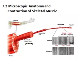
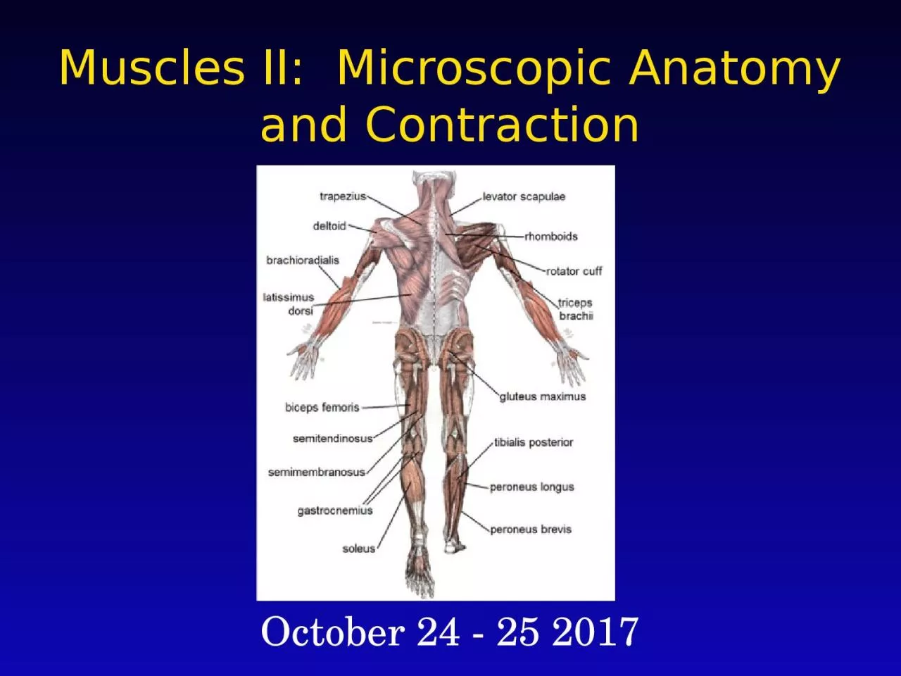
October 24 25 2017 Muscle Structure Muscle Fascicle bundle of fibers Muscle Fiber single cell Myofibril organelle Sarcomere unit of contraction Microscopic Anatomy of Skeletal Muscle ID: 1032571

myosin muscle contraction actin muscle myosin actin contraction filaments cell light heads ca2 sarcomere bands attach fiber skeletal neuron

Share:

Link:

Embed:

Download Presentation from below link

Download Presentation The PPT/PDF document "Muscles II: Microscopic Anatomy and Con..." is the property of its rightful owner. Permission is granted to download and print the materials on this web site for personal, non-commercial use only, and to display it on your personal computer provided you do not modify the materials and that you retain all copyright notices contained in the materials. By downloading content from our website, you accept the terms of this agreement.


Presentation Transcript

1. Muscles II: Microscopic Anatomy and ContractionOctober 24 - 25 2017

2. Muscle StructureMuscleFascicle (bundle of fibers)Muscle Fiber (single cell)Myofibril (organelle)Sarcomere (unit of contraction)

3. Microscopic Anatomy of Skeletal MuscleLarge, cylindrical, multinucleate cellsContain many mitochondria; nearly filled with myofibrilsSome organelles have unique vocabulary:Sarcolemma: cell membraneSarcoplasm: cytoplasmSarcoplasmic reticulum: modified ER, surrounds each myofibril; store Ca2+

4. Each myofibril can be divided into contractile units called sarcomeres. Sarcomeres consist of overlapping protein filaments of actin and myosin.Regular arrangement of dark and light bands. Dark bands occur where myosin is present. Microscopic Anatomy of Skeletal Muscle

5. The M line is where the myosin attachesZ discs (a membrane) mark the edge of each sarcomere; serve as attachment site for actin Microscopic Anatomy of Skeletal Muscle

6. Use the picture to come up with a definition of the following:I bandA bandH zone Microscopic Anatomy of Skeletal Muscle

7. Use the picture to come up with a definition of the following:I band – area without myosin fibers; aka light bandA band – area with myosin fibers; aka dark bandH zone – area without actin fibers Microscopic Anatomy of Skeletal Muscle

8. First match the words … actin cell myofibril group of cells sarcomere cell membrane fascicle protein muscle fiber organelle sarcolemma contractile unit Then, write a paragraph that uses all the words in both columns above and explains that structure of the muscle.Turn & Talk

9. Contraction OverviewGlobular heads of myosin filaments attach to actin filaments.Myosin pulls actin filaments : “Sliding filament theory” Causes sarcomere to shorten, particularly the light bands

10. Contraction OverviewGlobular heads of myosin filaments attach to actin filaments.Myosin pulls actin filaments : “Sliding filament theory” Causes sarcomere to shorten, particularly the light bands light dark light light dark light

11. Contraction OverviewWhich shows contracted muscle fibers?How can you tell?

12. Contraction Overview Relaxed muscle Contracted muscle (large light bands) (small light bands)

13. Contraction Overview What are these? Are the myofibrils organized left to right or top to bottom? See animation!MitochondriaLeft to right

14. Contraction DetailsA motor neuron stimulates the muscle cell by releasing the neurotransmitter acetylcholine ACh into the synaptic cleft between the neuron and muscle cell. Note:A motor unit is a single motor neuron and all the muscle fibers it activates

15. Contraction DetailsA motor neuron stimulates the muscle cell by releasing the neurotransmitter acetylcholine ACh into the synaptic cleft between the neuron and muscle cell.ACh causes an electric current called an action potential to move through the muscle cell.

16. Contraction DetailsA motor neuron stimulates the muscle cell by releasing the neurotransmitter acetylcholine ACh into the synaptic cleft between the neuron and muscle cell.ACh causes an electric current called an action potential to move through the muscle cell. The action potential causes the release of Ca2+ from the sarcoplasmic reticulum.

17. Contraction DetailsCa2+ exposes myosin-binding sites on actin filaments. Myosin heads (& ADP) attach to actin binding sites, forming cross-bridges. Muscle relaxed. Ca2+ present. No Ca2+ present. Cross-bridge formed. myosin actin ADP + P myosin head

18. Contraction DetailsMyosin heads release ADP, move the actin filament in “power stroke” Power stroke, ADP + P released myosin actin

19. Contraction DetailsMyosin heads release ADP, move the actin filament in “power stroke”ATP binds to myosin head. The crosslink between actin and myosin breaks. ATP becomes ADP + P, readying the myosin head to reattach to actin. Power stroke, ATP binds,ADP + P released cross-links break myosin actin

20. Contraction Details If Ca2+ is still present, cycle will repeat, with myosin heads reattaching and contracting the muscle even more. Once the action potential is over, the Ca2+ is reabsorbed into the sarcoplasmic reticulum. Without Ca2+, myosin cannot attach to actin. Watch me!

21. Contraction DetailsNOTE: ATP is required to breakcross-links, not to form them. Explains rigor mortisWhy then do muscles needATP?To reset head so it can contract further -- contraction is a series of sliding motions.

22. Turn & TalkDescribe the role of each of the following in muscle contractionScholar with more siblings….AChCa2+Scholar with less siblings … ATPAction potentialChallenge question: We discussed the process of contraction in skeletal muscles. How do you expect it differs for cardiac and smooth muscles?

23. Exit TicketIn comparing electron micrographs of a relaxed skeletal muscle fiber and a fully contracted muscle fiber, which would be seen only in the relaxed fiber?Z discsTriadsI bandsA bandsH zones

24. Exit Ticket2. Which word describes the unit of contraction of a muscle?MyofibrilSarcomereA bandH band

25. Exit Ticket3. Which of the following correctly lists the order of structure of the muscle from largest to smallest?fascicle, myofibril, sarcomere, muscle fibermyofibril, fascicle, sarcomere, muscle fiber fascicle, muscle fiber, myofibril, sarcomere muscle fiber, fascicle, myofibril, sarcomere

26. Exit TicketWhich of these stores calcium ion?Sarcoplasmic reticulumSarcomereSarcolemmamitochondria

27. Exit TicketWhich of these best describe the process of muscle contraction?The actin filaments shortenThe myosin filaments shortenThe light bands shortenThe dark bands shorten

28. Exit Ticket6. Which of these best describe the process of muscle contraction?Myosin heads attach to actin filaments that are exposed by the presence of ATPMyosin heads attach to actin filaments that are exposed by the presence of Ca2+ Actin heads attach to myosin filaments that are exposed by the presence of ATPActin heads attach to myosin filaments that are exposed by the presence of Ca2+