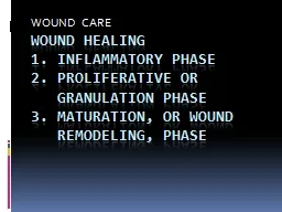
MUDr Martin Petráš Internal environmenthomeostasis The human body is surrounded by external environment that provides nutrients and oxygen that are necessary for life ID: 921019
Download Presentation The PPT/PDF document "Internal environment, care in ICU, wound..." is the property of its rightful owner. Permission is granted to download and print the materials on this web site for personal, non-commercial use only, and to display it on your personal computer provided you do not modify the materials and that you retain all copyright notices contained in the materials. By downloading content from our website, you accept the terms of this agreement.
Slide1
Internal environment, care in ICU, wound healing
MUDr.
Martin Petráš
Slide2Internal environment-homeostasis
The human body is surrounded by
external environment
,
that provides nutrients and oxygen that are necessary for life.
The human body has an
internal environment
, which is maintained by dynamic processes
of biological regulatory-mechanisms.
Dynamic constancy of internal environment is defined as
HOMEOSTASIS,
and
is carried out by organ systems working together
Slide3Internal environment
Internal environment is maintained more or less constant, and within narrow
range of limits
.
Disturbance
or breach of these limits
can cause disease,
or can prove fatal.
More specifically, internal environment is described as
extracellular fluids –
blood plasma and interstitial fluids
Slide4Slide5Body fluids
circa
60%
body weight
Intracellular – 40%
Extracellular
– 20%
Interstitial
Intravasal
Transcelular
–
cerebral spinal fluid (CSF), joint fluid, fluids of GI tract
*
plus „third space“ fluids - patologic state
Slide6Fluid balance/24 hours:
Intake:
Fluids (water)–
1500ml
Water in food–
700ml
Endogenous (metabolic) water–
300ml
Excretion:
Diuresis
– 1500ml
Breath
– 400ml
Sweat–
200ml
Stool
– 200ml
Slide7Internal environment
There are many factors that need to be maintained within narrow limits. Some of the most important are:
temperature - 35,5 37 ° C
w
ater and electrolyte concentration – Na+,K+,Cl-
p
H
of body fluids – pH 7,36 7,44
b
lood glucose level - 3,9 7,2 (ideally 5,5)
b
lood pressure - 120/80 129/89
b
lood and tissue oxygen and carbon dioxide levelssat. 95-100 % O2, paCO higher less than 75 mmHg
Slide8Basic concept
Majority of regulation processes work on „negative feedback“ system.
e.g.
b
ody temperature should range from 35-37°C
Slide9Slide10In general, negative feedback system decreases the stimulus
Slide11Another example?
Slide12Is there any „possitive feedback
“ system?
Oh yes there is
Positive feedback systems are not that common in humans.
In possitive feedback systems, the output enhances or exaggerates the original stimulus.
Can you think of at least one example?
Slide13Positive feedback
Slide14Thank you for your attention
Slide15BREAK
Slide16ICU
Definition?
Intensive care units
cater to patients with
severe and life-threatening illnesses and injuries
, which require
constant, close monitoring and support
from
special
equipment and
medications
,
in order to ensure normal bodily functions. They are staffed by highly trained doctors and nurses who specialise in caring for critically ill patients.
Patients may be transferred directly to an intensive care unit from an emergency department if required, or from a ward if they rapidly deteriorate, or immediately after surgery if the surgery is very invasive and the patient is at high risk of
complications, or might be unstable
Slide17Care in ICU
-critical care is a term used to describe as the care of patients who are extremely ill, and whose clinical condition is unstable, or potentially unstable, and may lead to death
Slide18ICU equipmentICU equipment includes patients monitoring devices, respiratory and cardiac support, pain management applicators, emergency resuscitation devices, and other life-support equipment, designed to care for patients who are seriously injured, have a critical, or life- threatening illness, or have undergone a major surgical procedure, and require 24 hour monitoring
Slide19Slide20ICU design
Difference in structure between ICU and standard department
Slide21Example
Central station- place for bureaucracy, doctors,nurses
Slide22Size of the ICU
Average size is 6-8 beds
less than 6 beds is considered uneconomical
it should not exceed 12 beds
It is wiser to create several ICU units with less beds than vice-versa
Slide23Classification of ICU care units
Level - I unit:
-provides monitoring, observation and short term ventilation.Nurse patient ratio is 1:3, and the medical staff are not present in the unit all the time.
Level – II unit:
-provides monitoring, observation, and long –term ventilation with resident doctors. The nurse patient ratio is 1:2, and usually the junior medical staff is available all the time (with consultations if needed)
Slide24Level III – unit:
-provides all aspects of intensive care, including invasive haemodynamic monitoring, dialysis,etc...
Nurse patient ratio is 1:1.
Slide25Types of ICU unitsNeonatal intensive care unit (NICU)
Paediatric intensive care unit (PICU)
Coronary care unit (CCU)
Cardiovascular intensive care unit (CICU)
Surgical intensive care unit (SICU)
Trauma intensive care unit (TICU)
.....
........
...........
Slide26Care in ICU
Regardless of the underlying cause of the illness, the provision of meticulous supportive care is essential to the management of any critically ill patient. Back in 2005, Jean Louis Vincent popularised the
FAST
HUG
G
S
mnemonic for recalling the key issues to review when looking after a critically ill patient
.
or you can extend it a little:
FAST HUGGS IN BED PLS
F
eeding/fluids
I
ndwelling catheter care/removal
A
nalgesia
N
asogastric tube
S
sedation
T
hromboprophylaxis
B
owel care
E
nvironment
H
ead –up position
D
e-escalation of treatment/support
U
lcer prophylaxis
G
lycemic control
P
sychosocial help
G
ood infection control
S
pontaneus breathing trial
Slide27Fluids:
it is very easy to accidentaly overdose patient with fluids ! That can impact on morbitidy and mortality-be cautious
Feeding:
always use p.o.
w
ay if possible
Analgesia:
critically ill patients are prone to intensive pain – better pain management, sooner release
Sedation:
important in ventilated patients . In non-ventilated patients, helps to treat anxiousness and delirium
Tromboprophylaxis:
patients are bed-bound-risk of DVT/PE
Head –up position:
easier breathing, low risk of food aspiration
Ulcers prophylaxis:
both gastric and pressure ulcers – prevent them, document them, treat them
Glycemic control:
even in non-diabetic patients, blood glucose can change from hypoglycemia to hyperglycemia-measure, treat
Good infection control:
proper antiseptic technique, ATBs – prophylaxis of VAP, CAUTI, CVCAS
Spontaneous breathing trial:
or so called „sedative vacation“
Slide28Daily review sheet
Slide29Daily progress note
Subjective: short narrative on how patient feel/did
Objective: vital signs and at least general physical overview – lung,cardio,abdomen, wounds,signs of TED, labs, control X-ray,etc...
A/P (actions and plans): write down what has been done with a patient, and what is planned for him/her
NEVER!!! NEVER just COPY and PASTE!!! – can result in a big f**k up
(for the patient, and also for you)
Slide30Role of physiotherapy in ICU
Physiotherapist role in the ICU can be separated into two key areas – respiratory and rehabilitation
Physiotherapist plays an important role during „weaning“ – gradual process leading to extubation of the patient, leaving him to breath spontaneously
Slide31Respiratory physiotherapyPatients in ICU may require mechanical ventilation to help them breath. However, this stops patient from coughing and clearing the daily load of sputum ( cca 100ml/day).
In case of respiratory infection, this amount can be increased
significantly
.
Physiotherapists with their techniques help patient to clear the airways of built up sputum collection, and decrease the possible complications.
Slide32Repiratory physiotherapyEarly activity:
such as getting into the chair or verticalisation/walk training – these encourage deep breaths and coughing
Positioning:
to allow gravity to help sputum to drain from the lungs
Manual techniques (shaking, vibrations):
t
hese are applied to the ribs to try to loosen and clear the sputum
Slide33Each position optimally for 10 minutes
Slide34Vibrations consist of a fine oscillation of the hands, directed inwards, against the chest, performed on exhalation after deep inhalation.
Helps move loosened mucus towards larger airways
Slide35Shaking is a coarser movement
i
n which the chest wall is rhytmically
c
ompressed.
D
rainage, and also stimulates cough
Slide36Rehabilitation in ICU
Gradual practice of
walk
Slide37Prophylaxis or pressure ulcers
Pressure ulcers are avoidable by proper positioning
Slide38Pressure ulcers
Patients turning schedule
Slide39Pressure ulcers, decubitus ulcer, bed sore
Slide40Slide41Slide42ICU rehabilitation
Cycling in ICU
Slide43Short brake....really, a short one
Slide44WOUND HEALING
What is a
wound?
Wound is a breach in continuity of skin, mucosa or surface of an organ.
There are many different categories of wounds, and they can be classified according to different aspects.
Slide45Wound types and classification
1.According to depth:
superficial
and
deep
2.According to complexity:
simple
and
complicated
3.According to their penetrance:
(non) –penetrant
Division according to possible ways of treatment:
clean
or
mechanically contaminated
aseptic or septic (biologically clean or infected)p
oisoned (animal or chemical poisons)
Slide46Slide47Wound types
vulnus scissum (incision wound)
v
ulnus sectum (cut wound)
v
ulnus punctum (stab wound)
v
ulnus sclopetarium (shot wound –GSW)
v
ulnus morsum (bite wound)
v
ulnus lacerum (tear wound)
v
ulnus contusum ( contusion wound)
Slide48Wound healing
Two main types:
primary
,
secondary
or
tertiary
wound healing
Primary wound healing (sanatio per primam intentionem):
Ideal way of wound healing – when wound edges are in close contact, and inflammation is minimal.
Primary healing occurs in six steps:
1.coagulation and inflammation
– fibrin connection of w.edges
-elevation of CO2 concentration, decrease in O2
-leucocytes and macropages migrate to wound site
-hemostasis, production of sterile inflammation, angiogenesis, accumulation of colagene
Slide492.Fibroplasia and matrix storage:
-replication of fibroblasts, stimulated by several cell agents – IGF,TGF,PDGF – released from thrombocytes
macrophage - cytokine production
newly proliferated fibroblasts excrete proteoglycans and colagene – these form the base matrix for wound connection
Slide503. Angiogenesis:
- 2nd to 4th day after trauma
-in PPI healing, blood vessels create anastomosis – they start to grow due to released cytokines and cell factors
4.Epitalisation:
-replication of epithelial cells thanks to TGF factors
5. Colagene fiber maturation:
-fibroblasts and leukocytes produce colagenasis, which reduce the formation of primary colagene – this takes up to 18 months
6. End of healing:
- if growth factors and cytokines are not balanced, hypertrophy may occur – this can happen due to chronic or repeated inflammation, corpus alienum in wound, irritation
Slide51Slide52Secondary wound healing
Secondary intention
is implemented when primary intention is not possible.
This is due to wounds being created by major trauma in which there has been a significant loss in tissue or tissue
damage
, etc...
The wound is allowed to granulate.
Surgeon may pack the wound with a gauze or use a drainage system.
Granulation results in a broader scar.
Healing process can be slow due to presence of drainage from infection.
Wound care must be performed daily to encourage wound debris removal to allow for granulation tissue formation.
Using antibiotics or antiseptics for the surgical wound healing by secondary intention is controversial
.
Examples:
gingivectomy
,
gingivoplasty
,
toot
h
extraction
,
poorly reduced fractures, burns, severe lacerations, pressure
ulcers
, infected wounds, diabetic ulcers
Slide53Slide54Slide55Not infected wound is quickly covered in layer of fibrine, surrounding tissue is blood soaked, immigration and exsudation of cell elements starts.
Around blood vessels, thin and delicate layer of granulation tissue is formed
Whole granulation tissue produces liquid of yellow colour-larger collection of this fluid forms
seroma.
In combination with blood elements, it forms
scab.
Granulation and epitelisation process continues under crust
Epitelisation stops in case of overproduction of granulation tissue from beneath – „caro luxurians“ image (proud flesh)
Slide56SEROMA
SCAB
Slide57Caro luxurians – over-production of granulation tissue. Epitelisation from sides is limited or not possible.
Wound healed by secondary
healing.
proud flesh
Slide58Tertiary healing
(Delayed primary closure/secondary closure)
1.Initially the wound is cleaned, debrided and observed for 4-5 days, before closure.
2. Wound remains unclosed on purpose.
3. Basically its secondary tissue healing, followed by suture.
Slide59Complications of wound healing
De
f
ficient
scar formation: Results in wound dehiscence or rupture of the wound due to inadequate formation of granulation tissue
.
Excessive scar formation: Hypertrophic scar, keloid,
desmoid
.
Exuberant granulation (proud flesh).
De
f
ficient
contraction (in skin grafts) or excessive contraction (in burns).
Others: Dystrophic calcification, pigmentary changes, painful scars, incisional herniaMarjolin's
ulcerInfection
Slide60Marjolins ulcer
(squamous carcinoma)
Dehiscent wound
Slide61Keloid wound
Slide62Skin contracture in burn
victim – inability to flex
cubital joint due to huge
scar
Slide63NPWT
N
egative
P
ressure
W
ound
T
herapy –
therapeutic technique using vacuum with dressing, allowing promotion of healing in acute, chronic or burn wounds
The
use of this technique in wound management increased dramatically over the
1990s and
2000s
and a large number of studies have been published examining NPWT
. NPWT appears to be useful for diabetic ulcers and management of the open abdomen (
laparotomy), and other, especially chronic and inflammated wounds.
Slide64Slide65NPWT system -coverage of extensive musculo-cutaneous defect
Slide66Slide67Slide68Umbilical wound treated by NPWT
Slide69Slide70Contraindications
Malignancy in the wound
Untreated osteomyelitis
Non enteric and unexplored fistulas
Necrotic tissue with
eschar
present
Exposed blood vessels, anastomotic sites, organs and nerves in the
periwound
area (must avoid direct foam contact with these structures
Slide71QUESTIONS?
Slide72Thank you for your attention,and have a nice day