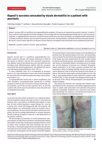
/
An expanded histopathologic spectrum of psoriasis:  review of 51 clinically confirmed An expanded histopathologic spectrum of psoriasis:  review of 51 clinically confirmed

An expanded histopathologic spectrum of psoriasis: review of 51 clinically confirmed - PowerPoint Presentation

ellena-manuel
ellena-manuel . @ellena-manuel
Follow
342 views
Uploaded On 2019-11-03

An expanded histopathologic spectrum of psoriasis: review of 51 clinically confirmed - PPT Presentation

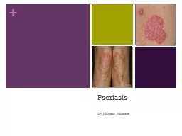
An expanded histopathologic spectrum of psoriasis review of 51 clinically confirmed cases Thinh Chau Kory K Parsi Toru Ogawa Maija Kiuru Thomas Konia Maxwell A Fung Introduction Introduction ID: 762783

dermal psoriasis clinical features psoriasis dermal features clinical histopathologic classic spongiosis acanthosis figure table hypergranulosis findings atypical vulgaris irregular

Share:

Link:

Embed:

Download Presentation from below link

Download Presentation The PPT/PDF document "An expanded histopathologic spectrum of ..." is the property of its rightful owner. Permission is granted to download and print the materials on this web site for personal, non-commercial use only, and to display it on your personal computer provided you do not modify the materials and that you retain all copyright notices contained in the materials. By downloading content from our website, you accept the terms of this agreement.


Presentation Transcript

An expanded histopathologic spectrum of psoriasis: review of 51 clinically confirmed casesThinh Chau, Kory K. Parsi, Toru Ogawa, Maija Kiuru, Thomas Konia, Maxwell A. Fung Introduction Introduction: Psoriasis is a common, chronic inflammatory skin disease affecting 2-3% of the US population. Although the classical histopathologic features of psoriasis have been well-documented for decades, the clinical manifestations of psoriasis are distinctive enough that the diagnosis is generally made on clinical features alone, both in routine practice and for clinical trials. Mirroring their clinical counterparts, the histopathologic features considered classical for psoriasis comprise a well-documented spectrum but have remained largely unchanged and unvalidated. Furthermore , there are no criteria for establishing a diagnosis of psoriasis in patients lacking classic clinical and/or histologic findings. Likewise, the range of non-classical findings that may be seen in psoriasis has not been well-documented. Consequently, when non-classical or atypical histopathologic findings are encountered, there may be uncertainty as to whether such findings are acceptable for psoriasis or indicative of an alternate diagnosis. Thus, there is a need to update the histopathologic features encompassed by psoriasis. Findings Acknowledgements Table 1. Patient Demographics and Clinical Characteristics University of California Davis, Departments of Dermatology and Pathology, Sacramento, CA Table 2. Typical and Atypical Histopathologic Features Figure 1. Irregular Acanthosis, Hypergranulosis, and Compact Orthokeratosis Figure 4. Eosinophilic Spongiosis and Dermal Eosinophils Conclusion Conclusion : Our study highlights atypical features such as irregular acanthosis, vacuolar interface changes, hypergranulosis, necrotic keratinocytes, neutrophilic spongiosis, dermal neutrophils and eosinophils that may occur in clinically confirmed psoriasis. Our study reveals that the histopathologic spectrum of psoriasis is broader than currently documented in the literature and may diminish the ability of dermatopathologists to rule out psoriasis based solely on histology. Typical histopathologic features included regular acanthosis, hypogranulosis, dermal papillae capillary ectasia, club-shaped rete ridges, suprapapillary plate thinning, Munro microabscess , and spongiform pustule of Kogoj. Atypical histopathologic features included irregular acanthosis, compact orthokeratosis, hypergranulosis, lichenoid and vacuolar interface alterations, stratum spinosum and stratum basale necrotic keratinocytes, spongiosis, eosinophilic spongiosis, dermal neutrophils, eosinophils, and plasma cells, and papillary dermal fibrosis.Minors and pregnant women were excluded from the study. Approval was obtained from the University of California Davis Institutional Review Board. The following individuals provided helpful comments for our clinical diagnostic criteria: Christine Carroll (Woodland, CA), Steve Feldman (Winston-Salem, NC), John Koo (San Francisco, CA), Marc Silverstein (Sacramento, CA), Emil Tanghetti (Sacramento, CA). Methods Methods: Clinical diagnostic criteria for psoriasis were developed, informally validated, and applied to a consecutive series of patients whose biopsies received a microscopic diagnosis of psoriasis or most likely psoriasis between 2010 and 2014, inclusive. Inclusion criteria comprised at least two of the following as documented in electronic medical records: 1. classic morphology, 2. classic distribution, 3. nail pitting, and 4. family history of psoriasis, requiring either classic morphology or distribution. Classic morphology was defined as well-circumscribed, sharply-demarcated red papules or plaques with silver-white, micaceous scale. Classic distribution was defined as involvement of the scalp, elbows, knees, or gluteal region for psoriasis vulgaris; axillary, inguinal, or intergluteal folds for inverse psoriasis; and palmoplantar for pustular psoriasis. Table 1A. Patient Demographics (n = 46, %)Age in years (median, range)53, 16-91Male (n, %)20, 43%Female (n, %)26, 57% Table 1B. Clinical Characteristic (n = 46, %)Classic Morphology42, 91%Classic Distribution 39, 84%Family History12, 26%Nail Pitting 10, 22%Psoriasis typeVulgaris30, 65%Inverse5, 11%Guttate3, 7%Palmoplantar2, 4%Erythrodermic6, 13% Table 2A. Typical Features (n=51, %)Hypogranulosis 49, 96%Club-shaped rete ridges 49, 96%Dermal papillae capillary ectasia 46 ,90%Munro microabscess 40, 78%Suprapapillary plate thinning 32, 63%Spongiform pustule of Kogoj27, 53%Regular acanthosis 7, 14% Table 2B. Atypical Features (n=51, %)Irregular acanthosis 43, 84%Junctional vacuolar alteration39, 76%Spongiosis 39, 76%Dermal neutrophils 35, 69%Hypergranulosis 33, 65%Stratum spinosum necrotic keratinocytes 31, 61%Neutrophilic spongiosis 31, 61% Dermal eosinophils25, 49%Compact orthokeratosis 19, 37%Stratum basale nectrotic keratinocytes18, 35%Papillary dermal fibrosis 18, 35%Lichenoid interface 13, 25%Dermal plasma cells 8, 16%Eosinophilic spongiosis 4, 8% Figure 1. Psoriasis vulgaris. Biopsy from the right forearm of a 37 year old woman shows irregular acanthosis, interspersed hypogranulosis and hypergranulosis, and compact orthokeratosis (arrows). Figure 2. Spongiotic Microvesicle and Junctional Vacuolar Alteration Figure 2. Psoriasis vulgaris. Biopsy from the left hand of a 75 year old woman shows a non-neutrophilic, non-eosinophilic spongiotic microvesicle and junctional vacuolar alteration (arrows). Figure 3. Lichenoid Interface Alteration and Necrotic Keratinocytes Figure 3. Psoriasis vulgaris. Biopsy from the back of a 50 year old man shows lichenoid interface alteration (arrows) and necrotic keratinocytes in the spinous and basal layers (insets). Figures 4. Psoriasis vulgaris. Biopsy from the right upper back of a 37 year old man shows eosinophilic spongiosis and dermal eosinophils ( arrows).