PPT-How t o look at bacteria &
Author : ellena-manuel | Published Date : 2019-06-26
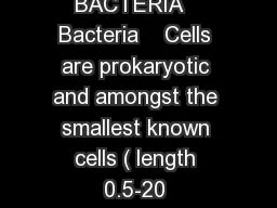
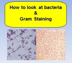
Gram Staining There are a couple pieces of data to collect from bacteria 1 how many bacteria grew on your plate count colonies 2 surface area of plate covered with
Presentation Embed Code
Download Presentation
Download Presentation The PPT/PDF document "How t o look at bacteria &" is the property of its rightful owner. Permission is granted to download and print the materials on this website for personal, non-commercial use only, and to display it on your personal computer provided you do not modify the materials and that you retain all copyright notices contained in the materials. By downloading content from our website, you accept the terms of this agreement.
How t o look at bacteria &: Transcript
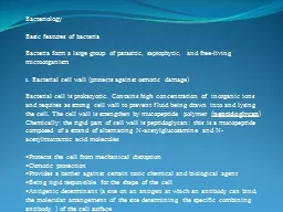
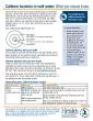
Gram Staining There are a couple pieces of data to collect from bacteria 1 how many bacteria grew on your plate count colonies 2 surface area of plate covered with growth estimate using graph paper. Mrs. Stewart. Medical Interventions. Central Magnet School. 2 Classes of Bacteria. Gram (-) . Thin layer of . peptidoglycan. Lipopolysaccharides. (. endotoxins. ). Stain red. Gram (+). Thick layer of . Helpful Bacteria. Most bacteria are harmless to humans.. Many are useful:. Aid in digestion. Provide nutrients for plants by breaking down dead material by decomposition. Provide drugs and hormones. Provide some types of food.. Bacterial Taxonomy: How are these unicellular organisms classified?. complex system of classification. based on shape & size; oxygen, pH, and temperature requirements; laboratory characteristics, biochemical . Biology and Biotechnology department. A large number of bacteria . are . motile. . . . Most possess one or more . flagella on their surface . that allow . them to swim. .. The . pattern of . flagellation. Dr. Yasir . A. Hussein, MD, Microbiology & Pathology. . Update, September 2016. Overview. . Introduction to anaerobic bacteria.. Types of . anaerobic . . 19, . Ch. 23, and more . Bacteria & Virus Overview. Interesting stuff!. We live with bacteria and viruses constantly, but most people don’t know much about them.. In this chapter we learn both the good and bad of each. . Bacteria form a large group of parasitic, saprophytic, and free-living microorganism . . 1. Bacterial cell wall (protects against osmotic damage). . Bacterial cell is prokaryotic. Contains high concentration . CHAPTER 27. PROKARYOTIC ADAPTATIONS. typical prokaryote:. 0.5 -5 microns. unicellular. variety of shapes. cocci. (spherical). bacilli (rods). spirochetes (corkscrews). Cell-Surface Structures. nearly all have cell wall. What are the characteristics of viruses? Bacteria? What kingdom do each of these belong? Are they living? Why or why not. ?. What do you already know?. What are the differences between viruses and bacteria?. Internal Structures:. cytoplasm. nucleoid. ribosomes. Boundaries:. cell membrane. cell wall. capsule. Appendages:. flagellum. pili. Shapes of Bacteria. Bacteria Identification Criteria. Shapes. Lack a nucleus. DNA is naked, a single loop not bound in a chromosome. May contain plasmids (small circular fragments of DNA). Have ribosomes, but no other organelles.. DNA present as a long circular molecule.. and . Function. Bacterial . Form . and Function. A. . Structures common to . all. bacterial cells. 1. . Cell membrane. 2. . Cytoplasm. 3. . Ribosomes. 4. . One (or a few) . chromosomes. Bacterial Form . Simple, one-celled organisms. Multiply rapidly. Killed by antibiotics. Some have become antibiotic resistant. Classified by shape & arrangement. Shapes of Bacteria. Bacteria Classes. Cocci. =round or spherical. E. coliTotal coliformTotal coliform = Environmental contaminationFecal coliformFecal coliform and E. coli = Fecal contaminationE. coli O157:H7 You should test for coliform bacteria at least once a yea
Download Rules Of Document
"How t o look at bacteria &"The content belongs to its owner. You may download and print it for personal use, without modification, and keep all copyright notices. By downloading, you agree to these terms.
Related Documents