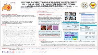
A CASE REPORT I GANZOUI Y AROUS R AOUINI M LANDOLSI S KOUKI H BOUJEMAA N BEN ABDALLAH Radiology Department Military Hospital of Tunis Montfleury Tunis Tunisia HN4 ID: 918871
Download Presentation The PPT/PDF document "CT and MRI FINDINGS IN LOCALIZED NASOPHA..." is the property of its rightful owner. Permission is granted to download and print the materials on this web site for personal, non-commercial use only, and to display it on your personal computer provided you do not modify the materials and that you retain all copyright notices contained in the materials. By downloading content from our website, you accept the terms of this agreement.
Slide1
CT and MRI FINDINGS IN LOCALIZED NASOPHARYNGEAL AMYLODOSIS : A CASE REPORT
I. GANZOUI, Y. AROUS, R. AOUINI, M. LANDOLSI, S. KOUKI, H. BOUJEMAA, N. BEN ABDALLAHRadiology Department, Military Hospital of Tunis, Montfleury, Tunis, Tunisia
HN4
Slide2INTRODUCTION
Amyloidosis is a group of disorders that result from the deposition of amyloid proteins in organs and tissues.
Localized form of amyloidosis in the head and neck is extremely rare.
the involvement of the nasopharynx is still more uncommon.
Slide3A 66-year-old woman
presented with : breathing and food swallowing difficulties
bilateral hypoacousia
chronic bilateral nasal obstruction
bilateral enlarging neck masses.
OBSERVATION
Slide4Physical examination revealed : yellowish polypoid mass developing in the rhinopharynx completely obstructing nasal fossa, bulging behind the uvula and the soft palate.
bilateral cervical lymph nodes
Biopsy under nasopharyngoscopy wasperformed.The histological findings concluded
to a type AL amyloidosis with no evidence of malignancy.
Slide6CT and MRI were performed in order to determine the local extent before surgery.
The possibility of a partial representation of apotential systemic amyloidosis was excluded.
Slide7CT FINDINGSHomogeneous significant thickening of the naso and oropharynx walls bulging into the nasal fossa.
Bilateral enlarged cervical lymph nodes.No bone erosion was seen.
Slide8Slide9MRI FINDINGSLow intensity heterogeneous thickening and masses in T2 weighted images.
Slightly hypointense in T1 weighted images.Heteregeneous enhancement.
Slide10Axial T1 weighted images
Slide11Axial and coronal T2 weighted images
Slide12Axial T1 weighted with fat saturation images after Gadolinium injection
Slide13Coronal and sagittalT1 weighted images after Gadolinium injection
Slide14DISCUSSIONLocalized amyloidosis has been reported in major and minor salivary glands, oral cavity, pharynx, sinonasal cavities, larynx, trachea, bronchi and lungs.
The larynx is by far the most common site of localized amyloid disease in the respiratory tract.
Localized amyloidosis in the nasopharynx is rare.
Amyloid deposits appear as relatively well-defined submucosal masses in CT scans with no evidence of bone destruction and intracranial extension.
Such deposits usually appear homogeneous, except for the presence of calcification.Slight contrast enhancement.
Slide16The presence of calcification helps to narrow the differential diagnosis.
In nasopharyngeal amyloidosis with neck calcification on CT scans, the possibility of neck amyloid deposition should be considered.
Slide17It is well known that such a lesion shows low intensity on a T2-weighted image of MRI.
Nasopharyngeal amyloidosis shows slight or no enhancement.
Being a hypocellular lesion, it should show delayed
enhancement rather than early enhancement on dynamic contrast enhanced examination.
Slide18Some reports state that plasmacytes are occasionally observed within a mass of amyloid deposit and might produce amyloid-related proteins.It is important to know that plasmacyte infiltration, which occasionally occurs with amyloidosis, shows high intensity on T2 weighted images and remarkable early enhancement on dynamic MRI.
Slide19CONCLUSION Although rare, localized
amyloidosis should be considered in the differential diagnosis of head and neck masses.CT and MRI help in delineating the local extent.
Calcification on CT and low intensity on T2 weighted images are suggestive of the diagnostic