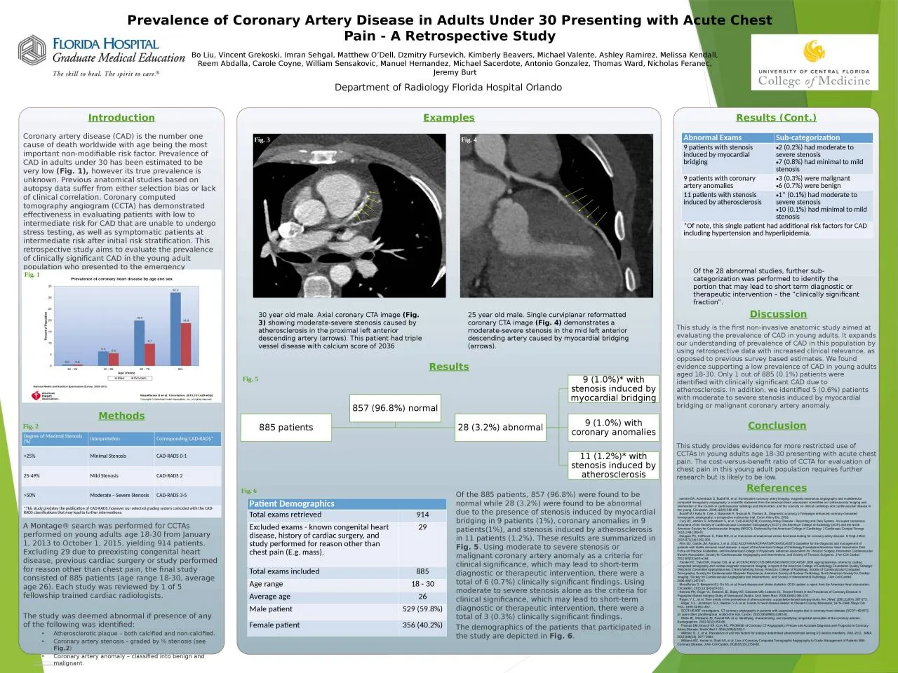
/
Coronary artery disease (CAD) is the number one cause of death Coronary artery disease (CAD) is the number one cause of death

Coronary artery disease (CAD) is the number one cause of death - PowerPoint Presentation

emery
emery . @emery
Follow
342 views
Uploaded On 2022-02-14

Coronary artery disease (CAD) is the number one cause of death - PPT Presentation

worldwide with age being the most important nonmodifiable risk factor Prevalence of CAD in adults under 30 has been estimated to be very low Fig 1 however its true prevalence is unknown ID: 908844

patients coronary artery disease coronary patients disease artery study stenosis cad american fig heart society prevalence cardiovascular severe moderate

Share:

Link:

Embed:

Download Presentation from below link

Download Presentation The PPT/PDF document "Coronary artery disease (CAD) is the num..." is the property of its rightful owner. Permission is granted to download and print the materials on this web site for personal, non-commercial use only, and to display it on your personal computer provided you do not modify the materials and that you retain all copyright notices contained in the materials. By downloading content from our website, you accept the terms of this agreement.


Presentation Transcript

Slide1

Coronary artery disease (CAD) is the number one cause of death worldwide with age being the most important non-modifiable risk factor. Prevalence of CAD in adults under 30 has been estimated to be very low (Fig. 1), however its true prevalence is unknown. Previous anatomical studies based on autopsy data suffer from either selection bias or lack of clinical correlation. Coronary computed tomography angiogram (CCTA) has demonstrated effectiveness in evaluating patients with low to intermediate risk for CAD that are unable to undergo stress testing, as well as symptomatic patients at intermediate risk after initial risk stratification. This retrospective study aims to evaluate the prevalence of clinically significant CAD in the young adult population who presented to the emergency department for chest pain as determined by findings on CCTA.

Introduction

This study is the first non-invasive anatomic study aimed at evaluating the prevalence of CAD in young adults. It expands our understanding of prevalence of CAD in this population by using retrospective data with increased clinical relevance, as opposed to previous survey based estimates. We found evidence supporting a low prevalence of CAD in young adults aged 18-30. Only 1 out of 885 (0.1%) patients were identified with clinically significant CAD due to atherosclerosis. In addition, we identified 5 (0.6%) patients with moderate to severe stenosis induced by myocardial bridging or malignant coronary artery anomaly. This study provides evidence for more restricted use of CCTAs in young adults age 18-30 presenting with acute chest pain. The cost-versus-benefit ratio of CCTA for evaluation of chest pain in this young adult population requires further research but is likely to be low.

References

luemke DA, Achenbach S, Budoff M, et al. Noninvasive coronary artery imaging: magnetic resonance angiography and multidetector computed tomography angiography: a scientific statement from the american heart association committee on cardiovascular imaging and intervention of the council on cardiovascular radiology and intervention, and the councils on clinical cardiology and cardiovascular disease in the young. Circulation. 2008;118(5):586-606.Budoff MJ, Kalia N, Cole J, Nakanishi R, Nezarat N, Thomas JL. Diagnostic accuracy of Visipaque enhanced coronary computed tomographic angiography: a prospective multicenter trial. Coron Artery Dis. 2016;Cury RC, Abbara S, Achenbach S, et al. CAD-RADS(TM) Coronary Artery Disease - Reporting and Data System. An expert consensus document of the Society of Cardiovascular Computed Tomography (SCCT), the American College of Radiology (ACR) and the North American Society for Cardiovascular Imaging (NASCI). Endorsed by the American College of Cardiology. J Cardiovasc Comput Tomogr. 2016;10(4):269-81.Douglas PS, Hoffmann U, Patel MR, et al. Outcomes of anatomical versus functional testing for coronary artery disease. N Engl J Med. 2015;372(14):1291-300.Fihn SD, Gardin JM, Abrams J, et al. 2012 ACCF/AHA/ACP/AATS/PCNA/SCAI/STS Guideline for the diagnosis and management of patients with stable ischemic heart disease: a report of the American College of Cardiology Foundation/American Heart Association Task Force on Practice Guidelines, and the American College of Physicians, American Association for Thoracic Surgery, Preventive Cardiovascular Nurses Association, Society for Cardiovascular Angiography and Interventions, and Society of Thoracic Surgeons. J Am Coll Cardiol. 2012;60(24):e44-e164.Hendel RC, Patel MR, Kramer CM, et al. ACCF/ACR/SCCT/SCMR/ASNC/NASCI/SCAI/SIR 2006 appropriateness criteria for cardiac computed tomography and cardiac magnetic resonance imaging: a report of the American College of Cardiology Foundation Quality Strategic Directions Committee Appropriateness Criteria Working Group, American College of Radiology, Society of Cardiovascular Computed Tomography, Society for Cardiovascular Magnetic Resonance, American Society of Nuclear Cardiology, North American Society for Cardiac Imaging, Society for Cardiovascular Angiography and Interventions, and Society of Interventional Radiology. J Am Coll Cardiol. 2006;48(7):1475-97.Mozaffarian D, Benjamin EJ, Go AS, et al. Heart disease and stroke statistics--2015 update: a report from the American Heart Association. Circulation. 2015;131(4):e29-322.Nemetz PN, Roger VL, Ransom JE, Bailey KR, Edwards WD, Leibson CL. Recent Trends in the Prevalence of Coronary Disease: A Population-Based Autopsy Study of Nonnatural Deaths. Arch Intern Med. 2008;168(3):264-270.Roger, V. L., et al. Time trends in the prevalence of atherosclerosis: a population-based autopsy study. Am J Med. 2001;110(4): 267-273. Roger, V.L., Jacobsen, S.J., Weston, S.A. et al, Trends in heart disease deaths in Olmsted County, Minnesota, 1979–1994. Mayo Clin Proc. 1999;74:651–657.SCOT-HEART investigators. CT coronary angiography in patients with suspected angina due to coronary heart disease (SCOT-HEART): an open-label, parallel-group, multicentre trial. Lancet. 2015;385(9985):2383-91.Shriki JE, Shinbane JS, Rashid MA, et al. Identifying, characterizing, and classifying congenital anomalies of the coronary arteries. Radiographics. 2012;32(2):453-68.Thomas DM, Branch KR, Cury RC. PROMISE of Coronary CT Angiography: Precise and Accurate Diagnosis and Prognosis in Coronary Artery Disease. South Med J. 2016;109(4):242-7.Webber, B. J., et al. Prevalence of and risk factors for autopsy-determined atherosclerosis among US service members, 2001-2011. JAMA. 2012;308(24): 2577-2583.Williams MC, Hunter A, Shah AS, et al. Use of Coronary Computed Tomographic Angiography to Guide Management of Patients With Coronary Disease. J Am Coll Cardiol. 2016;67(15):1759-68.

Department of Radiology Florida Hospital Orlando

Bo Liu, Vincent

Grekoski, Imran Sehgal, Matthew O’Dell, Dzmitry Fursevich, Kimberly Beavers, Michael Valente, Ashley Ramirez, Melissa Kendall, Reem Abdalla, Carole Coyne, William Sensakovic, Manuel Hernandez, Michael Sacerdote, Antonio Gonzalez, Thomas Ward, Nicholas Feranec, Jeremy Burt

Prevalence of Coronary Artery Disease in Adults Under 30 Presenting with Acute Chest Pain - A Retrospective Study

Fig. 1

Degree of Maximal Stenosis (%)

Interpretation

Corresponding CAD-RADS*<25%Minimal StenosisCAD-RADS 0-125-49%Mild StenosisCAD-RADS 2>50%Moderate – Severe StenosisCAD-RADS 3-5*This study predates the publication of CAD-RADS, however our selected grading system coincided with the CAD-RADS classifications that may lead to further interventions.

Abnormal ExamsSub-categorization9 patients with stenosis induced by myocardial bridging2 (0.2%) had moderate to severe stenosis7 (0.8%) had minimal to mild stenosis9 patients with coronary artery anomalies3 (0.3%) were malignant6 (0.7%) were benign11 patients with stenosis induced by atherosclerosis1* (0.1%) had moderate to severe stenosis10 (0.1%) had minimal to mild stenosis*Of note, this single patient had additional risk factors for CAD including hypertension and hyperlipidemia.

Patient DemographicsTotal exams retrieved914Excluded exams - known congenital heart disease, history of cardiac surgery, and study performed for reason other than chest pain (E.g. mass).29Total exams included885Age range18 - 30Average age 26Male patient529 (59.8%)Female patient356 (40.2%)

A Montage® search was performed for CCTAs performed on young adults age 18-30 from January 1, 2013 to October 1, 2015, yielding 914 patients. Excluding 29 due to preexisting congenital heart disease, previous cardiac surgery or study performed for reason other than chest pain, the final study consisted of 885 patients (age range 18-30, average age 26). Each study was reviewed by 1 of 5 fellowship trained cardiac radiologists.The study was deemed abnormal if presence of any of the following was identified:Atherosclerotic plaque – both calcified and non-calcified.Coronary artery stenosis – graded by % stenosis (see Fig.2)Coronary artery anomaly – classified into benign and malignant.

30 year old male. Axial coronary CTA image (Fig. 3) showing moderate-severe stenosis caused by atherosclerosis in the proximal left anterior descending artery (arrows). This patient had triple vessel disease with calcium score of 2036

25 year old male. Single curviplanar reformatted coronary CTA image (Fig. 4) demonstrates a moderate-severe stenosis in the mid left anterior descending artery caused by myocardial bridging (arrows).

Discussion

Results

Methods

Examples

Fig. 2

Of the 885 patients, 857 (96.8%) were found to be normal while 28 (3.2%) were found to be abnormal due to the presence of stenosis induced by myocardial bridging in 9 patients (1%), coronary anomalies in 9 patients(1%), and stenosis induced by atherosclerosis in 11 patients (1.2%). These results are summarized in Fig. 5. Using moderate to severe stenosis or malignant coronary artery anomaly as a criteria for clinical significance, which may lead to short-term diagnostic or therapeutic intervention, there were a total of 6 (0.7%) clinically significant findings. Using moderate to severe stenosis alone as the criteria for clinical significance, which may lead to short-term diagnostic or therapeutic intervention, there were a total of 3 (0.3%) clinically significant findings. The demographics of the patients that participated in the study are depicted in Fig. 6.

Fig.

3

Fig. 4

Fig.

5

Fig.

6

Results (Cont.)

Of the 28 abnormal studies, further sub-categorization was performed to identify the portion that may lead to short term diagnostic or therapeutic intervention – the “clinically significant fraction”.

Conclusion