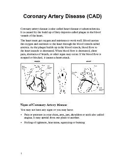
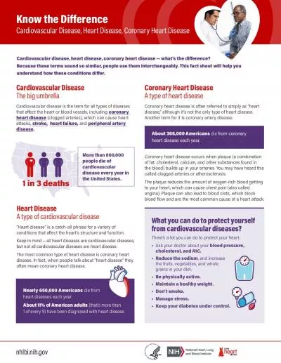
Most common form of heart disease Can develop to become Chronic stable angina Acute coronary syndrome Unstable angina Myocardial infarction Can occur in any artery Prefers coronary arteries ID: 775387
Download Presentation The PPT/PDF document " Coronary artery disease And nursing imp..." is the property of its rightful owner. Permission is granted to download and print the materials on this web site for personal, non-commercial use only, and to display it on your personal computer provided you do not modify the materials and that you retain all copyright notices contained in the materials. By downloading content from our website, you accept the terms of this agreement.
Slide1
Coronary artery disease
And nursing implications
Slide2Most common form of heart diseaseCan develop to become:Chronic stable angina Acute coronary syndromeUnstable anginaMyocardial infarctionCan occur in any arteryPrefers coronary arteries
Etiology & Pathophysiology
Slide3Barrier between the vessel wall and the lumen of the vesselNonreactive to platelets and leukocytes Nonreactive to coagulation, fibrinolytic, and complement factorsEndothelial wall alters as a result of inflammation and injury
Normal Endothelium & Injury Response
Slide4Fatty Streak
Earliest lesionsCharacterized by lipid-filled smooth muscle cellsCan begin as early as age 15Potentially reversible with lowering LDL cholesterol
Slide5Fibrous Plaque
Beginning of progressive changes in the arterial wallCan appear by age 30 and increases with ageLipoproteins transport cholesterol and other lipids into the arterial intimaFatty streak is covered by collagen, forming a fibrous plaque Narrows the artery and a reduces blood flow to the distal tissues
Slide6Complicated Lesion
Continued inflammation resulting in plaque instability, ulceration, and rupturePlatelets accumulate and thrombus formsIncrease narrowing or total occlusion of lumen
Slide7Factors contributing to the growth and extent of collateral circulationInherited predisposition to develop new blood vesselsPresence of chronic ischemiaWhen occlusion occurs slowly there is an increased chance of adequate collateral circulation and adequate myocardial blood flowWith rapid onset CAD or coronary spasm, there is not enough time to establish collateral vesselsDiminished arterial flow results in more severe ischemia or infarction
Collateral Circulation
Slide8Risk factors for CAD
Slide9AgeRisk increases with ageGenderIncidence is highest in white middle-aged menAfter age 65, incidence in men and women similarWomen tend to manifest CAD 10 years later than menWhen symptoms develop, women experience symptoms of angina rather than MI – opposite is true in men
Nonmodifiable Risk Factors
Slide10EthnicityWhite middle-aged men have highest incidence African American women have a higher incidence and death rate compared to white womenNative Americans have mortality rates 2x high as other AmericansFamily historyFamilial hypercholesterolemiaGenetic predispositionAutosomal dominant disorder
Nonmodifiable
Risk Factors
Slide11Elevated serum lipidsCholesterol >200 mg/dlTriglycerides >150 mg/dlLipoproteinsHDLs - high density high levels desirablelow levels associated with risk for CAD LDLs - low densityElevated levels correlate most closely with increased incidence of CAD
Modifiable Risk Factors
Slide12Elevated blood pressureHypertension: BP > 140/90 Increases risk of atherosclerosisTobacco useNicotine can cause catecholamine release HR, peripheral vasoconstriction, BPPlatelet adhesion leading to emboli formation
Modifiable Risk Factors
Slide13Physical inactivityPeople who are active have higher HDLsExercise increases collateral circulation & lowers BPObesityBMI > 30 kg/m2Leads to increased LDLs and triglyceridesAssociated with hypertensionPeople who are apple-shaped (store fat in abdomen) have higher incidence of CAD
Modifiable Risk Factors
Slide14Diabetes & Metabolic syndromeIncidence of CAD 2-4x higher ObesityHypertensionElevated triglycerides, abnormal serum lipids, elevated fasting blood glucoseInsulin resistance
Modifiable Risk Factors
Slide15Psychological statesIncrease risk of CADInclude depression, hopelessness, anxiety, hostility, & angerStress correlated with CADElevated homocysteine levelDamage the inner lining of blood vesselsPromote plaque build-upAlter the clotting mechanism to make clots more likely to occur
Modifiable Risk Factors
Slide16Prevention & Treatment
Slide17Health Promotion
Hypertension
Have
regular blood pressure
checkups
Take
prescribed medications for blood pressure
control
Reduce
salt
intake
Stop
tobacco use; avoid exposure to environmental tobacco (secondhand)
smoke
Control
or reduce
weight
Perform
physical activity
regularly
Slide18Health Promotion
Elevated serum lipids
Reduce
total fat
intake
Reduce
animal (saturated) fat
intake
Take
prescribed medications for lipid
reduction
Adjust
total caloric intake to achieve and maintain ideal body
weight
Engage
in regular physical
activity
Increase
amount of complex carbohydrates and vegetable proteins in
diet
Slide19Health Promotion
Tobacco use
Enroll
in a smoking cessation
program
Change
daily routines associated with smoking to reduce desire to
smoke
Substitute
other activities for
smoking
Ask
caregivers to support efforts to stop
smoking
Avoid
exposure to environmental tobacco
smoke
Slide20Health Promotion
Physical Inactivity
Develop
and maintain at least 30 minutes of moderate physical activity on most days of the
week
Increase
activities to a fitness
level
Slide21Health Promotion
Psychological State
Increase
awareness of behaviors that are detrimental to
health
Alter
patterns that are conducive to
stress
Set
realistic goals for
self
Reassess
priorities in light of health
needs
Learn
effective stress management
strategies
Seek
professional help if feeling depressed, angry
, or anxious
Plan
time for adequate rest and
sleep
Slide22Health Promotion
Obesity
Change
eating patterns and
habits
Reduce
caloric intake to achieve
BMI of <25
Increase
physical activity to increase caloric
expenditure
Avoid
fad and crash diets, which are not effective over
time
Avoid
large, heavy
meals
Slide23Health Promotion
Diabetes
Follow
the recommended
diet
Control
or reduce
weight
Take
prescribed
anti-diabetic medications
Monitor
blood glucose levels
regularly
Slide24Calorie restrictionsDecrease dietary fat/cholesterolLimit saturated fats and cholesterol and emphasize complex carbohydratesFats – only about 30% of caloriesReduce or omit red meats, eggs, whole milkOmega 3 fatty acids
Nutritional Therapy
Slide25Thorough health historyAccurate medication listPrescriptionOver the counterPresence of cardiovascular symptomsEnvironmental patterns like diet and activityValues and beliefs about health and illness
Identify people at risk
Slide26Drugs that restrict lipoprotein productionStatinsInhibit synthesis of cholesterol in the liverNeed to monitor liver enzymesCreatine kinase assessed if myopathy suspectedNiacinInterferes with the synthesis of LDL and triglyceridesIncreases HDLCan cause flushing, pruritus, GI complaints, orthostatic hypotensionFlushing may be prevented by aspirin or NSAIDs LopidLower triglyceridesSide effects may include rashes and mild GI disturbances
Cholesterol Lowering Drug Therapy
Slide27Drugs that increase lipoprotein removalBile acid sequestrants (e.g., cholestyramine [Questran])Increase conversion of cholesterol to bile acidsDecrease hepatic cholesterolSide effects include complaints of palatability and upper and lower GI symptomsDrugs that decrease cholesterol absorptionZetia
Cholesterol Lowering Drug Therapy
Slide28ASA , PlavixLow dose ASA (81mg)Studies show decrease in first MI’sCan cause GI bleedUse if benefit outweighs riskPlavix has decreased risk of bleed
Antiplatelet Drug
Therapy
Slide29Clinical Manifestations of CAD
Slide30Temporary myocardial ischemia = Angina (chest pain)Happens when oxygen supply is less than oxygen demandCardiacNon cardiacPrimary reason for insufficient blood flow is narrowing of coronary arteries by atherosclerosis
Chronic Stable Angina
Slide31Physical exertionTemperature extremesStrong emotionsConsumption of heavy mealTobacco use or environmental tobacco smokeSexual activityStimulantsCircadian rhythm patterns
Precipitating Factors of Angina
Slide32Referred pain in left shoulder and arm is from transmission of the pain message to the cardiac nerve rootsPain usually lasts 3 to 5 minutesPain subsides when precipitating factor relievedPain at rest is unusualECG reveals ST-segment depression and/or T-wave inversion
Chronic Stable Angina
Slide33Intermittent chest pain that occurs over a long period with the same pattern of onset, duration, and intensity of symptomsConstrictiveSqueezingHeavyChokingSuffocating sensation
Chronic Stable Angina
Slide34Angina Pain Sites
Most of the pain experienced in angina appears substernally, it may also occur in the neck May radiate to:JawShouldersDown the armsPain between the shoulder blades often is dismissed as not being heart related
Slide35Silent IschemiaIschemia that occurs in the absence of any subjective symptoms Associated with diabetic neuropathyConfirmed by ECG changesUp to 80% of patients with ischemia are asymptomaticIschemia with or without pain has same prognosis
Types of Stable Angina
Slide36Nocturnal anginaOccurs only at night but not necessarily during sleep or in recumbent positionAngina decubitusChest pain that occurs only while lying downRelieved by standing or sitting
Types of Stable Angina
Slide37Prinzmetal’s AnginaOccurs at rest usually in response to spasm of major coronary arterySeen in patients with a history of migraine headaches and Raynaud’s phenomenonSpasm may occur in the absence of CAD.When spasm occursChest painMarked, transient ST-segment elevationMay occur during REM sleepMay be relieved by moderate exercise or may disappear spontaneously
Types of Stable Angina
Slide38Microvascular AnginaMay occur in the absence of significant coronary atherosclerosis or coronary spasmSeen especially in womenPain is related to myocardial ischemia associated with abnormalities of the coronary microcirculation.
Types of Stable Angina
Slide39Acute Coronary Syndrome
Slide40Progression of Cardiovascular disease
Slide41When ischemia is prolonged and is not immediately reversible, acute coronary syndrome (ACS) develops.ACS encompassesUnstable angina (UA)Non–ST-segment-elevation myocardial infarction (NSTEMI)ST-segment-elevation MI (STEMI)
Acute Coronary Syndrome
Slide42Result Partial occlusion of coronary artery: USA or NSTEMITotal occlusion of coronary artery: STEMI
Progression of CAD
Slide43Chest pain that is:New in onsetOccurs at restHas a worsening patternChronic stable angina can develop to unstableSignificant change in the pattern of painIncreasing frequencyEasily provoked by minimal or no exertionUnpredictable and represents a medical emergency
Unstable Angina
Slide44Result of sustained ischemia (>20 minutes), causing irreversible myocardial cell death (necrosis)Necrosis of entire thickness of myocardium takes 4 to 6 hours. Eighty percent to 90% of all acute MIs are secondary to thrombus formation
Myocardial Infarction
Slide45Infarctions are usually described according to the location of damageAnteriorInferiorlateralposterior wall infarction
Myocardial Infarction
Slide46The degree of altered function depends on the area of the heart involved and the size of the infarct.Contractile function of the heart is disrupted in areas of myocardial necrosis.Most MIs involve the left ventricle (LV).
Myocardial Infarction
Slide47Pain Total occlusion → Anaerobic metabolism and lactic acid accumulation → Severe, immobilizing chest pain not relieved by rest, position change, or nitrate administrationDescribed as heaviness, constriction, tightness, burning, pressure, or crushing Common locations: Substernal, retrosternal, or epigastric areas; pain may radiate to neck, jaw, arms
Clinical Manifestations of ACS
Slide48Stimulation of sympathetic nervous system results in Release of glycogenDiaphoresisVasoconstriction of peripheral blood vesselsSkin: Ashen, clammy, and/or cool to touch
Clinical Manifestations of ACS
Slide49CardiovascularInitially, ↑ HR and BP, then ↓ BP (secondary to ↓ in cardiac output) Crackles Jugular venous distentionAbnormal heart soundsS3 or S4New murmur
Clinical Manifestations of ACS
Slide50Nausea and vomitingCan result from reflex stimulation of the vomiting center by severe painFeverSystemic manifestation of the inflammatory process caused by cell death
Clinical Manifestations of ACS
Slide5112-lead ECGCan rule out or confirm MIChanges in QRS complex, ST segment, & T waveNot always evident shortly after infarctSerum cardiac markersReleased into blood from necrotic heart muscle after MITroponinCK-MBMyoglobinSerial
Diagnostic Studies
Slide52ECG Changes
Slide53ECG Changes
Slide54Coronary angiographyCardiac catheterizationPercutaneous coronary intervention (PCI)StentBalloon angioplastyStress testingExerciseDrugPersantineAdenocardEchocardiogram
Diagnostic Studies
Slide55DysrhythmiasAbnormal heart rhythmsMost common complicationPresent in 80% of MI patientsMost common cause of death in the pre-hospital periodLife-threatening dysrhythmias seen most often with anterior MI, heart failure, or shock
Complications
Slide56Heart failureA complication that occurs when the pumping power of the heart has diminishedSymptomsmild dyspnearestlessnessagitationslight tachycardia. pulmonary congestion, crackles jugular vein distention
Complications
Slide57Cardiogenic shockOccurs when inadequate oxygen and nutrients are supplied to the tissues because of severe LV failureMortality rate is highSigns and symptomsLow BPConfusionpale, clammy skinDecreased urine outputRequires aggressive managementcontrol of dysrhythmiasIntra-aortic balloon pump (IABP) therapyvasoactive drugs
Complications
Slide58Papillary muscle dysfunctionCauses mitral valve regurgitationCondition aggravates an already compromised LVNew systolic murmur at the cardiac apexConfirmed with echocardiogramRequires surgical treatment
Complications
Slide59Ventricular aneurysmResults when the infarcted myocardial wall becomes thinned and bulges out during contractionSigns and symptomsChest painVentricular arrhythmiasHeart failureTreatmentAnticoagulationSurgery
Complications
Slide60Acute pericarditisAn inflammation of visceral and/or parietal pericardiumOccurs 2-3 days after MIMay result in cardiac compression, ↓ LV filling and emptying, heart failureSigns and symptomsaggravated by inspiration, coughing, and movement of the upper bodyPericardial friction rub may be heard on auscultationChest pain different from MI pain Treatment includes pain relief with aspirin or corticosteroids
Complications
Slide61Dressler syndromeMay also occur after cardiac surgeryCharacterized by pericarditis with effusion and fever that develop 4 to 6 weeks after MIArthralgiaThought to be caused by an antigen-antibody reaction to the necrotic myocardiumTreatmentShort-term corticosteroids
Complications
Slide62Collaborative Care of ACS
Slide63Maintain cardiac outputTreat painAssess for complicationsIncrease activity toleranceRelieve anxietyOngoing and discharge teaching
Nursing Goals: AMI
Slide64Emergency managementInitial interventionsRapid assessment and diagnosisAdminister oxygen therapy12 lead ECGVS, pulse oximetry Assess lung and heart soundsEstablish IV Pain managementAdminister sublingual nitroglycerinAdminister aspirin (chewable)Morphine sulfate (if nitro not effective)
Collaborative Care of ACS
Slide65Ongoing interventionsFrequent VS and pulse oximetryBedrest for 12 – 24 hoursclear liquid dietNSTEMIASA, heparin, LovenoxPercutaneous coronary intervention (PCI) – once stabilizedSTEMIThrombolytic therapy
Collaborative Care of ACS
Slide66Myocardial Revascularization – Emergent PCITreatment of choice for confirmed STEMI90 minute door to table timeAmbulatory 24 hours after the procedureVariety of proceduresPercutaneous transluminal coronary angioplasty (PTCA)Intracoronary stentingDrug-eluting stents
Collaborative Care of ACS
Slide67Advantages of PCIProvide alternative to surgical interventionPerformed under local anestheticPatient ambulatory 24 hours after procedureLength of hospital stay decreased1-3 days vs. 4–6 days after CABG Rapid return to work Return to work 5–7 days vs. 2–8 weeks after CABG
Collaborative Care of ACS
Slide68Nursing care post PCIMonitoring for signs of recurrent anginaMonitor VSEvaluating groin site for signs of bleedingMonitor for infectionAssess CMS of extremity usedAssess for hematoma formationBed rest
Collaborative Care of ACS
Slide69Thrombolytic therapyIndications and contraindications History of or current major bleeding problem (hemorrhagic stroke, GI bleed) prevents administration2nd choice for revascularizationAdministered at facilities that do not have PCI capabilitiesMay still need PCI after thrombolytics if not fully effectiveMarker of reperfusion: Return of ST segment to baselineMajor complication: BleedingFFP is used to reverse thrombolytics
Collaborative Care of ACS
Slide70Time is muscle; 6-hour windowSeveral thrombolytic agents available, such as:Tissue plasminogen activator (t-PA)StreptokinaseReteplaseHeparin and Lovenox
Collaborative Care of ACS
Slide71Coronary Artery Bypass GraftingConstruction of new conduitsRequiresSternotomyCardiopulmonary bypassCommon artery used:Internal mammary artery (IMA)Saphenous vein
Collaborative Care of ACS
Slide72Indications for CABGUnstable anginaAMIFailure of percutaneous interventionsGoals of CABGIncrease blood flow to myocardiumRelieve symptomsProlong survivalImprove quality of life
Collaborative Care of ACS
Slide73Antiplatelet aggregation therapyChewable ASA 160 to 325 mg Nitroglycerin SLGiven q 5 minutes 3 doses total IV nitroglycerinGoal: reduce anginal pain and improve coronary blood flowImmediate onset of actionTitrated for pain reliefCommon side effects:HYPOTENSIONHEADACHE
Drug Therapy
Slide74Morphine sulfateGiven if chest pain not relieved with nitro SLAlso acts as vasodilator and decreases myocardial oxygen consumption, reduces contractility, and decreased BP and HRAlso reduces anxiety and fearβ-adrenergic blockersDecrease myocardial oxygen demand by reducing HR, BP, and contractilityIV administration in acute phasei.e. metoprolol (Lopressor)
Drug Therapy
Slide75Angiotensin-converting enzyme (ACE) inhibitors Prevent ventricular remodeling and prevent or slow progression of HFi.e. captopril (Capoten), enalapril (Vasotec)Antidysrhythmia drugsDysrhythmias are only treated in life threatening
Drug Therapy
Slide76Cholesterol-lowering drugsMust first obtain fasting lipid panel If LDL is elevated, cholesterol-lowering drugs are initiatedStool softenersPromote bowel movementPrevents straining and vagal stimulation from Valsalva maneuver
Drug Therapy
Slide77Acute PainDecreased Cardiac OutputAnxietyActivity intoleranceRisk for ineffective tissue perfusion
Nursing Diagnoses