/
Inflammatory conditions of the pulp Inflammatory conditions of the pulp

Inflammatory conditions of the pulp - PowerPoint Presentation

essencemessage
essencemessage . @essencemessage
Follow
342 views
Uploaded On 2020-06-17

Inflammatory conditions of the pulp - PPT Presentation

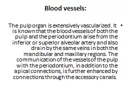
Several factors make the pulp unique The pulp is almost totally surrounded by a hard tissue dentin The pulp has almost a total lack of collateral circulation The pulp possesses unique set of cells the ID: 779921

pulpitis pulp irreversible pain pulp pulpitis pain irreversible tissue cells percussion necrosis inflammatory inflammation canal reversible root treatment test

Share:

Link:

Embed:

Download Presentation from below link

Download The PPT/PDF document "Inflammatory conditions of the pulp" is the property of its rightful owner. Permission is granted to download and print the materials on this web site for personal, non-commercial use only, and to display it on your personal computer provided you do not modify the materials and that you retain all copyright notices contained in the materials. By downloading content from our website, you accept the terms of this agreement.


Presentation Transcript

Slide1

Inflammatory conditions of the pulp

Slide2

Several factors make the pulp

unique

The pulp is almost totally surrounded by a hard tissue (dentin)

The pulp has almost a total lack of collateral circulation

The pulp possesses unique set of cells, the

odontoblasts

as well as

undif

.

Mesenchymal

cells.

Slide3

Pulpitis

caries, microleakage

irritants

(

bac.toxin

&

caious

dentin)

Direct

destroy to

odontoblast

osmotic effect

Chemical mediators

e.g. Histamine, bradykinin(mediators of vascular changes)

Initiate inflammation

Slide4

Increased vascular permeability &

extravasation

(edema) causing

elevation in local pressure later

distraction to

odontoblasts.

Chemical modification evidences by increasing eosinophil, Dilation of vessels lead to slowing of erythrocytes , migration of

leukocytes around the dilated vessels.

Slide5

Slide6

Acute cells

is variable. Leukocytes of all forms are present

Chronic

cells

(lymphocytes, macrophages, and plasma cells) quickly dominate

Mast cell is rarely seen in healthy pulps but appears in large numbers with inflammation Immune and inflammatory reactions may destroy adjacent normal cellular and extracellular components The ability of the pulp to withstand injury is related to severity of inflammation &blood supply.

In general, the density of inflammatory cells and the size of the pulp lesion increase as the caries progresses in depth and width.

Slide7

Pain is often caused by different factors.

Release of mediators of inflammation causes pain directly

by

lowering the sensory nerve threshold

.

These substances also cause pain indirectly by increasing both vasodilatation and permeability of vessels, resulting in edema and elevation of tissue pressure. This pressure acts directly on sensory nerve receptors.

Slide8

The pathway of the pulp and periapical pathosis set out from caries

Slide9

Slide10

Slide11

Classification of pulpal

disease:

1.Soft tissue change:

Reversible

pulpitis

Irreversible

pulpitis Hyperplastic pulpitis Pulp necrosis 2.Hard tissue change

Pulp calcification

Internal resorption

Slide12

Normal Pulp

Symptoms: None

Radiograph No

periapical

change

Pulp tests

mild to moderate transient response to thermal & electrical stimuliPeriapical tests Not tender to percussion or palpation

Slide13

Vitality test

cold

test

: ethyl

chloride

Ice stick

Slide14

Electric Pulp Test

Slide15

Percussion Test

Vertical percussion

Horizontal percussion

Slide16

Palpation

Slide17

Reversible

Pulpitis

Is a reactive inflammatory process resolves or diminishes with removal of irritant.

Irritants

Caries ,

microleakage, unbased restorationsPeriodontal scaling, root planningCervical erosion, occlusal attritionMost operative procedure and enamel fractures resulting in exposure of dentinal tubules.

Slide18

Slide19

Reversible

Pulpitis

( Pulp Hyperemia)

Mild, transient, localized inflammatory response.

CLINICAL FEATURES

: Tooth is sensitive to thermal changes, especially cold (

sharp transient pain) for a short duration, disappears on withdrawal of thermal irritant.Responds to stimulation of electric pulp tester at lower level of current indicating low pain threshold.

Teeth usually show deep caries, metallic restoration with defective margins.

Slide20

Reversible

Pulpitis

In this stage there is a condensed inflammatory reaction of chronic type with no sign of necrosis

That mean once we remove the cause it will return back to healing process.

Reactionary dentin may continue to form after the onset of pulpitis, providing the pulp has not been irreversibly damaged.

Slide21

Reversible

Pulpitis

Prognosis

Irritant remains

Symptoms persist

+

more widespread

Irreversible

pulpitis

Irritant

removed

+appropriately treatedAsymptomatic

uninflammed

pulp

Slide22

Reversible

pulpitis

Distinguished from irreversible

pulpitis

Thermal test

Reversible pulpitis

momentary, painful response subsides as soon as the stimulus removed

Irreversible

pulpitis

painful, lasts longer, linger after the stimulus removed, pain may come even without a stimulus.

Slide23

Irreversible

pulpitis


The pulp is damaged beyond repair and even with the removal of the irritant it will not heal.

Irreversible pulpitis

is often a sequel to and progression from reversible pulpitis. Irreversible pulpitis may be acute,

subacute, or chronic, partial or total and the pulp may be infected or sterile. symptomatic or asymptomatic.

Slide24

TREATMENT & PROGNOSIS:

Drainage of exudates from pulp chamber.

Pulpotomy

.

Root canal treatment. Extraction of tooth.

Slide25

Symptomatic pulpitis

Characterized by intermittent or continuous episodes of spontaneous pain (with no external stimuli) pain tends to be moderate to sever depending on the severity of inflammation, and it may be sharp or dull. Localized or referred.

Pain may last only minutes or for hours.

Application of thermal stimuli to teeth may produce an immediate response, and the response does not disappear and is prolonged after removal of stimuli.

Occasionally, application of cold in-patients with acute painful irreversible

pulpitis

causes vasoconstriction, drop in pulpal pressure, and

subsequent pain relief.

Slide26

Asymptomatic pupitis

May

be developed from a symptomatic

pulpitis

or

from

a low-grade pulp irritant. It may develop from any type of injury, but is usually caused by a large carious exposure or by previous traumatic injury that resulted in painless pulp exposure of long duration.

Slide27

Asymptomatic

pulpitis

The infection leads to the development of micro abscess begins as

tiny zones

of necrosis with dense inflammatory cell

Commonly an abscess contains of necrotic and degenerating cells, cellular elements and microorganisms.

The important inflammatory cell in the abscess is the

neutrophil. Immediately surrounding the abscess may be a dense infiltration of lymphocytes, plasma cells and macrophages

Slide28

Asymptomatic

pulpitis

It may end up with complete necrosis of the pulp.

It is proceeded further to involve

radicular

pulp part (chronic total pulpitis with partial necrosis), we can see more than one pulp abscess in the coronal and radicular

pulp and it is more sever. And it may end up with complete necrosis of the pulp.

Slide29

Some pulps respond to carious exposure by surface ulceration that exposes the pulp to the oral cavity (chronic ulcerative

pulpitis

). And this is considering as safety valve that delays the spread of injury

Slide30

Clinical tips:

Elicited by thermal stimuli or

referred

On mastication/tooth contact and

well localized

Pulpal

Peri-radicular

Throbbing constant reacts to heat

Dull short reacts to cold&sweets

Irreversible pulpitis

Reversible pulpitis

Pain

Slide31

Percussion

Lateral

Periodontal

Apical

Pulpal

Slide32

Test

:if inflammation is confined and has not extended

periapically

, teeth respond within normal limits to palpation and percussion.

Extension of inflammation to the periodontal ligament causes percussion sensitivity and better localization of pain.

Treatment: Root canal treatment or extraction is indicated for teeth with signs and symptoms of irreversible pulpitis.

Slide33

Hyperplastic

pulpitis

(pulp polyp)

A form of irreversible

pulpitis, is the result of growth of chronically inflamed young pulp into

occlusal surfaces Usually asymptomatic. reddish cauliflower-like growth of connective tissue.Attributed to a low- grade chronic irritation

Slide34

Hyperplastic

pulpitis

(pulp polyp)

It is occasionally associated with signs of irreversible

pulpitis

such as spontaneous pain. The teeth respond within normal limits when palpated or percussion. The threshold to electrical stimulation is similar to that found with normal pulp.

Slide35

Hyperplastic

pulpitis

(pulp polyp)

Microscopically:

A complex of new capillaries

proliferating fibroblasts & inflammatory cells. Sensory nerve elements are almost totally absent near the surface,

in contrast to the rich innervations of an exposed non-hyperplastic pulp.Treatment: requires pulpectomy, R.C.T or extraction.

Slide36

Pulp necrosis

Slide37

Pulp necrosis

Death of the pulp may result from an untreated

irreversible

pulpitis

or from traumatic injury that disrupts the blood supply to the pulp. It can be partial N. which gives similar symptoms to

irreversable pulpitis

or total N. which is asymptomatic.If not treated it will spread beyond the apical foramen causing inflammation of the periodontal ligament; result in thickening of periodontal ligament, which may be quit sensitive to percussion

Slide38

Pulp necrosis of 2 types:

Liquefaction N

: A complete liquefaction of cellular elements surrounded by disintegrated

polymorphonuclear

leukocytes and chronic inflammatory cells and usually leads to an apical abscess (liquefaction necrosis and pus).

Coagulation N: as a result of protein denaturation

that followed hypoxic tissue death. Under microscope we see cellular boundaries of the cells, but no intracellular content. Pulp can remain necrotic in that manner for many years.

Slide39

Hard tissue changes due to

pulpal

inflammation:

Internal

resorption

Is another type of asymptomatic irreversible pulpitis

. The term internal resorption is applied to the destruction of predentin and dentin.

visible on radiographscan be seen as a pink area through the intact enamel. The pink color is due to the granulation tissue in the coronal dentin undermining the enamel.

Slide40

Internal resorption

It may begin in the pulp chamber, or the root canal.

If allowed to continue untreated, it can perforate either above bone or into the periodontal ligament within bone. Such communication of the pulp and

periodontium

creates sever, irreversible pathosis

.

Slide41

Clinically

Pain may be a presenting symptom if perforation occurs and the

metaplastic

tissue is exposed to the oral fluid.

Teeth with

intracanal resorpative lesions usually respond within normal limits to pulpal and

periapical test.

Slide42

Radiographs

reveal

radiolucency

with irregular enlargement of root canal compartment.

Histologically

: granulation tissue with multinucleated giant cell, an area of necrotic pulp is found coronal to granulation area.Treatment: Immediate removal of inflamed tissue and institution of root canal treatment is recommended

Slide43

Canal calcification

Deposition of abnormally large amount of reparative dentin through out the canal system.

Related to various forms of injury (Restorative procedures, attrition, abrasion, and trauma), where as others regard it as a natural phenomenon.

As irritation increases, the amount of calcification may also increases leading to partial or complete radiographic obliteration of the pulp chamber and root canal. A yellowish discoloration of the crown is often a manifestation of

calcific

metamorphosis.

Slide44

Clinically

: pain threshold to thermal and electrical stimuli usually increases, or often the teeth are unresponsive.

Palpation and percussion are usually within normal limits.

Radiographically

:

calcification of pulp tissue is associated with various degrees of pulp space radiographic obliteration. A reduction of coronal pulp space followed by a gradual narrowing of the root canal is the first signs.

Tt: Calcification of itself is not pathosis and dose not require treatment

Slide45

Subjective History

Pain History

Location

Intensity

Duration

StimulusReliefSpontaneity

Slide46

Objective Testing

Visual Examination

Percussion

Palpation

Mobility, Occlusion

Periodontal probingSelective anesthesiaTest cavity

TransilluminationThermal tests, Electric Pulp TestRadiographs

Slide47

Discoloration

Slide48

Thank you